Biological membrane

Last updated
Cross-sectional view of the structures that can be formed by phospholipids in an aqueous solution Phospholipids aqueous solution structures.svg
Cross-sectional view of the structures that can be formed by phospholipids in an aqueous solution

A biological membrane, biomembrane or cell membrane is a selectively permeable membrane that separates the interior of a cell from the external environment or creates intracellular compartments by serving as a boundary between one part of the cell and another. Biological membranes, in the form of eukaryotic cell membranes, consist of a phospholipid bilayer with embedded, integral and peripheral proteins used in communication and transportation of chemicals and ions. The bulk of lipids in a cell membrane provides a fluid matrix for proteins to rotate and laterally diffuse for physiological functioning. Proteins are adapted to high membrane fluidity environment of the lipid bilayer with the presence of an annular lipid shell, consisting of lipid molecules bound tightly to the surface of integral membrane proteins. The cell membranes are different from the isolating tissues formed by layers of cells, such as mucous membranes, basement membranes, and serous membranes.

Contents

Composition

Asymmetry

A fluid membrane model of the phospholipid bilayer. 0303 Lipid Bilayer With Various Components.jpg
A fluid membrane model of the phospholipid bilayer.

The lipid bilayer consists of two layers- an outer leaflet and an inner leaflet. [1] The components of bilayers are distributed unequally between the two surfaces to create asymmetry between the outer and inner surfaces. [2] This asymmetric organization is important for cell functions such as cell signaling. [3] The asymmetry of the biological membrane reflects the different functions of the two leaflets of the membrane. [4] As seen in the fluid membrane model of the phospholipid bilayer, the outer leaflet and inner leaflet of the membrane are asymmetrical in their composition. Certain proteins and lipids rest only on one surface of the membrane and not the other.

• Both the plasma membrane and internal membranes have cytosolic and exoplasmic faces • This orientation is maintained during membrane trafficking – proteins, lipids, glycoconjugates facing the lumen of the ER and Golgi get expressed on the extracellular side of the plasma membrane. In eucaryotic cells, new phospholipids are manufactured by enzymes bound to the part of the endoplasmic reticulum membrane that faces the cytosol. [5] These enzymes, which use free fatty acids as substrates, deposit all newly made phospholipids into the cytosolic half of the bilayer. To enable the membrane as a whole to grow evenly, half of the new phospholipid molecules then have to be transferred to the opposite monolayer. This transfer is catalyzed by enzymes called flippases. In the plasma membrane, flippases transfer specific phospholipids selectively, so that different types become concentrated in each monolayer. [5]

Using selective flippases is not the only way to produce asymmetry in lipid bilayers, however. In particular, a different mechanism operates for glycolipids—the lipids that show the most striking and consistent asymmetric distribution in animal cells. [5]

Lipids

The biological membrane is made up of lipids with hydrophobic tails and hydrophilic heads. [6] The hydrophobic tails are hydrocarbon tails whose length and saturation is important in characterizing the cell. [7] Lipid rafts occur when lipid species and proteins aggregate in domains in the membrane. These help organize membrane components into localized areas that are involved in specific processes, such as signal transduction.

Red blood cells, or erythrocytes, have a unique lipid composition. The bilayer of red blood cells is composed of cholesterol and phospholipids in equal proportions by weight. [7] Erythrocyte membrane plays a crucial role in blood clotting. In the bilayer of red blood cells is phosphatidylserine. [8] This is usually in the cytoplasmic side of the membrane. However, it is flipped to the outer membrane to be used during blood clotting. [8]

Proteins

Phospholipid bilayers contain different proteins. These membrane proteins have various functions and characteristics and catalyze different chemical reactions. Integral proteins span the membranes with different domains on either side. [6] Integral proteins hold strong association with the lipid bilayer and cannot easily become detached. [9] They will dissociate only with chemical treatment that breaks the membrane. Peripheral proteins are unlike integral proteins in that they hold weak interactions with the surface of the bilayer and can easily become dissociated from the membrane. [6] Peripheral proteins are located on only one face of a membrane and create membrane asymmetry.

SOME EXAMPLES OF PLASMA MEMBRANE PROTEINS AND THEIR FUNCTIONS
FUNCTIONAL CLASSPROTEIN EXAMPLESPECIFIC FUNCTION
Transporters Na+ Pump actively pumps Na+ out of cells and K+ in
Anchors integrins link intracellular actin filaments to extracellular matrix proteins
Receptors platelet-derived growth factor receptorbinds extracellular PDGF and, as a consequence, generates intracellular signals that cause the cell to grow and divide
Enzymes adenylyl cyclase catalyzes the production of intracellular signaling molecule cyclic AMP in response to extracellular signals

Oligosaccharides

Oligosaccharides are sugar containing polymers. In the membrane, they can be covalently bound to lipids to form glycolipids or covalently bound to proteins to form glycoproteins. Membranes contain sugar-containing lipid molecules known as glycolipids. In the bilayer, the sugar groups of glycolipids are exposed at the cell surface, where they can form hydrogen bonds. [9] Glycolipids provide the most extreme example of asymmetry in the lipid bilayer. [10] Glycolipids perform a vast number of functions in the biological membrane that are mainly communicative, including cell recognition and cell-cell adhesion. Glycoproteins are integral proteins. [2] They play an important role in the immune response and protection. [11]

Formation

The phospholipid bilayer is formed due to the aggregation of membrane lipids in aqueous solutions. [4] Aggregation is caused by the hydrophobic effect, where hydrophobic ends come into contact with each other and are sequestered away from water. [6] This arrangement maximises hydrogen bonding between hydrophilic heads and water while minimising unfavorable contact between hydrophobic tails and water. [10] The increase in available hydrogen bonding increases the entropy of the system, creating a spontaneous process.

Function

Biological molecules are amphiphilic or amphipathic, i.e. are simultaneously hydrophobic and hydrophilic. [6] The phospholipid bilayer contains charged hydrophilic headgroups, which interact with polar water. The layers also contain hydrophobic tails, which meet with the hydrophobic tails of the complementary layer. The hydrophobic tails are usually fatty acids that differ in lengths. [10] The interactions of lipids, especially the hydrophobic tails, determine the lipid bilayer physical properties such as fluidity.

Membranes in cells typically define enclosed spaces or compartments in which cells may maintain a chemical or biochemical environment that differs from the outside. For example, the membrane around peroxisomes shields the rest of the cell from peroxides, chemicals that can be toxic to the cell, and the cell membrane separates a cell from its surrounding medium. Peroxisomes are one form of vacuole found in the cell that contain by-products of chemical reactions within the cell. Most organelles are defined by such membranes, and are called membrane-bound organelles.

Selective permeability

Probably the most important feature of a biomembrane is that it is a selectively permeable structure. This means that the size, charge, and other chemical properties of the atoms and molecules attempting to cross it will determine whether they succeed in doing so. Selective permeability is essential for effective separation of a cell or organelle from its surroundings. Biological membranes also have certain mechanical or elastic properties that allow them to change shape and move as required.

Generally, small hydrophobic molecules can readily cross phospholipid bilayers by simple diffusion. [12]

Particles that are required for cellular function but are unable to diffuse freely across a membrane enter through a membrane transport protein or are taken in by means of endocytosis, where the membrane allows for a vacuole to join onto it and push its contents into the cell. Many types of specialized plasma membranes can separate cell from external environment: apical, basolateral, presynaptic and postsynaptic ones, membranes of flagella, cilia, microvillus, filopodia and lamellipodia, the sarcolemma of muscle cells, as well as specialized myelin and dendritic spine membranes of neurons. Plasma membranes can also form different types of "supramembrane" structures such as caveolae, postsynaptic density, podosome, invadopodium, desmosome, hemidesmosome, focal adhesion, and cell junctions. These types of membranes differ in lipid and protein composition.

Distinct types of membranes also create intracellular organelles: endosome; smooth and rough endoplasmic reticulum; sarcoplasmic reticulum; Golgi apparatus; lysosome; mitochondrion (inner and outer membranes); nucleus (inner and outer membranes); peroxisome; vacuole; cytoplasmic granules; cell vesicles (phagosome, autophagosome, clathrin-coated vesicles, COPI-coated and COPII-coated vesicles) and secretory vesicles (including synaptosome, acrosomes, melanosomes, and chromaffin granules). Different types of biological membranes have diverse lipid and protein compositions. The content of membranes defines their physical and biological properties. Some components of membranes play a key role in medicine, such as the efflux pumps that pump drugs out of a cell.

Fluidity

The hydrophobic core of the phospholipid bilayer is constantly in motion because of rotations around the bonds of lipid tails. [13] Hydrophobic tails of a bilayer bend and lock together. However, because of hydrogen bonding with water, the hydrophilic head groups exhibit less movement as their rotation and mobility are constrained. [13] This results in increasing viscosity of the lipid bilayer closer to the hydrophilic heads. [6]

Below a transition temperature, a lipid bilayer loses fluidity when the highly mobile lipids exhibits less movement becoming a gel-like solid. [14] The transition temperature depends on such components of the lipid bilayer as the hydrocarbon chain length and the saturation of its fatty acids. Temperature-dependence fluidity constitutes an important physiological attribute for bacteria and cold-blooded organisms. These organisms maintain a constant fluidity by modifying membrane lipid fatty acid composition in accordance with differing temperatures. [6]

In animal cells, membrane fluidity is modulated by the inclusion of the sterol cholesterol. This molecule is present in especially large amounts in the plasma membrane, where it constitutes approximately 20% of the lipids in the membrane by weight. Because cholesterol molecules are short and rigid, they fill the spaces between neighboring phospholipid molecules left by the kinks in their unsaturated hydrocarbon tails. In this way, cholesterol tends to stiffen the bilayer, making it more rigid and less permeable. [5]

For all cells, membrane fluidity is important for many reasons. It enables membrane proteins to diffuse rapidly in the plane of the bilayer and to interact with one another, as is crucial, for example, in cell signaling. It permits membrane lipids and proteins to diffuse from sites where they are inserted into the bilayer after their synthesis to other regions of the cell. It allows membranes to fuse with one another and mix their molecules, and it ensures that membrane molecules are distributed evenly between daughter cells when a cell divides. If biological membranes were not fluid, it is hard to imagine how cells could live, grow, and reproduce. [5]

The fluidity property is at the center of the Helfrich model which allows for calculating the energy cost of an elastic deformation to the membrane.

See also

Related Research Articles

<span class="mw-page-title-main">Lipid</span> Substance of biological origin that is soluble in nonpolar solvents

Lipids are a broad group of organic compounds which include fats, waxes, sterols, fat-soluble vitamins, monoglycerides, diglycerides, phospholipids, and others. The functions of lipids include storing energy, signaling, and acting as structural components of cell membranes. Lipids have applications in the cosmetic and food industries, and in nanotechnology.

<span class="mw-page-title-main">Phospholipid</span> Class of lipids

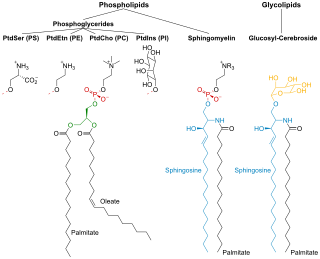
Phospholipids are a class of lipids whose molecule has a hydrophilic "head" containing a phosphate group and two hydrophobic "tails" derived from fatty acids, joined by an alcohol residue. Marine phospholipids typically have omega-3 fatty acids EPA and DHA integrated as part of the phospholipid molecule. The phosphate group can be modified with simple organic molecules such as choline, ethanolamine or serine.

<span class="mw-page-title-main">Lipoprotein</span> Biochemical assembly whose purpose is to transport hydrophobic lipid molecules

A lipoprotein is a biochemical assembly whose primary function is to transport hydrophobic lipid molecules in water, as in blood plasma or other extracellular fluids. They consist of a triglyceride and cholesterol center, surrounded by a phospholipid outer shell, with the hydrophilic portions oriented outward toward the surrounding water and lipophilic portions oriented inward toward the lipid center. A special kind of protein, called apolipoprotein, is embedded in the outer shell, both stabilising the complex and giving it a functional identity that determines its role.

<span class="mw-page-title-main">Lipid bilayer</span> Membrane of two layers of lipid molecules

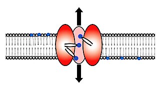
The lipid bilayer is a thin polar membrane made of two layers of lipid molecules. These membranes are flat sheets that form a continuous barrier around all cells. The cell membranes of almost all organisms and many viruses are made of a lipid bilayer, as are the nuclear membrane surrounding the cell nucleus, and membranes of the membrane-bound organelles in the cell. The lipid bilayer is the barrier that keeps ions, proteins and other molecules where they are needed and prevents them from diffusing into areas where they should not be. Lipid bilayers are ideally suited to this role, even though they are only a few nanometers in width, because they are impermeable to most water-soluble (hydrophilic) molecules. Bilayers are particularly impermeable to ions, which allows cells to regulate salt concentrations and pH by transporting ions across their membranes using proteins called ion pumps.

<span class="mw-page-title-main">Peripheral membrane protein</span> Membrane proteins that adhere temporarily to membranes with which they are associated

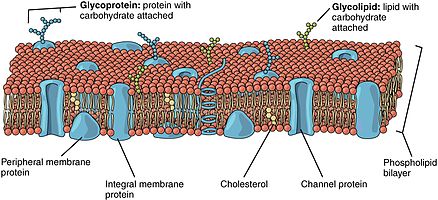
Peripheral membrane proteins, or extrinsic membrane proteins, are membrane proteins that adhere only temporarily to the biological membrane with which they are associated. These proteins attach to integral membrane proteins, or penetrate the peripheral regions of the lipid bilayer. The regulatory protein subunits of many ion channels and transmembrane receptors, for example, may be defined as peripheral membrane proteins. In contrast to integral membrane proteins, peripheral membrane proteins tend to collect in the water-soluble component, or fraction, of all the proteins extracted during a protein purification procedure. Proteins with GPI anchors are an exception to this rule and can have purification properties similar to those of integral membrane proteins.

<span class="mw-page-title-main">Semipermeable membrane</span> Membrane which will allow certain molecules or ions to pass through it by diffusion

Semipermeable membrane is a type of biological or synthetic, polymeric membrane that will allow certain molecules or ions to pass through it by osmosis. The rate of passage depends on the pressure, concentration, and temperature of the molecules or solutes on either side, as well as the permeability of the membrane to each solute. Depending on the membrane and the solute, permeability may depend on solute size, solubility, properties, or chemistry. How the membrane is constructed to be selective in its permeability will determine the rate and the permeability. Many natural and synthetic materials which are rather thick are also semipermeable. One example of this is the thin film on the inside of the egg.

<span class="mw-page-title-main">Fluid mosaic model</span> Describe the fluid mosaic model of plasma membrane

The fluid mosaic model explains various observations regarding the structure of functional cell membranes. According to this biological model, there is a lipid bilayer in which protein molecules are embedded. The phospholipid bilayer gives fluidity and elasticity to the membrane. Small amounts of carbohydrates are also found in the cell membrane. The biological model, which was devised by Seymour Jonathan Singer and Garth L. Nicolson in 1972, describes the cell membrane as a two-dimensional liquid that restricts the lateral diffusion of membrane components. Such domains are defined by the existence of regions within the membrane with special lipid and protein cocoon that promote the formation of lipid rafts or protein and glycoprotein complexes. In addition, it came before the other model that introduced the first bilayer. Another way to define membrane domains is the association of the lipid membrane with the cytoskeleton filaments and the extracellular matrix through membrane proteins. The current model describes important features relevant to many cellular processes, including: cell-cell signaling, apoptosis, cell division, membrane budding, and cell fusion. The fluid mosaic model is the most acceptable model of the plasma membrane. Its main function is to separate the contents of the cell from the exterior.

<span class="mw-page-title-main">Glycolipid</span> Class of chemical compounds

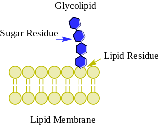
Glycolipids are lipids with a carbohydrate attached by a glycosidic (covalent) bond. Their role is to maintain the stability of the cell membrane and to facilitate cellular recognition, which is crucial to the immune response and in the connections that allow cells to connect to one another to form tissues. Glycolipids are found on the surface of all eukaryotic cell membranes, where they extend from the phospholipid bilayer into the extracellular environment.

<span class="mw-page-title-main">Sphingomyelin</span>

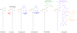
Sphingomyelin is a type of sphingolipid found in animal cell membranes, especially in the membranous myelin sheath that surrounds some nerve cell axons. It usually consists of phosphocholine and ceramide, or a phosphoethanolamine head group; therefore, sphingomyelins can also be classified as sphingophospholipids. In humans, SPH represents ~85% of all sphingolipids, and typically make up 10–20 mol % of plasma membrane lipids.

<span class="mw-page-title-main">Glycerophospholipid</span> Class of lipids

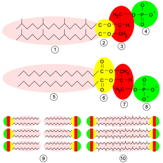
Glycerophospholipids or phosphoglycerides are glycerol-based phospholipids. They are the main component of biological membranes. Two major classes are known: those for bacteria and eukaryotes and a separate family for archaea.

<span class="mw-page-title-main">Amphiphile</span> Hydrophilic and lipophilic chemical compound

An amphiphile, or amphipath, is a chemical compound possessing both hydrophilic and lipophilic (fat-loving) properties. Such a compound is called amphiphilic or amphipathic. Amphiphilic compounds include surfactants. The phospholipid amphiphiles are the major structural component of cell membranes.

<span class="mw-page-title-main">Phospholipid scramblase</span> Protein

Scramblase is a protein responsible for the translocation of phospholipids between the two monolayers of a lipid bilayer of a cell membrane. In humans, phospholipid scramblases (PLSCRs) constitute a family of five homologous proteins that are named as hPLSCR1–hPLSCR5. Scramblases are not members of the general family of transmembrane lipid transporters known as flippases. Scramblases are distinct from flippases and floppases. Scramblases, flippases, and floppases are three different types of enzymatic groups of phospholipid transportation enzymes. The inner-leaflet, facing the inside of the cell, contains negatively charged amino-phospholipids and phosphatidylethanolamine. The outer-leaflet, facing the outside environment, contains phosphatidylcholine and sphingomyelin. Scramblase is an enzyme, present in the cell membrane, that can transport (scramble) the negatively charged phospholipids from the inner-leaflet to the outer-leaflet, and vice versa.

Lipid metabolism is the synthesis and degradation of lipids in cells, involving the breakdown and storage of fats for energy and the synthesis of structural and functional lipids, such as those involved in the construction of cell membranes. In animals, these fats are obtained from food and are synthesized by the liver. Lipogenesis is the process of synthesizing these fats. The majority of lipids found in the human body from ingesting food are triglycerides and cholesterol. Other types of lipids found in the body are fatty acids and membrane lipids. Lipid metabolism is often considered as the digestion and absorption process of dietary fat; however, there are two sources of fats that organisms can use to obtain energy: from consumed dietary fats and from stored fat. Vertebrates use both sources of fat to produce energy for organs such as the heart to function. Since lipids are hydrophobic molecules, they need to be solubilized before their metabolism can begin. Lipid metabolism often begins with hydrolysis, which occurs with the help of various enzymes in the digestive system. Lipid metabolism also occurs in plants, though the processes differ in some ways when compared to animals. The second step after the hydrolysis is the absorption of the fatty acids into the epithelial cells of the intestinal wall. In the epithelial cells, fatty acids are packaged and transported to the rest of the body.

<span class="mw-page-title-main">Surfactin</span> Chemical compound

Surfactin is a cyclic lipopeptide, commonly used as an antibiotic for its capacity as a surfactant. It is an amphiphile capable of withstanding hydrophilic and hydrophobic environments. The Gram-positive bacterial species Bacillus subtilis produces surfactin for its antibiotic effects against competitors. Surfactin showcases antibacterial, antiviral, antifungal, and hemolytic effects.

In biology, membrane fluidity refers to the viscosity of the lipid bilayer of a cell membrane or a synthetic lipid membrane. Lipid packing can influence the fluidity of the membrane. Viscosity of the membrane can affect the rotation and diffusion of proteins and other bio-molecules within the membrane, there-by affecting the functions of these things.

<span class="mw-page-title-main">Membrane lipid</span> Lipid molecules on cell membrane

Membrane lipids are a group of compounds which form the lipid bilayer of the cell membrane. The three major classes of membrane lipids are phospholipids, glycolipids, and cholesterol. Lipids are amphiphilic: they have one end that is soluble in water ('polar') and an ending that is soluble in fat ('nonpolar'). By forming a double layer with the polar ends pointing outwards and the nonpolar ends pointing inwards membrane lipids can form a 'lipid bilayer' which keeps the watery interior of the cell separate from the watery exterior. The arrangements of lipids and various proteins, acting as receptors and channel pores in the membrane, control the entry and exit of other molecules and ions as part of the cell's metabolism. In order to perform physiological functions, membrane proteins are facilitated to rotate and diffuse laterally in two dimensional expanse of lipid bilayer by the presence of a shell of lipids closely attached to protein surface, called annular lipid shell.

One property of a lipid bilayer is the relative mobility (fluidity) of the individual lipid molecules and how this mobility changes with temperature. This response is known as the phase behavior of the bilayer. Broadly, at a given temperature a lipid bilayer can exist in either a liquid or a solid phase. The solid phase is commonly referred to as a “gel” phase. All lipids have a characteristic temperature at which they undergo a transition (melt) from the gel to liquid phase. In both phases the lipid molecules are constrained to the two dimensional plane of the membrane, but in liquid phase bilayers the molecules diffuse freely within this plane. Thus, in a liquid bilayer a given lipid will rapidly exchange locations with its neighbor millions of times a second and will, through the process of a random walk, migrate over long distances.

<span class="mw-page-title-main">Cell membrane</span> Biological membrane that separates the interior of a cell from its outside environment

The cell membrane is a biological membrane that separates and protects the interior of a cell from the outside environment. The cell membrane consists of a lipid bilayer, made up of two layers of phospholipids with cholesterols interspersed between them, maintaining appropriate membrane fluidity at various temperatures. The membrane also contains membrane proteins, including integral proteins that span the membrane and serve as membrane transporters, and peripheral proteins that loosely attach to the outer (peripheral) side of the cell membrane, acting as enzymes to facilitate interaction with the cell's environment. Glycolipids embedded in the outer lipid layer serve a similar purpose. The cell membrane controls the movement of substances in and out of a cell, being selectively permeable to ions and organic molecules. In addition, cell membranes are involved in a variety of cellular processes such as cell adhesion, ion conductivity, and cell signalling and serve as the attachment surface for several extracellular structures, including the cell wall and the carbohydrate layer called the glycocalyx, as well as the intracellular network of protein fibers called the cytoskeleton. In the field of synthetic biology, cell membranes can be artificially reassembled.

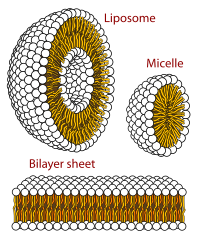
A unilamellar liposome is a spherical liposome, a vesicle, bounded by a single bilayer of an amphiphilic lipid or a mixture of such lipids, containing aqueous solution inside the chamber. Unilamellar liposomes are used to study biological systems and to mimic cell membranes, and are classified into three groups based on their size: small unilamellar liposomes/vesicles (SUVs) that with a size range of 20–100 nm, large unilamellar liposomes/vesicles (LUVs) with a size range of 100–1000 nm and giant unilamellar liposomes/vesicles (GUVs) with a size range of 1-200 µm. GUVs are mostly used as models for biological membranes in research work. Animal cells are 10–30 µm and plant cells are typically 10–100 µm. Even smaller cell organelles such as mitochondria are typically 1-2 µm. Therefore, a proper model should account for the size of the specimen being studied. In addition, the size of vesicles dictates their membrane curvature which is an important factor in studying fusion proteins. SUVs have a higher membrane curvature and vesicles with high membrane curvature can promote membrane fusion faster than vesicles with lower membrane curvature such as GUVs.

Before the emergence of electron microscopy in the 1950s, scientists did not know the structure of a cell membrane or what its components were; biologists and other researchers used indirect evidence to identify membranes before they could actually be visualized. Specifically, it was through the models of Overton, Langmuir, Gorter and Grendel, and Davson and Danielli, that it was deduced that membranes have lipids, proteins, and a bilayer. The advent of the electron microscope, the findings of J. David Robertson, the proposal of Singer and Nicolson, and additional work of Unwin and Henderson all contributed to the development of the modern membrane model. However, understanding of past membrane models elucidates present-day perception of membrane characteristics. Following intense experimental research, the membrane models of the preceding century gave way to the fluid mosaic model that is accepted today.

References

  1. Murate, Motohide; Kobayashi, Toshihide (2016). "Revisiting transbilayer distribution of lipids in the plasma membrane". Chemistry and Physics of Lipids. 194: 58–71. doi:10.1016/j.chemphyslip.2015.08.009. PMID   26319805.
  2. 1 2 Nickels, Jonathan D.; Smith, Jeremy C.; Cheng, Xiaolin (2015). "Lateral organization, bilayer asymmetry, and inter-leaflet coupling of biological membranes". Chemistry and Physics of Lipids. 192: 87–99. doi: 10.1016/j.chemphyslip.2015.07.012 . PMID   26232661.
  3. Chong, Zhi-Soon; Woo, Wei-Fen; Chng, Shu-Sin (2015-12-01). "Osmoporin OmpC forms a complex with MlaA to maintain outer membrane lipid asymmetry in Escherichia coli". Molecular Microbiology. 98 (6): 1133–1146. doi: 10.1111/mmi.13202 . PMID   26314242.
  4. 1 2 Forrest, Lucy R. (2015-01-01). "Structural Symmetry in Membrane Proteins". Annual Review of Biophysics. 44 (1): 311–337. doi:10.1146/annurev-biophys-051013-023008. PMC   5500171 . PMID   26098517.
  5. 1 2 3 4 5 Alberts, Bray, Hopkin, Johnson, Lewis, Raff, Roberts, Walter, Bruce, Dennis, Karen, Alexander, Julian, Martin, Keith, Peter (2010). Essential Cell Biology third edition. New York: Garland Science, Taylor & Francis Group, LLC, an informa business. p. 370. ISBN   978-0815341291.{{cite book}}: CS1 maint: multiple names: authors list (link)
  6. 1 2 3 4 5 6 7 Voet, Donald (2012). Fundamentals of Biochemistry: Life at the Molecular Level (4 ed.). Wiley. ISBN   978-1118129180.
  7. 1 2 Dougherty, R. M.; Galli, C.; Ferro-Luzzi, A.; Iacono, J. M. (1987). "Lipid and phospholipid fatty acid composition of plasma, red blood cells, and platelets and how they are affected by dietary lipids: a study of normal subjects from Italy, Finland, and the USA". The American Journal of Clinical Nutrition. 45 (2): 443–455. doi:10.1093/ajcn/45.2.443. PMID   3812343. S2CID   4436467.
  8. 1 2 Lentz, Barry R. (2003). "Exposure of platelet membrane phosphatidylserine regulates blood coagulation". Progress in Lipid Research. 42 (5): 423–438. doi:10.1016/s0163-7827(03)00025-0. PMID   12814644.
  9. 1 2 Lein, Max; deRonde, Brittany M.; Sgolastra, Federica; Tew, Gregory N.; Holden, Matthew A. (2015-11-01). "Protein transport across membranes: Comparison between lysine and guanidinium-rich carriers". Biochimica et Biophysica Acta (BBA) – Biomembranes. 1848 (11, Part A): 2980–2984. doi:10.1016/j.bbamem.2015.09.004. PMC   4704449 . PMID   26342679.
  10. 1 2 3 Alberts, Bruce; Johnson, Alexander; Lewis, Julian; Raff, Martin; Roberts, Keith; Walter, Peter (2002-01-01). "The Lipid Bilayer".{{cite journal}}: Cite journal requires |journal= (help)
  11. Daubenspeck, James M.; Jordan, David S.; Simmons, Warren; Renfrow, Matthew B.; Dybvig, Kevin (2015-11-23). "General N-and O-Linked Glycosylation of Lipoproteins in Mycoplasmas and Role of Exogenous Oligosaccharide". PLOS ONE. 10 (11): e0143362. Bibcode:2015PLoSO..1043362D. doi: 10.1371/journal.pone.0143362 . PMC   4657876 . PMID   26599081.
  12. Brown, Bernard (1996). Biological Membranes (PDF). London, U.K.: The Biochemical Society. p. 21. ISBN   978-0904498325. Archived from the original (PDF) on 2015-11-06. Retrieved 2014-05-01.
  13. 1 2 Vitrac, Heidi; MacLean, David M.; Jayaraman, Vasanthi; Bogdanov, Mikhail; Dowhan, William (2015-11-10). "Dynamic membrane protein topological switching upon changes in phospholipid environment". Proceedings of the National Academy of Sciences. 112 (45): 13874–13879. Bibcode:2015PNAS..11213874V. doi: 10.1073/pnas.1512994112 . PMC   4653158 . PMID   26512118.
  14. Rojko, Nejc; Anderluh, Gregor (2015-12-07). "How Lipid Membranes Affect Pore Forming Toxin Activity". Accounts of Chemical Research. 48 (12): 3073–3079. doi:10.1021/acs.accounts.5b00403. PMID   26641659.