Blue baby syndrome

Last updated

Blue baby syndrome
Other namesBlue baby, cyanotic infant, cyanotic baby, cyanotic newborn
Cyanotic neonate.jpg
A cyanotic newborn, or "blue baby".
Note the blue coloration of the fingertips.
Specialty Pediatrics, cardiac surgery

Blue baby syndrome can refer to conditions that cause cyanosis, or blueness of the skin, in babies as a result of low oxygen levels in the blood. This term has traditionally been applied to cyanosis as a result of:. [1]

Contents

  1. Cyanotic heart disease, which is a category of congenital heart defect that results in low levels of oxygen in the blood. [2] This can be caused by either reduced blood flow to the lungs or mixing of oxygenated and deoxygenated blood. [2]
  2. Methemoglobinemia, which is a disease defined by high levels of methemoglobin in the blood. Increased levels of methemoglobin prevent oxygen from being released into the tissues and result in hypoxemia. [3]

Although these are the most common causes of cyanosis, there are other potential factors that can cause a blue tint to a baby's skin or mucous membranes. These factors include hypoventilation, perfusion or ventilation differences in the lungs, and poor cardiac output of oxygenated blood, among others. The blue baby syndrome or cyanosis occurs when absolute amount of deoxygenated hemoglobin > 3g/dL which is typically reflected with an O2 saturation of < 85 %. [1]

Both of these conditions cause cyanosis, or a bluish discoloration of skin or mucous membranes. [4] Normally, oxygenated blood appears red and deoxygenated blood has more of a blue appearance. [5] In babies with low levels of oxygen or mixing of oxygenated and deoxygenated blood, the blood can have a blue or purple color, causing cyanosis. [6]

Signs and symptoms

The main sign of blue baby syndrome is cyanosis. Depending on the underlying cause of the cyanosis, additional symptoms may be: [7]

Causes

Blue baby syndrome has been attributed to cyanotic congenital heart diseases and methemoglobinemia, however there are additional causes that could result in a baby becoming cyanotic, such as: [8]

Mechanism

Cyanotic heart disease

Specific types of congenital heart defects that cause blood to pass directly from the right side of the heart to the left side result in cyanosis. [10] In these defects, some of the blood that is pumped to the body has not been oxygenated by the lungs and therefore will appear more blue. Infants with these types of heart defects may have a constant bluish tint to their skin, or they may have temporary episodes of cyanosis. The degree of cyanosis is dependent on how much deoxygenated blood is mixed with oxygenated blood before being pumped to the body.

Cardiac conditions in which there is decreased blood flow to the lungs such as, tetralogy of Fallot or pulmonary valve atresia, result in less blood becoming oxygenated. There are also cardiac conditions such as transposition of the great arteries or truncus arteriosus, that results in an overall increase in blood flow to the lungs but with limited flow of the oxygenated blood to the rest of the body. Conditions in which there is poor blood flow to the systemic circulation, such as coarctation of the aorta suggests that the body does not receive the oxygenated blood it requires with resultant cyanosis. [11]

The five most common cyanotic heart defects that may result in Blue Baby Syndrome include the following:

PathologyBrief DescriptionEpidemiologyDiagram
1. Persistent (or patent) truncus arteriosus Instead of two separate major blood vessels leaving the heart, there is one common outgoing vessel. [12] 3-5/100,000 births [13] Truncus arteriosus.jpg
2. Transposition of the great vessels The positions of the pulmonary artery and the aorta are switched, with the aorta connecting to the right ventricle and the pulmonary artery connecting to the left ventricle. [14] 1-5/10,000 births [15] D-tga-575px.jpg
3. Tricuspid atresia The heart valve connecting the right atrium to the right ventricle does not form properly, disrupting blood flow in the heart. [16] 1-9/100,000 births [17] Tricuspid atresia.svg
4. Tetralogy of Fallot A heart defect with 4 features including a narrow pulmonary artery, a thick right ventricle, an aorta that connects to both the right and left ventricles, and a hole in the ventricular wall. [18] 1-5/10,000 births [19] Teralogy web.jpg
5. Anomalous pulmonary venous connection The pulmonary veins returning oxygenated blood from the lungs do not connect properly to the heart. [20] 6-12/100,000 births [21] Tapv-575px.jpg

Methemoglobinemia

Hemoglobin oxygenation dissociation curve. In the case of methemoglobinemia the curve is shifted to the left given the higher affinity for oxygen. Oxyhaemoglobin dissociation curve.png
Hemoglobin oxygenation dissociation curve. In the case of methemoglobinemia the curve is shifted to the left given the higher affinity for oxygen.

Methemoglobinemia can be acquired or congenital. It occurs when the iron in hemoglobin is oxidized from Fe2+ to Fe3+, leading to poor binding of oxygen. Additionally, the oxygen that is already bound is held more tightly to the hemoglobin due to a higher affinity, resulting in less oxygen delivery. A methemoglobin level > 1.5 g/dL causes cyanosis. The most common congenital cause is a deficiency in the enzyme cytochrome b5 reductase which reduces methemoglobin in the blood. [22]

However, in infants the most common cause of methemoglobinemia is acquired through the ingestion of nitrates (NO3) through well water or foods. Nitrites (NO2) produced by the microbial reduction of nitrate (directly in the drinkwater, or after ingestion by the infant, in his digestive system) are more powerful oxidizers than nitrates and are the chemical agent really responsible of the oxidation of Fe2+ into Fe3+ in the tetrapyrrole heme of hemoglobin. Infants younger than 4 months are at greater risk given that they drink more water per body weight, they have a lower NADH- cytochrome b5 reductase activity, and they have a higher level of fetal hemoglobin which converts more easily to methemoglobin. Additionally, infants are at an increased risk after an episode of gastroenteritis due to the production of nitrites by bacteria. [22] The sources of nitrate can include fertilizers used in agricultural lands, waste dumps or pit latrines. [23] For example, nitrate levels are subject to monitoring to comply with drinking water quality standards in the United States and other countries. [24] [25] The link between blue baby syndrome and nitrates in drinking water is widely accepted, but as of 2006 some studies indicated that other contaminants or dietary nitrate sources, might also play a role in the syndrome. [26] [27] [28]

Diagnosis

When diagnosing blue baby syndrome, it is important to perform a thorough history and physical exam. When obtaining the history, it is important to determine the timing of symptoms and to ask about risk factors/exposures, such as prenatal history or access to well-water. [29]

Pulse oximeter on infant's foot. Pulse Oximeter.jpg
Pulse oximeter on infant's foot.

On physical exam it is important to visualize where the cyanosis is present to differentiate between peripheral and central cyanosis. Central cyanosis is typically visible as a blueish discoloration over the entire body and mucous membranes. In contrast, peripheral cyanosis typically has a blueish discoloration over the extremities. Cyanosis can be noted in babies around the lips, tongue, and sublingual area, where the skin is thinnest. [30] In addition, it is important to observe the infant for signs of respiratory distress, visualized as nasal flaring, subcostal retractions, etc. Examination should include a respiratory and cardiac assessment. [29]

One of the key tools in diagnosing is a pulse oximeter to determine oxygen saturation. While severe cyanosis can be easily noticed, an oxygen saturation as low as 80 % causes only mild clinical cyanosis that is difficult to see. [6] Additionally an arterial blood gas is useful, for example in the case of methemoglobinemia the PO2 can be expected to be normal even with a low oxygen saturation. Additional work up includes a complete blood count, blood glucose, blood culture, chest X-ray, and an echocardiography. [29]

Babies with cyanosis due to congenital heart disease usually present with symptoms hours to days after birth. In addition to cyanosis, they often show signs of tachypnea (fast breathing), a heart murmur, and decreased peripheral pulses. [6] [31] If congenital heart disease is suspected in a newborn, doctors will likely perform several tests to evaluate the heart, including a chest x-ray, echocardiogram, and electrocardiogram. [32] In tetralogy of Fallot, episodes in which infants become cyanotic are called tet spells, typically occurring during feeding or crying. [18] When older, children may squat to feel relief since this increases the systemic vascular resistance causing more blood to go towards the lungs, resulting in increased oxygenation. [18]

Babies with cyanosis due to methemoglobinemia also usually present with cyanosis in the neonatal period, but pulse oximetry may be falsely elevated and does not always reveal low blood oxygen saturation. A CO-oximeter can be used to detect levels of methemoglobin in the blood if methemoglobinemia is suspected, by seeing the difference between oxygen saturation on an arterial blood gas and the measurement on a co-oximeter. Additionally, a direct methemoglobin level can be obtained. [33]

Prevention/Screening

As of 22 May 2007, the United States Environmental Protection Agency has established a maximum contaminant level of 10 mg/L for nitrate and 1 mg/L for nitrite in drinking water due to the potential harmful effects in infants. [34]

A screening tool has been developed to screen for critical cardiac defects, which refers to cardiac lesions that require surgery or intervention in the 1st year of life. Screening for critical congenital heart defects should be done on all newborns after 24hours or shortly before discharge. Oxygen saturation is measured in the right hand and either foot. [35]

A screening is considered positive if:

Management

Treatment for blue baby syndrome will depend on the underlying cause.

When evaluating a patient for cyanosis or respiratory distress, vital signs should be monitored, especially the patient's heart rate and oxygen saturation. It is beneficial to have vascular access established. In newborns, the pulse oximeter is typically placed on the right hand to determine pre-ductal oxygenation, referring to oxygenation before the ductus arteriosus (connection between aorta and pulmonary artery). [36] This gives the oxygenation level the heart and brain receive. Traditionally, supplemental oxygen is given in an escalating manner beginning with free-flowing oxygen, progressing to positive pressure ventilation or continuous positive airway pressure, and ending with mechanical intubation. The goal oxygen saturation is between 85 and 95 %. If an infant requires supplemental oxygen for a prolonged time it should be heated and humidified to avoid heat loss. [36]

Cyanotic heart disease

Some babies born with cyanotic heart disease are treated with prostaglandin E1 after birth to keep the ductus arteriosus open and allow for more oxygenated blood to be pumped to the body. Many also receive oxygen therapy to increase the percentage of oxygen in the blood. Most of these babies will require surgery during infancy to correct their structural heart defect. [31]

Severe methemoglobinemia

The first-line treatment for severe methemoglobinemia is methylene blue, a medication that will reduce methemoglobin in the blood. This is possible because methylene blue oxidizes NADPH, which in turn can convert methemoglobin back to hemoglobin. [33]

Epidemiology

Out of all the babies born with congenital heart defects, about 25 % have cyanosis as a result. Tetralogy of Fallot is the most common cyanotic cardiac heart defect. [37]

Methemoglobinemia is considered to be rare, with acquired methemoglobinemia encountered more than the congenital form. [38]

Outcomes

In the case of cyanotic causing heart defects, about 75 % of infants survive to 1 year of age and 69 % survive to 18 years of age. These individuals have an increased risk of developmental delay, heart failure, or heart rhythm disorders. [37]

Methemoglobinemia responds well to treatment, its prognosis is associated with the level of methemoglobinemia and the degree of end organ damage it can cause. Death can occur when levels reach 70 %. [39]

History

The first successful operation to treat blue baby syndrome caused by tetralogy of Fallot occurred at Johns Hopkins University in 1944. Through a collaboration between pediatric cardiologist Helen Taussig, surgeon Alfred Blalock, and surgical technician Vivien Thomas, the Blalock-Thomas-Taussig shunt was created. Dr. Taussig had recognized that children with Tetralogy of Fallot who also had a patent ductus arteriosus (PDA) typically lived longer, so the trio tried to create the same effect as a PDA by joining the subclavian artery to the pulmonary artery, relieving the child's cyanosis. [40] The operation was published in the Journal of the American Medical Association in 1945 and impacted management of blue babies around the world. [41]

Anna was the name of the dog who was the first survivor of the surgery, considered an experimental procedure at the time. Anna survived the first pulmonary bypass after having been operated on twice. The second operation was required to replace the original stitches with flexible ones. After their success with Anna, Blalock and Thomas had the courage to perform the very first open heart surgery on Eileen Saxon in 1944. In 1950, Anna's story was made into a movie, and the film has been shown to various schools and other groups. [42]

Related Research Articles

<span class="mw-page-title-main">Tetralogy of Fallot</span> Type of congenital heart defect

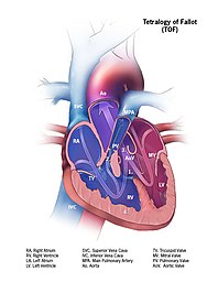
Tetralogy of Fallot (TOF), formerly known as Steno-Fallot tetralogy, is a congenital heart defect characterized by four specific cardiac defects. Classically, the four defects are:

<span class="mw-page-title-main">Methemoglobinemia</span> Condition of elevated methemoglobin in the blood

Methemoglobinemia, or methaemoglobinaemia, is a condition of elevated methemoglobin in the blood. Symptoms may include headache, dizziness, shortness of breath, nausea, poor muscle coordination, and blue-colored skin (cyanosis). Complications may include seizures and heart arrhythmias.

<span class="mw-page-title-main">Cyanosis</span> Decreased oxygen in the blood

Cyanosis is the change of body tissue color to a bluish-purple hue, as a result of decrease in the amount of oxygen bound to the hemoglobin in the red blood cells of the capillary bed. Cyanosis is apparent usually in the body tissues covered with thin skin, including the mucous membranes, lips, nail beds, and ear lobes. Some medications may cause discoloration such as medications containing amiodarone or silver. Furthermore, mongolian spots, large birthmarks, and the consumption of food products with blue or purple dyes can also result in the bluish skin tissue discoloration and may be mistaken for cyanosis. Appropriate physical examination and history taking is a crucial part to diagnose cyanosis. Management of cyanosis involves treating the main cause, as cyanosis isn’t a disease, it is a symptom.

<span class="mw-page-title-main">Patent ductus arteriosus</span> Condition wherein the ductus arteriosus fails to close after birth

Patent ductus arteriosus (PDA) is a medical condition in which the ductus arteriosus fails to close after birth: this allows a portion of oxygenated blood from the left heart to flow back to the lungs through the aorta, which has a higher blood pressure, to the pulmonary artery, which has a lower blood pressure. Symptoms are uncommon at birth and shortly thereafter, but later in the first year of life there is often the onset of an increased work of breathing and failure to gain weight at a normal rate. With time, an uncorrected PDA usually leads to pulmonary hypertension followed by right-sided heart failure.

dextro-Transposition of the great arteries Medical condition

dextro-Transposition of the great arteries is a potentially life-threatening birth defect in the large arteries of the heart. The primary arteries are transposed.

<span class="mw-page-title-main">Congenital heart defect</span> Defect in the structure of the heart that is present at birth

A congenital heart defect (CHD), also known as a congenital heart anomaly, congenital cardiovascular malformation, and congenital heart disease, is a defect in the structure of the heart or great vessels that is present at birth. A congenital heart defect is classed as a cardiovascular disease. Signs and symptoms depend on the specific type of defect. Symptoms can vary from none to life-threatening. When present, symptoms are variable and may include rapid breathing, bluish skin (cyanosis), poor weight gain, and feeling tired. CHD does not cause chest pain. Most congenital heart defects are not associated with other diseases. A complication of CHD is heart failure.

A cyanotic heart defect is any congenital heart defect (CHD) that occurs due to deoxygenated blood bypassing the lungs and entering the systemic circulation, or a mixture of oxygenated and unoxygenated blood entering the systemic circulation. It is caused by structural defects of the heart such as right-to-left or bidirectional shunting, malposition of the great arteries, or any condition which increases pulmonary vascular resistance. The result may be the development of collateral circulation.

<span class="mw-page-title-main">Methemoglobin</span> Type of hemoglobin

Methemoglobin (British: methaemoglobin, shortened MetHb) (pronounced "met-hemoglobin") is a hemoglobin in the form of metalloprotein, in which the iron in the heme group is in the Fe3+ (ferric) state, not the Fe2+ (ferrous) of normal hemoglobin. Sometimes, it is also referred to as ferrihemoglobin. Methemoglobin cannot bind oxygen, which means it cannot carry oxygen to tissues. It is bluish chocolate-brown in color. In human blood a trace amount of methemoglobin is normally produced spontaneously, but when present in excess the blood becomes abnormally dark bluish brown. The NADH-dependent enzyme methemoglobin reductase (a type of diaphorase) is responsible for converting methemoglobin back to hemoglobin.

<span class="mw-page-title-main">Eisenmenger syndrome</span> Medical condition

Eisenmenger syndrome or Eisenmenger's syndrome is defined as the process in which a long-standing left-to-right cardiac shunt caused by a congenital heart defect causes pulmonary hypertension and eventual reversal of the shunt into a cyanotic right-to-left shunt. Because of the advent of fetal screening with echocardiography early in life, the incidence of heart defects progressing to Eisenmenger syndrome has decreased.

<span class="mw-page-title-main">Blalock–Thomas–Taussig shunt</span> Cardiac surgery procedure

The Blalock–Thomas–Taussig shunt, previously known as the Blalock-Taussig Shunt, is a surgical procedure used to increase blood flow to the lungs in some forms of congenital heart disease such as pulmonary atresia and tetralogy of Fallot, which are common causes of blue baby syndrome. The procedure involves connecting a branch of the subclavian artery or carotid artery to the pulmonary artery. In modern practice, this procedure is temporarily used to direct blood flow to the lungs and relieve cyanosis while the infant is waiting for corrective or definitive surgery when their heart is larger. The BTT shunt is used in the first step of the three-stage palliation.

<span class="mw-page-title-main">Helen B. Taussig</span> American cardiologist (1898–1986)

Helen Brooke Taussig was an American cardiologist, working in Baltimore and Boston, who founded the field of pediatric cardiology. She is credited with developing the concept for a procedure that would extend the lives of children born with Tetralogy of Fallot. This concept was applied in practice as a procedure known as the Blalock-Thomas-Taussig shunt. The procedure was developed by Alfred Blalock and Vivien Thomas, who were Taussig's colleagues at the Johns Hopkins Hospital.

<span class="mw-page-title-main">Hypoplastic left heart syndrome</span> Type of congenital heart defect

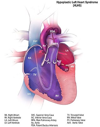
Hypoplastic left heart syndrome (HLHS) is a rare congenital heart defect in which the left side of the heart is severely underdeveloped and incapable of supporting the systemic circulation. It is estimated to account for 2-3% of all congenital heart disease. Early signs and symptoms include poor feeding, cyanosis, and diminished pulse in the extremities. The etiology is believed to be multifactorial resulting from a combination of genetic mutations and defects resulting in altered blood flow in the heart. Several structures can be affected including the left ventricle, aorta, aortic valve, or mitral valve all resulting in decreased systemic blood flow.

<span class="mw-page-title-main">Transposition of the great vessels</span> Group of congenital heart defects

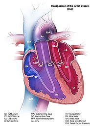
Transposition of the great vessels (TGV) is a group of congenital heart defects involving an abnormal spatial arrangement of any of the great vessels: superior and/or inferior venae cavae, pulmonary artery, pulmonary veins, and aorta. Congenital heart diseases involving only the primary arteries belong to a sub-group called transposition of the great arteries (TGA), which is considered the most common congenital heart lesion that presents in neonates.

<span class="mw-page-title-main">Tricuspid atresia</span> Medical condition

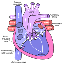
Tricuspid atresia is a form of congenital heart disease whereby there is a complete absence of the tricuspid valve. Therefore, there is an absence of right atrioventricular connection. This leads to a hypoplastic (undersized) or absent right ventricle. This defect is contracted during prenatal development, when the heart does not finish developing. It causes the systemic circulation to be filled with relatively deoxygenated blood. The causes of tricuspid atresia are unknown.

<span class="mw-page-title-main">Atrioventricular septal defect</span> Medical condition

Atrioventricular septal defect (AVSD) or atrioventricular canal defect (AVCD), also known as "common atrioventricular canal" or "endocardial cushion defect" (ECD), is characterized by a deficiency of the atrioventricular septum of the heart that creates connections between all four of its chambers. It is a very specific combination of 3 defects:

<span class="mw-page-title-main">Fetal circulation</span> Circulatory system of fetuses

In humans, the circulatory system is different before and after birth. The fetal circulation is composed of the placenta, umbilical blood vessels encapsulated by the umbilical cord, heart and systemic blood vessels. A major difference between the fetal circulation and postnatal circulation is that the lungs are not used during the fetal stage resulting in the presence of shunts to move oxygenated blood and nutrients from the placenta to the fetal tissue. At birth, the start of breathing and the severance of the umbilical cord prompt various changes that quickly transform fetal circulation into postnatal circulation.

A right-to-left shunt is a cardiac shunt which allows blood to flow from the right heart to the left heart. This terminology is used both for the abnormal state in humans and for normal physiological shunts in reptiles.

<span class="mw-page-title-main">Hypoplastic right heart syndrome</span> Type of congenital heart disease

Hypoplastic right heart syndrome or HRHS is a congenital heart defect in which the structures on the right side of the heart, particularly the right ventricle, are underdeveloped. This defect causes inadequate blood flow to the lungs, and thus a cyanotic infant.

<span class="mw-page-title-main">Hemoglobin M disease</span> Medical condition

Hemoglobin M disease is a rare form of hemoglobinopathy, characterized by the presence of hemoglobin M (HbM) and elevated methemoglobin (metHb) level in blood. HbM is an altered form of hemoglobin (Hb) due to point mutation occurring in globin-encoding genes, mostly involving tyrosine substitution for proximal (F8) or distal (E7) histidine residues. HbM variants are inherited as autosomal dominant disorders and have altered oxygen affinity. The pathophysiology of hemoglobin M disease involves heme iron autoxidation promoted by heme pocket structural alteration.

Raghib syndrome is rare a congenital heart defect where the left superior vena cava (LSVC) is draining into the left atrium in addition to an absent coronary sinus and an atrial septal defect. This can be considered a dangerous heart condition because it puts the individual at a high risk of stroke. Other defects that are often associated with Raghib syndrome can include ventricular septal defects, enlargement of the tricuspid annulus, and pulmonary stenosis. While this is considered an extremely rare developmental complex, cases regarding a persistent left superior vena cava (PLSVC) are relatively common among congenital heart defects. It is also important to note that the PLSVC often drains into the right atrium, and only drains into the left atrium in approximately 10 to 20% of individuals with the defect.

References

  1. 1 2 Pahal, Parul; Goyal, Amandeep (2024). "Central and Peripheral Cyanosis". StatPearls. StatPearls Publishing. PMID   32644593.
  2. 1 2 MedlinePlus Encyclopedia : Cyanotic heart disease
  3. MedlinePlus Encyclopedia : Methemoglobinemia
  4. Snider, H. L. (1990). "Cyanosis". Clinical Methods: The History, Physical, and Laboratory Examinations. Butterworths. ISBN   978-0-409-90077-4. PMID   21250208.
  5. MedlinePlus Encyclopedia : Blue discoloration of the skin
  6. 1 2 3 Silove, E D (1994). "Assessment and management of congenital heart disease in the newborn by the district paediatrician". Archives of Disease in Childhood - Fetal and Neonatal Edition. 70 (1): F71–F74. doi:10.1136/fn.70.1.f71. PMC   1060995 . PMID   8117134.
  7. Enlow, Elizabeth; Greenberg, James M. (2019). "Clinical Manifestations of Diseases in the Newborn Period". In Kliegman, Robert M.; St Geme, Joseph W. (eds.). Nelson Textbook of Pediatrics. Elsevier Health Sciences. pp. 910–913.e4. ISBN   978-0-323-56888-3.
  8. Stack, Anne M (2006). "Cyanosis". In Fleisher, Gary Robert; Ludwig, Stephen; Henretig, Fred M (eds.). Textbook of Pediatric Emergency Medicine. Lippincott Williams & Wilkins. ISBN   978-0-7817-5074-5.
  9. MedlinePlus Encyclopedia : Neonatal respiratory distress syndrome
  10. Lin, Pei-Yi; Hagan, Katherine; Fenoglio, Angela; Grant, P. Ellen; Franceschini, Maria Angela (16 May 2016). "Reduced cerebral blood flow and oxygen metabolism in extremely preterm neonates with low-grade germinal matrix- intraventricular hemorrhage". Scientific Reports. 6 (1): 25903. Bibcode:2016NatSR...625903L. doi:10.1038/srep25903. PMC   4867629 . PMID   27181339.
  11. Bernstein, Daniel. Nelson Textbook of Pediatrics. Elsevier Inc. pp. 2371–2373.
  12. "Facts about Truncus Arteriosus". Congenital Heart Defects. Centers for Disease Control and Prevention. 22 November 2019.
  13. Reserved, INSERM US14 – All Rights. "Orphanet: Truncus arteriosus". www.orpha.net. Retrieved 20 November 2019.{{cite web}}: CS1 maint: numeric names: authors list (link)
  14. CDC (15 November 2019). "Congenital Heart Defects – dextro-Transposition of the Great Arteries". Centers for Disease Control and Prevention. Retrieved 20 November 2019.
  15. Reserved, INSERM US14 – All Rights. "Orphanet: Transposition of the great arteries". www.orpha.net. Retrieved 20 November 2019.{{cite web}}: CS1 maint: numeric names: authors list (link)
  16. CDC (19 November 2019). "Congenital Heart Defects – Facts about Tricuspid Atresia | CDC". Centers for Disease Control and Prevention. Retrieved 20 November 2019.
  17. Reserved, INSERM US14 – All Rights. "Orphanet: Tricuspid atresia". www.orpha.net. Retrieved 20 November 2019.{{cite web}}: CS1 maint: numeric names: authors list (link)
  18. 1 2 3 "Facts about Tetralogy of Fallot". Congenital Heart Defects. Centers for Disease Control and Prevention. 3 February 2023.
  19. Reserved, INSERM US14 – All Rights. "Orphanet: Tetralogy of Fallot". www.orpha.net. Retrieved 20 November 2019.{{cite web}}: CS1 maint: numeric names: authors list (link)
  20. CDC (19 November 2019). "Congenital Heart Defects – Facts about TAVPR | CDC". Centers for Disease Control and Prevention. Retrieved 20 November 2019.
  21. "Total Anomalous Pulmonary Venous Return". The Pediatric Cardiac Anesthesia Handbook. 2017. pp. 107–111. doi:10.1002/9781119095569.ch16. ISBN   978-1-119-09553-8.
  22. 1 2 3 Smith-Whitley, Kwiatkowski. Nelson Textbook of Pediatrics. Elsevier Inc. pp. 2540–2558.
  23. Majumdar Deepanjan (2003). "The Blue Baby Syndrome". Resonance. 8 (10): 20–30. doi:10.1007/BF02840703. S2CID   117099503.
  24. "National Primary Drinking Water Regulations". EPA: United States Environmental Protection Agency. 30 November 2015. Retrieved 14 July 2018.
  25. "Water-related diseases". World Health Organization. Archived from the original on 21 April 2017. Retrieved 14 July 2018.
  26. Fewtrell, Lorna (22 July 2004). "Drinking-Water Nitrate, Methemoglobinemia, and Global Burden of Disease: A Discussion". Environmental Health Perspectives. 112 (14): 1371–1374. doi:10.1289/ehp.7216. PMC   1247562 . PMID   15471727.
  27. van Grinsven, Hans JM; Ward, Mary H = 2006 (2006). "Does the evidence about health risks associated with nitrate ingestion warrant an increase of the nitrate standard for drinking water?". Environ Health. 5 (1): 26. Bibcode:2006EnvHe...5...26V. doi: 10.1186/1476-069X-5-26 . PMC   1586190 . PMID   16989661.{{cite journal}}: CS1 maint: numeric names: authors list (link)
  28. Ward, Mary H.; deKok, Theo M.; Levallois, Patrick; Brender, Jean; Gulis, Gabriel; Nolan, Bernard T.; VanDerslice, James (23 June 2005). "Workgroup Report: Drinking-Water Nitrate and Health—Recent Findings and Research Needs". Environmental Health Perspectives. 113 (11): 1607–1614. doi:10.1289/ehp.8043. PMC   1310926 . PMID   16263519.
  29. 1 2 3 Bearl, David W.; Hill, Kevin (2019), Jain, Lucky; Suresh, Gautham K. (eds.), "Approach to the Cyanotic Infant", Clinical Guidelines in Neonatology, New York, NY: McGraw-Hill Education, retrieved 21 January 2022
  30. McMullen, Sarah M.; Patrick, Ward (March 2013). "Cyanosis". The American Journal of Medicine. 126 (3): 210–212. doi:10.1016/j.amjmed.2012.11.004. PMID   23410559. S2CID   244083635.
  31. 1 2 Khalil, Markus; Jux, Christian; Rueblinger, Lucie; Behrje, Johanna; Esmaeili, Anoosh; Schranz, Dietmar (April 2019). "Acute therapy of newborns with critical congenital heart disease". Translational Pediatrics. 8 (2): 114–126. doi: 10.21037/tp.2019.04.06 . PMC   6514285 . PMID   31161078.
  32. "Symptoms and Diagnosis of Congenital Heart Defects". www.heart.org. Retrieved 13 November 2019.
  33. 1 2 Da-Silva, Shonola S.; Sajan, Imran S.; Underwood, Joseph P. (August 2003). "Congenital Methemoglobinemia: A Rare Cause of Cyanosis in the Newborn—A Case Report". Pediatrics. 112 (2): e158–e161. doi: 10.1542/peds.112.2.e158 . PMID   12897322.
  34. "US EPA Archive Document- Nitrates and Nitrites" (PDF). US EPA. 22 May 2007. Archived (PDF) from the original on 30 April 2022. Retrieved 2 March 2023.
  35. "Critical Congenital Heart Defects". CDC. Archived from the original on 11 September 2014.
  36. 1 2 American Academy of Pediatrics and American Heart Association (2016). Textbook of Neonatal Resuscitation. American Academy of Pediatrics. pp. 33–65.
  37. 1 2 Ossa Galvis, Maria M.; Bhakta, Rupal T.; Tarmahomed, Abdulla; Mendez, Magda D. (2024). "Cyanotic Heart Disease". StatPearls. StatPearls Publishing. PMID   29763177.
  38. Ludlow, John T.; Wilkerson, Richard G.; Nappe, Thomas M. (2024). "Methemoglobinemia". StatPearls. StatPearls Publishing. PMID   30726002.
  39. Methemoglobinemia at eMedicine
  40. Thomas, Vivien T. (1998). Partners of the Heart: Vivien Thomas and His Work with Alfred Blalock. University of Pennsylvania Press, Incorporated. ISBN   978-0-8122-1634-9.[ page needed ]
  41. "That First Operation". Johns Hopkins Medical Institutions. Archived from the original on 12 June 2020. Retrieved 13 November 2019.
  42. Smith, Chris. "Information on Anna". Archived from the original on 31 March 2016. Retrieved 22 July 2016.