Patent ductus arteriosus

Last updated

Patent ductus arteriosus
Other namesPersistent ductus arteriosus
Patent ductus arteriosus.svg
Diagram of a cross-section through a heart with PDA
Specialty Cardiac surgery, paediatrics
Symptoms Shortness of breath, failure to thrive, tachycardia, heart murmur
Complications Heart failure, Eisenmenger's syndrome, pulmonary hypertension
Causes Idiopathic
Risk factors Preterm birth, congenital rubella syndrome, chromosomal abnormalities, genetic conditions
Diagnostic method Echocardiography, Doppler, X-ray
Prevention Screening at birth, high index of suspicion in neonates at risk
Treatment Nonsteroidal anti-inflammatory drugs (NSAIDs), surgery

Patent ductus arteriosus (PDA) is a medical condition in which the ductus arteriosus fails to close after birth: this allows a portion of oxygenated blood from the left heart to flow back to the lungs through the aorta, which has a higher blood pressure, to the pulmonary artery, which has a lower blood pressure. Symptoms are uncommon at birth and shortly thereafter, but later in the first year of life there is often the onset of an increased work of breathing and failure to gain weight at a normal rate. With time, an uncorrected PDA usually leads to pulmonary hypertension followed by right-sided heart failure.

Contents

The ductus arteriosus is a fetal blood vessel that normally closes soon after birth. This closure is caused by vessel constriction immediately after birth as circulation changes occur, followed by the occlusion of the vessel’s lumen in the following days. [1] In a PDA, the vessel does not close, but remains patent (open), resulting in an abnormal transmission of blood from the aorta to the pulmonary artery. PDA is common in newborns with persistent respiratory problems such as hypoxia, and has a high occurrence in premature newborns. Premature newborns are more likely to be hypoxic and have PDA due to underdevelopment of the heart and lungs.

If the congenital defect transposition of the great vessels is present in addition to a PDA, the PDA is not surgically closed since it is the only way that oxygenated blood can mix with deoxygenated blood. In these cases, prostaglandins are used to keep the PDA open, and NSAIDs are not administered until surgical correction of the two defects is completed.

In full-term newborns, PDA occurs in 1 in 2,000 births, and accounts for 5–10% of congenital heart disease cases. PDA occurs in 20–60% of all premature newborns, where its incidence inversely linked with gestational age and weight. [2]

Signs and symptoms

Common symptoms include:[ citation needed ]

Signs include:[ citation needed ]

People with patent ductus arteriosus typically present in good health, with normal respirations and heart rate. If the PDA is moderate or large, widened pulse pressure and bounding peripheral pulses are frequently present, reflecting increased left ventricular stroke volume and diastolic run-off of blood into the (initially lower-resistance) pulmonary vascular bed. [4] Eisenmenger physiology is pulmonary hypertension due to a left-to-right shunt. Prominent suprasternal and carotid pulsations may be noted secondary to increased left ventricular stroke volume. [5]

Risk factors

Known risk factors include: [6]

Diagnosis

Phonocardiograms from normal and abnormal heart sounds Phonocardiograms from normal and abnormal heart sounds.svg
Phonocardiograms from normal and abnormal heart sounds

PDA is usually diagnosed using noninvasive techniques. Echocardiography (in which sound waves are used to capture the motion of the heart) and associated Doppler studies are the primary methods of detecting PDA. Electrocardiography (ECG), in which electrodes are used to record the electrical activity of the heart, is not particularly helpful as no specific rhythms or ECG patterns can be used to detect PDA. [7]

A chest X-ray may be taken, which reveals overall heart size (as a reflection of the combined mass of the cardiac chambers) and the appearance of blood flow to the lungs. A small PDA most often accompanies a normal-sized heart and normal blood flow to the lungs. A large PDA generally accompanies an enlarged cardiac silhouette and increased blood flow to the lungs.[ citation needed ]

Prevention

Some evidence suggests that intravenous NSAIDs, such as indomethacin, administration on the first day of life to all preterm infants reduces the risk of developing a PDA and the complications associated with PDA. [8] Intravenous Indomethacin treatment in premature infants also may reduce the need for surgical intervention. [8] Administering ibuprofen probably helps to prevent PDA and reduce the need for surgery but it also likely increases the risk of kidney complications. [9]

Treatment

Symptomatic PDA can be treated with both surgical and non-surgical methods. [10]

Conservative

Neonates without adverse symptoms may simply be monitored as outpatients.[ citation needed ]

Surgery

Surgically, the DA may be closed by ligation (though support in premature infants is mixed). [11] This can either be performed manually and be tied shut, or with intravascular coils or plugs that leads to formation of a thrombus in the DA.[ citation needed ]

Devices developed by Franz Freudenthal block the blood vessel with woven structures of nitinol wire. [12] Newer procedures performed effectively in older, bigger children include catheter PDA occlusion and video-assisted thoracoscopic PDA clipping. [13]

Prostaglandin inhibitors

Because prostaglandin E2 is responsible for keeping the DA open, NSAIDs (which can inhibit prostaglandin synthesis) such as indomethacin or a special form of ibuprofen have been suggested as therapy to initiate PDA closure. [3] [14] [15] Findings from a 2015 systematic review concluded that, for closure of a PDA in preterm and/or low birth weight infants, ibuprofen is as effective as indomethacin. It also causes fewer side effects (such as transient acute kidney injury) and reduces the risk of necrotising enterocolitis. [16] The evidence supporting the effectiveness and safety of paracetamol (acetaminophen) is less clear. [17] A review and meta-analysis showed that paracetamol may be effective for closure of a PDA in preterm infants. [18] A 2018 network meta-analysis that compared indomethacin, paracetamol and ibuprofen at different doses and administration schemes among them found that a high dose of oral ibuprofen may offer the highest likelihood of closure in preterm infants. [19] [20] [21] However, a 2020 systematic review found that early (≤7 days of life) or very early (≤72 hours of life) pharmacological treatment of symptomatic PDA does not reduce death or other poor clinical outcomes in preterm infants but instead increases their exposure to NSAIDS. [22] Vasodilator therapy is suitable for people with Eisenmenger physiology. To assess improvement in people with Eisenmenger physiology, close monitory of toe oxygen saturation is required, for there exists a chance of reversal after a successful right-to-left shunt [ citation needed ]

While indometacin can be used to close a PDA, some neonates require their PDA be kept open. Keeping a ductus arteriosus patent is indicated in neonates born with concurrent heart malformations, such as transposition of the great vessels. Drugs such as alprostadil, a PGE-1 analog, can be used to keep a PDA open until the primary defect is corrected surgically.[ citation needed ]

Prognosis

If left untreated, the disease may progress from left-to-right shunt (acyanotic heart) to right-to-left shunt (cyanotic heart), called Eisenmenger's syndrome. Pulmonary hypertension is a potential long-term outcome, which may require a heart and/or lung transplant. Another complication of PDA is intraventricular hemorrhage.[ citation needed ]

History

Robert Edward Gross, MD performed the first successful ligation of a patent ductus arteriosus on a seven-year-old girl at Children's Hospital Boston in 1938. [23]

Adult

Since PDA is usually identified in infants, it is less common in adults, but it can have serious consequences, and is usually corrected surgically upon diagnosis.[ citation needed ]

See also

Related Research Articles

<span class="mw-page-title-main">Paracetamol</span> Common medication for pain and fever

Paracetamol is a non-opioid analgesic and antipyretic agent used to treat fever and mild to moderate pain. It is a widely used over the counter medication. Common brand names include Tylenol and Panadol.

<span class="mw-page-title-main">Preterm birth</span> Birth at less than a specified gestational age

Preterm birth, also known as premature birth, is the birth of a baby at fewer than 37 weeks gestational age, as opposed to full-term delivery at approximately 40 weeks. Extreme preterm is less than 28 weeks, very early preterm birth is between 28 and 32 weeks, early preterm birth occurs between 32 and 34 weeks, late preterm birth is between 34 and 36 weeks' gestation. These babies are also known as premature babies or colloquially preemies or premmies. Symptoms of preterm labor include uterine contractions which occur more often than every ten minutes and/or the leaking of fluid from the vagina before 37 weeks. Premature infants are at greater risk for cerebral palsy, delays in development, hearing problems and problems with their vision. The earlier a baby is born, the greater these risks will be.

dextro-Transposition of the great arteries Medical condition

dextro-Transposition of the great arteries is a potentially life-threatening birth defect in the large arteries of the heart. The primary arteries are transposed.

<span class="mw-page-title-main">Ductus arteriosus</span> Blood vessel connecting the pulmonary artery to the proximal descending aorta

The ductus arteriosus, also called the ductus Botalli, named after the Italian physiologist Leonardo Botallo, is a blood vessel in the developing fetus connecting the trunk of the pulmonary artery to the proximal descending aorta. It allows most of the blood from the right ventricle to bypass the fetus's fluid-filled non-functioning lungs. Upon closure at birth, it becomes the ligamentum arteriosum.

<span class="mw-page-title-main">Blue baby syndrome</span> Two situations that lead to cyanosis in infants

Blue baby syndrome can refer to conditions that cause cyanosis, or blueness of the skin, in babies as a result of low oxygen levels in the blood. This term has traditionally been applied to cyanosis as a result of:.

  1. Cyanotic heart disease, which is a category of congenital heart defect that results in low levels of oxygen in the blood. This can be caused by either reduced blood flow to the lungs or mixing of oxygenated and deoxygenated blood.
  2. Methemoglobinemia, which is a disease defined by high levels of methemoglobin in the blood. Increased levels of methemoglobin prevent oxygen from being released into the tissues and result in hypoxemia.
<span class="mw-page-title-main">Infant respiratory distress syndrome</span> Human disease affecting newborns

Infant respiratory distress syndrome (IRDS), also called respiratory distress syndrome of newborn, or increasingly surfactant deficiency disorder (SDD), and previously called hyaline membrane disease (HMD), is a syndrome in premature infants caused by developmental insufficiency of pulmonary surfactant production and structural immaturity in the lungs. It can also be a consequence of neonatal infection and can result from a genetic problem with the production of surfactant-associated proteins.

<span class="mw-page-title-main">Congenital heart defect</span> Defect in the structure of the heart that is present at birth

A congenital heart defect (CHD), also known as a congenital heart anomaly, congenital cardiovascular malformation, and congenital heart disease, is a defect in the structure of the heart or great vessels that is present at birth. A congenital heart defect is classed as a cardiovascular disease. Signs and symptoms depend on the specific type of defect. Symptoms can vary from none to life-threatening. When present, symptoms are variable and may include rapid breathing, bluish skin (cyanosis), poor weight gain, and feeling tired. CHD does not cause chest pain. Most congenital heart defects are not associated with other diseases. A complication of CHD is heart failure.

<span class="mw-page-title-main">Eisenmenger syndrome</span> Medical condition

Eisenmenger syndrome or Eisenmenger's syndrome is defined as the process in which a long-standing left-to-right cardiac shunt caused by a congenital heart defect causes pulmonary hypertension and eventual reversal of the shunt into a cyanotic right-to-left shunt. Because of the advent of fetal screening with echocardiography early in life, the incidence of heart defects progressing to Eisenmenger syndrome has decreased.

<span class="mw-page-title-main">Indometacin</span> Anti-inflammatory drug

Indometacin, also known as indomethacin, is a nonsteroidal anti-inflammatory drug (NSAID) commonly used as a prescription medication to reduce fever, pain, stiffness, and swelling from inflammation. It works by inhibiting the production of prostaglandins, endogenous signaling molecules known to cause these symptoms. It does this by inhibiting cyclooxygenase, an enzyme that catalyzes the production of prostaglandins.

Tocolytics are medications used to suppress premature labor. Preterm birth accounts for 70% of neonatal deaths. Therefore, tocolytic therapy is provided when delivery would result in premature birth, postponing delivery long enough for the administration of glucocorticoids, which accelerate fetal lung maturity but may require one to two days to take effect.

<span class="mw-page-title-main">Neonatology</span> Medical care of newborns, especially the ill or premature

Neonatology is a subspecialty of pediatrics that consists of the medical care of newborn infants, especially the ill or premature newborn. It is a hospital-based specialty and is usually practised in neonatal intensive care units (NICUs). The principal patients of neonatologists are newborn infants who are ill or require special medical care due to prematurity, low birth weight, intrauterine growth restriction, congenital malformations, sepsis, pulmonary hypoplasia, or birth asphyxia.

<span class="mw-page-title-main">Hypoplastic left heart syndrome</span> Type of congenital heart defect

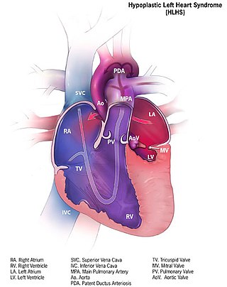
Hypoplastic left heart syndrome (HLHS) is a rare congenital heart defect in which the left side of the heart is severely underdeveloped and incapable of supporting the systemic circulation. It is estimated to account for 2-3% of all congenital heart disease. Early signs and symptoms include poor feeding, cyanosis, and diminished pulse in the extremities. The etiology is believed to be multifactorial resulting from a combination of genetic mutations and defects resulting in altered blood flow in the heart. Several structures can be affected including the left ventricle, aorta, aortic valve, or mitral valve all resulting in decreased systemic blood flow.

<span class="mw-page-title-main">Low birth weight</span>

Low birth weight (LBW) is defined by the World Health Organization as a birth weight of an infant of 2,499 g or less, regardless of gestational age. Infants born with LBW have added health risks which require close management, often in a neonatal intensive care unit (NICU). They are also at increased risk for long-term health conditions which require follow-up over time.

<span class="mw-page-title-main">Transposition of the great vessels</span> Group of congenital heart defects

Transposition of the great vessels (TGV) is a group of congenital heart defects involving an abnormal spatial arrangement of any of the great vessels: superior and/or inferior venae cavae, pulmonary artery, pulmonary veins, and aorta. Congenital heart diseases involving only the primary arteries belong to a sub-group called transposition of the great arteries (TGA), which is considered the most common congenital heart lesion that presents in neonates.

Necrotizing enterocolitis (NEC) is a devastating intestinal disease that affects premature or very low birth weight infants. Symptoms may include poor feeding, bloating, decreased activity, blood in the stool, vomiting of bile, bowel death, multiorgan failure, and even death.

<span class="mw-page-title-main">Persistent truncus arteriosus</span> Medical condition

Persistent truncus arteriosus (PTA), often referred to simply as truncus arteriosus, is a rare form of congenital heart disease that presents at birth. In this condition, the embryological structure known as the truncus arteriosus fails to properly divide into the pulmonary trunk and aorta. This results in one arterial trunk arising from the heart and providing mixed blood to the coronary arteries, pulmonary arteries, and systemic circulation. For the International Classification of Diseases (ICD-11), the International Paediatric and Congenital Cardiac Code (IPCCC) was developed to standardize the nomenclature of congenital heart disease. Under this system, English is now the official language, and persistent truncus arteriosus should properly be termed common arterial trunk.

<span class="mw-page-title-main">Tricuspid atresia</span> Medical condition

Tricuspid atresia is a form of congenital heart disease whereby there is a complete absence of the tricuspid valve. Therefore, there is an absence of right atrioventricular connection. This leads to a hypoplastic (undersized) or absent right ventricle. This defect is contracted during prenatal development, when the heart does not finish developing. It causes the systemic circulation to be filled with relatively deoxygenated blood. The causes of tricuspid atresia are unknown.

<span class="mw-page-title-main">Fetal circulation</span> Circulatory system of fetuses

In humans, the circulatory system is different before and after birth. The fetal circulation is composed of the placenta, umbilical blood vessels encapsulated by the umbilical cord, heart and systemic blood vessels. A major difference between the fetal circulation and postnatal circulation is that the lungs are not used during the fetal stage resulting in the presence of shunts to move oxygenated blood and nutrients from the placenta to the fetal tissue. At birth, the start of breathing and the severance of the umbilical cord prompt various changes that quickly transform fetal circulation into postnatal circulation.

<span class="mw-page-title-main">Bronchopulmonary dysplasia</span> Medical condition

Bronchopulmonary dysplasia is a chronic lung disease which affects premature infants. Premature (preterm) infants who require treatment with supplemental oxygen or require long-term oxygen are at a higher risk. The alveoli that are present tend to not be mature enough to function normally. It is also more common in infants with low birth weight (LBW) and those who receive prolonged mechanical ventilation to treat respiratory distress syndrome. It results in significant morbidity and mortality. The definition of bronchopulmonary dysplasia has continued to evolve primarily due to changes in the population, such as more survivors at earlier gestational ages, and improved neonatal management including surfactant, antenatal glucocorticoid therapy, and less aggressive mechanical ventilation.

Interrupted aortic arch is a very rare heart defect in which the aorta is not completely developed. There is a gap between the ascending and descending thoracic aorta. In a sense it is the complete form of a coarctation of the aorta. Almost all patients also have other cardiac anomalies, including a ventricular septal defect (VSD), aorto-pulmonary window, and truncus arteriosus. There are three types of interrupted aortic arch, with type B being the most common. Interrupted aortic arch is often associated with DiGeorge syndrome.

References

  1. Clyman, Ronald I. (2017), "Mechanisms Regulating Closure of the Ductus Arteriosus", Fetal and Neonatal Physiology, Elsevier, pp. 592–599.e4, doi:10.1016/b978-0-323-35214-7.00057-3, ISBN   978-0-323-35214-7 , retrieved 14 November 2023
  2. Dice, JE; Bhatia, J (July 2007). "Patent ductus arteriosus: an overview". The Journal of Pediatric Pharmacology and Therapeutics. 12 (3): 138–46. doi:10.5863/1551-6776-12.3.138. PMC   3462096 . PMID   23055849.
  3. 1 2 MedlinePlus - Patent ductus arteriosus Update Date: 21 December 2009
  4. "Medically Sound: Critical Blood Flow Redirection and a Fetus under Pressure – Fetal Heart Defects". Medically Sound. 6 October 2020. Retrieved 1 November 2020.
  5. Kumar, Prakash; Sinha, Santosh Kumar; Pandey, Umeshwar; Thakur, Ramesh; Varma, Chandra Mohan; Sachan, Mohit; Goel, Amit (2016). "Patent Ductus Arteriosus With Eisenmenger Syndrome: Difficult Diagnosis Made Easily With Saline Contrast Echocardiography". Cardiology Research. Elmer Press, Inc. 7 (3): 117–118. doi: 10.14740/cr447w . ISSN   1923-2829. PMC   5295518 .
  6. Anilkumar, Mehra (2013). "Patent Ductus Arteriosus". Cardiology Clinics. Elsevier BV. 31 (3): 417–430. doi:10.1016/j.ccl.2013.05.006. ISSN   0733-8651.
  7. "Tests and Diagnosis". Mayo Clinic. 16 December 2015. Retrieved 1 April 2015.
  8. 1 2 Fowlie, PW; Davis PG; McGuire W (19 May 2010). "Prophylactic intravenous indomethacin for preventing mortality and morbidity in preterm infants (Review)". The Cochrane Database of Systematic Reviews. 2010 (7): CD000174. doi:10.1002/14651858.CD000174.pub2. PMC   7045285 . PMID   20614421.
  9. Ohlsson, A; Shah, SS (27 January 2020). "Ibuprofen for the prevention of patent ductus arteriosus in preterm and/or low birth weight infants". The Cochrane Database of Systematic Reviews. 1 (1): CD004213. doi:10.1002/14651858.CD004213.pub5. PMC   6984616 . PMID   31985838.
  10. Zahaka, KG and Patel, CR. "Congenital defects'". Fanaroff, AA and Martin, RJ (eds.). Neonatal-perinatal medicine: Diseases of the fetus and infant. 7th ed. (2002):1120–1139. St. Louis: Mosby.
  11. Mosalli R, Alfaleh K, Paes B (July 2009). "Role of prophylactic surgical ligation of patent ductus arteriosus in extremely low birth weight infants: Systematic review and implications for clinical practice". Ann Pediatr Cardiol. 2 (2): 120–6. doi: 10.4103/0974-2069.58313 . PMC   2922659 . PMID   20808624.
  12. Alejandra Martins (2 October 2014). "The inventions of the Bolivian doctor who saved thousands of children". BBC Mundo. Retrieved 30 March 2015.
  13. Hines MH, Bensky AS, Hammon JW Jr et al. Video-assisted thoracoscopic ligation of patent ductus arteriosus: safe and outpatient. Ann Thorac Surg 1998;66:853–8.
  14. circ.ahajournals.org
  15. MayoClinic > Patent ductus arteriosus (PDA). 22 Dec. 2009
  16. Ohlsson A, Walia R, Shah SS (2015). "Ibuprofen for the treatment of patent ductus arteriosus in preterm or low birth weight (or both) infants". Cochrane Database Syst Rev (2): CD003481. doi:10.1002/14651858.CD003481.pub6. PMID   25692606.
  17. Ohlsson, Arne; Shah, Prakeshkumar S. (6 April 2018). "Paracetamol (acetaminophen) for patent ductus arteriosus in preterm or low birth weight infants". The Cochrane Database of Systematic Reviews. 4 (4): CD010061. doi:10.1002/14651858.CD010061.pub3. ISSN   1469-493X. PMC   6494526 . PMID   29624206.
  18. Ohlsson, Arne; Shah, Prakeshkumar S. (27 January 2020). "Paracetamol (acetaminophen) for patent ductus arteriosus in preterm or low birth weight infants". The Cochrane Database of Systematic Reviews. 1 (1): CD010061. doi:10.1002/14651858.CD010061.pub4. ISSN   1469-493X. PMC   6984659 . PMID   31985831.
  19. Mitra, Souvik; Florez, Ivan D.; Tamayo, Maria E.; Mbuagbaw, Lawrence; Vanniyasingam, Thuva; Veroniki, Areti Angeliki; Zea, Adriana M.; Zhang, Yuan; Sadeghirad, Behnam (27 March 2018). "Association of Placebo, Indomethacin, Ibuprofen, and Acetaminophen With Closure of Hemodynamically Significant Patent Ductus Arteriosus in Preterm Infants". JAMA. 319 (12): 1221–1238. doi:10.1001/jama.2018.1896. ISSN   0098-7484. PMC   5885871 . PMID   29584842.
  20. "Oral ibuprofen may be an option for closing patent ductus arteriosus in premature babies". NIHR Evidence (Plain English summary). 3 July 2018. doi:10.3310/signal-000611. S2CID   240438747.
  21. "Medically Sound: Diagnosing and treating congenital heart defects". Medically Sound. 6 October 2020. Retrieved 1 November 2020.
  22. Mitra, Souvik; Scrivens, Alexandra; von Kursell, Adelaide M; Disher, Tim (10 December 2020). "Early treatment versus expectant management of hemodynamically significant patent ductus arteriosus for preterm infants". Cochrane Database of Systematic Reviews. 2020 (12): CD013278. doi:10.1002/14651858.CD013278.pub2. ISSN   1465-1858. PMC   8812277 . PMID   33301630. S2CID   228100506.
  23. fa.hms.harvard.edu; Robert E. Gross, Harvard Medical School Office for Faculty Affairs.