The diagnostic tests in cardiology are methods of identifying heart conditions associated with healthy vs. unhealthy, pathologic heart function.

Medical ultrasound includes diagnostic techniques using ultrasound, as well as therapeutic applications of ultrasound. In diagnosis, it is used to create an image of internal body structures such as tendons, muscles, joints, blood vessels, and internal organs, to measure some characteristics or to generate an informative audible sound. The usage of ultrasound to produce visual images for medicine is called medical ultrasonography or simply sonography, or echography. The practice of examining pregnant women using ultrasound is called obstetric ultrasonography, and was an early development of clinical ultrasonography. The machine used is called an ultrasound machine, a sonograph or an echograph. The visual image formed using this technique is called an ultrasonogram, a sonogram or an echogram.

In cardiac physiology, cardiac output (CO), also known as heart output and often denoted by the symbols , , or , is the volumetric flow rate of the heart's pumping output: that is, the volume of blood being pumped by a single ventricle of the heart, per unit time. Cardiac output (CO) is the product of the heart rate (HR), i.e. the number of heartbeats per minute (bpm), and the stroke volume (SV), which is the volume of blood pumped from the left ventricle per beat; thus giving the formula:

Echocardiography, also known as cardiac ultrasound, is the use of ultrasound to examine the heart. It is a type of medical imaging, using standard ultrasound or Doppler ultrasound. The visual image formed using this technique is called an echocardiogram, a cardiac echo, or simply an echo.

Palpitations are perceived abnormalities of the heartbeat characterized by awareness of cardiac muscle contractions in the chest, which is further characterized by the hard, fast and/or irregular beatings of the heart.

A cardiac stress test is a cardiological examination that evaluates the cardiovascular system's response to external stress within a controlled clinical setting. This stress response can be induced through physical exercise or intravenous pharmacological stimulation of heart rate.

A transesophageal echocardiogram, or TEE, is an alternative way to perform an echocardiogram. A specialized probe containing an ultrasound transducer at its tip is passed into the patient's esophagus. This allows image and Doppler evaluation which can be recorded. It is commonly used during cardiac surgery and is an excellent modality for assessing the aorta, although there are some limitations.

Cardiac catheterization is the insertion of a catheter into a chamber or vessel of the heart. This is done both for diagnostic and interventional purposes.
A transthoracic echocardiogram (TTE) is the most common type of echocardiogram, which is a still or moving image of the internal parts of the heart using ultrasound. In this case, the probe is placed on the chest or abdomen of the subject to get various views of the heart. It is used as a non-invasive assessment of the overall health of the heart, including a patient's heart valves and degree of heart muscle contraction. The images are displayed on a monitor for real-time viewing and then recorded.

Atrioventricular block is a type of heart block that occurs when the electrical signal traveling from the atria, or the upper chambers of the heart, to ventricles, or the lower chambers of the heart, is impaired. Normally, the sinoatrial node produces an electrical signal to control the heart rate. The signal travels from the SA node to the ventricles through the atrioventricular node. In an AV block, this electrical signal is either delayed or completely blocked. When the signal is completely blocked, the ventricles produce their own electrical signal to control the heart rate. The heart rate produced by the ventricles is much slower than that produced by the SA node.
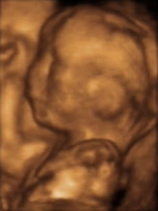
3D ultrasound is a medical ultrasound technique, often used in fetal, cardiac, trans-rectal and intra-vascular applications. 3D ultrasound refers specifically to the volume rendering of ultrasound data. When involving a series of 3D volumes collected over time, it can also be referred to as 4D ultrasound or real-time 3D ultrasound.
The American Society of Echocardiography (ASE) is a professional organization of physicians, cardiac sonographers, nurses and scientists involved in echocardiography, the use of ultrasound to image the heart and vascular system. The organization was founded in 1975 and has more than 17,000 members nationally and internationally. The American Society of Echocardiography promotes cardiovascular ultrasound and its application to patient care through education, advocacy, research, innovation and service. The society also provides research grants and scholarships to support advances in cardiovascular care.
The American Registry for Diagnostic Medical Sonography (ARDMS), incorporated in June 1975, is an independent nonprofit organization that administers examinations and awards credentials in the areas of diagnostic medical sonography, diagnostic cardiac sonography, vascular technology, physicians’ vascular interpretation, musculoskeletal sonography and midwifery ultrasound. ARDMS has over 90,000 certified individuals in the U.S., Canada and throughout the world. ARDMS provides certifications, resources, and career information to healthcare practitioners and students practicing medical sonography.
Fetal echocardiography, or Fetal echocardiogram, is the name of the test used to diagnose cardiac conditions in the fetal stage. Cardiac defects are amongst the most common birth defects. Their diagnosis is important in the fetal stage as it might help provide an opportunity to plan and manage the baby as and when the baby is born. Not all pregnancies need to undergo fetal echo.
Cardiothoracic anesthesiology is a subspeciality of the medical practice of anesthesiology, devoted to the preoperative, intraoperative, and postoperative care of adult and pediatric patients undergoing cardiothoracic surgery and related invasive procedures.

Coronary ischemia, myocardial ischemia, or cardiac ischemia, is a medical term for a reduced blood flow in the coronary circulation through the coronary arteries. Coronary ischemia is linked to heart disease, and heart attacks. Coronary arteries deliver oxygen-rich blood to the heart muscle. Reduced blood flow to the heart associated with coronary ischemia can result in inadequate oxygen supply to the heart muscle. When oxygen supply to the heart is unable to keep up with oxygen demand from the muscle, the result is the characteristic symptoms of coronary ischemia, the most common of which is chest pain. Chest pain due to coronary ischemia commonly radiates to the arm or neck. Certain individuals such as women, diabetics, and the elderly may present with more varied symptoms. If blood flow through the coronary arteries is stopped completely, cardiac muscle cells may die, known as a myocardial infarction, or heart attack.

A myocardial bridge (MB) is a congenital heart defect in which one of the coronary arteries tunnels through the heart muscle itself (myocardium). In normal patients, the coronary arteries rest on top of the heart muscle and feed blood down into smaller vessels which then take blood into the heart muscle itself. However, if a band of muscle forms around one of the coronary arteries during the fetal stage of development, then a myocardial bridge is formed – a "bridge" of heart muscle over the artery. Each time the heart squeezes to pump blood, the band of muscle exerts pressure and constricts the artery, reducing blood flow to the heart. This defect is present from birth. It is important to note that even a very thin ex. <1 mm and/or short ex. 20 mm MB can cause significant symptoms. MBs can range from a few mm in length to 10 cm or more. The overall prevalence of myocardial bridge is 19%, although its prevalence found by autopsy is much higher (42%).

Cardiac imaging refers to minimally invasive imaging of the heart using ultrasound, magnetic resonance imaging (MRI), computed tomography (CT), or nuclear medicine (NM) imaging with PET or SPECT. These cardiac techniques are otherwise referred to as echocardiography, Cardiac MRI, Cardiac CT, Cardiac PET and Cardiac SPECT including myocardial perfusion imaging.

Doppler ultrasonography is medical ultrasonography that employs the Doppler effect to perform imaging of the movement of tissues and body fluids, and their relative velocity to the probe. By calculating the frequency shift of a particular sample volume, for example, flow in an artery or a jet of blood flow over a heart valve, its speed and direction can be determined and visualized.
Harvey Feigenbaum is an American cardiologist known for his life-long work in the field of echocardiography. He wrote the first textbook on the subject in 1972, which is currently in its 8th edition, and has published over 300 articles. He has trained generations of cardiologists including many of the world's pioneers in the field through his numerous visitors, frequent workshops, annual courses in Indianapolis, Indiana beginning in 1968, the year when he started formal fellowship training He founded the field of cardiac sonography in 1965 and the American Society of Echocardiography in 1975. His seminal article on the diagnosis of pericardial effusions published in 1965 with his technique "brought echocardiography to the attention of thousands of practitioners".
