Related Research Articles
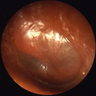
Otitis media is a group of inflammatory diseases of the middle ear. One of the two main types is acute otitis media (AOM), an infection of rapid onset that usually presents with ear pain. In young children this may result in pulling at the ear, increased crying, and poor sleep. Decreased eating and a fever may also be present. The other main type is otitis media with effusion (OME), typically not associated with symptoms, although occasionally a feeling of fullness is described; it is defined as the presence of non-infectious fluid in the middle ear which may persist for weeks or months often after an episode of acute otitis media. Chronic suppurative otitis media (CSOM) is middle ear inflammation that results in a perforated tympanic membrane with discharge from the ear for more than six weeks. It may be a complication of acute otitis media. Pain is rarely present. All three types of otitis media may be associated with hearing loss. If children with hearing loss due to OME do not learn sign language, it may affect their ability to learn.

A cleft lip contains an opening in the upper lip that may extend into the nose. The opening may be on one side, both sides, or in the middle. A cleft palate occurs when the palate contains an opening into the nose. The term orofacial cleft refers to either condition or to both occurring together. These disorders can result in feeding problems, speech problems, hearing problems, and frequent ear infections. Less than half the time the condition is associated with other disorders.

Treacher Collins syndrome (TCS) is a genetic disorder characterized by deformities of the ears, eyes, cheekbones, and chin. The degree to which a person is affected, however, may vary from mild to severe. Complications may include breathing problems, problems seeing, cleft palate, and hearing loss. Those affected generally have normal intelligence.

Conductive hearing loss (CHL) occurs when there is a problem transferring sound waves anywhere along the pathway through the outer ear, tympanic membrane (eardrum), or middle ear (ossicles). If a conductive hearing loss occurs in conjunction with a sensorineural hearing loss, it is referred to as a mixed hearing loss. Depending upon the severity and nature of the conductive loss, this type of hearing impairment can often be treated with surgical intervention or pharmaceuticals to partially or, in some cases, fully restore hearing acuity to within normal range. However, cases of permanent or chronic conductive hearing loss may require other treatment modalities such as hearing aid devices to improve detection of sound and speech perception.
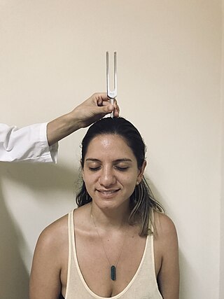
The Weber test is a screening test for hearing performed with a tuning fork. It can detect unilateral (one-sided) conductive hearing loss and unilateral sensorineural hearing loss. The test is named after Ernst Heinrich Weber (1795–1878). Conductive hearing ability is mediated by the middle ear composed of the ossicles: the malleus, the incus, and the stapes. Sensorineural hearing ability is mediated by the inner ear composed of the cochlea with its internal basilar membrane and attached cochlear nerve. The outer ear consisting of the pinna, ear canal, and ear drum or tympanic membrane transmits sounds to the middle ear but does not contribute to the conduction or sensorineural hearing ability save for hearing transmissions limited by cerumen impaction.

Hemifacial microsomia (HFM) is a congenital disorder that affects the development of the lower half of the face, most commonly the ears, the mouth and the mandible. It usually occurs on one side of the face, but both sides are sometimes affected. If severe, it may result in difficulties in breathing due to obstruction of the trachea—sometimes even requiring a tracheotomy. With an incidence in the range of 1:3500 to 1:4500, it is the second most common birth defect of the face, after cleft lip and cleft palate. HFM shares many similarities with Treacher Collins syndrome.

Microtia is a congenital deformity where the auricle is underdeveloped. A completely undeveloped pinna is referred to as anotia. Because microtia and anotia have the same origin, it can be referred to as microtia-anotia. Microtia can be unilateral or bilateral. Microtia occurs in 1 out of about 8,000–10,000 births. In unilateral microtia, the right ear is most commonly affected. It may occur as a complication of taking Accutane (isotretinoin) during pregnancy.

A bone-anchored hearing aid (BAHA) is a type of hearing aid based on bone conduction. It is primarily suited for people who have conductive hearing losses, unilateral hearing loss, single-sided deafness and people with mixed hearing losses who cannot otherwise wear 'in the ear' or 'behind the ear' hearing aids. They are more expensive than conventional hearing aids, and their placement involves invasive surgery which carries a risk of complications, although when complications do occur, they are usually minor.

Goldenhar syndrome is a rare congenital defect characterized by incomplete development of the ear, nose, soft palate, lip and mandible on usually one side of the body. Common clinical manifestations include limbal dermoids, preauricular skin tags and strabismus. It is associated with anomalous development of the first branchial arch and second branchial arch.
Abruzzo–Erickson syndrome is an extremely rare disorder characterized by deafness, protruding ears, coloboma, a cleft palate or palatal rugosity, radial synostosis, and short stature. It was first characterized by Abruzzo and Erickson in 1977 as a CHARGE like syndrome as variably expressed among a family of two brothers, their mother, and their maternal uncle. Members of this family exhibited many of the CHARGE symptoms, but notably did not have choanal atresia and the brothers experienced typical genital development. Due to the recent discovery of this disorder, its etiology is not fully known but it is understood that it arises from mutations on the TBX22 gene on the X-chromosome. The disorder is inherited in an X-linked recessive manner. There is currently no known cure but its symptoms can be treated.

Pierre Robin sequence is a congenital defect observed in humans which is characterized by facial abnormalities. The three main features are micrognathia, which causes glossoptosis, which in turn causes breathing problems due to obstruction of the upper airway. A wide, U-shaped cleft palate is commonly also present. PRS is not merely a syndrome, but rather it is a sequence—a series of specific developmental malformations which can be attributed to a single cause.

Ectrodactyly–ectodermal dysplasia–cleft syndrome, or EEC, and also referred to as EEC syndrome and split hand–split foot–ectodermal dysplasia–cleft syndrome is a rare form of ectodermal dysplasia, an autosomal dominant disorder inherited as a genetic trait. EEC is characterized by the triad of ectrodactyly, ectodermal dysplasia, and facial clefts. Other features noted in association with EEC include vesicoureteral reflux, recurrent urinary tract infections, obstruction of the nasolacrimal duct, decreased pigmentation of the hair and skin, missing or abnormal teeth, enamel hypoplasia, absent punctae in the lower eyelids, photophobia, occasional cognitive impairment and kidney anomalies, and conductive hearing loss.
Marshall syndrome is a genetic disorder of the connective tissue that can cause hearing loss. The three most common areas to be affected are the eyes, which are uncommonly large, joints and the mouth and facial structures. Marshall syndrome and Stickler syndrome closely resemble each other; in fact they are so similar, some say they are the same. The condition is named for D. Weber.
Marfanoid is a constellation of signs resembling those of Marfan syndrome, including long limbs, with an arm span that is at least 1.03 of the height of the individual, and a crowded oral maxilla, sometimes with a high arch in the palate, arachnodactyly, and hyperlaxity.

Miller syndrome, also known as Genée–Wiedemann syndrome, Wildervanck–Smith syndrome or postaxial acrofacial dysostosis, is an extremely rare genetic condition that manifests as craniofacial, limb and eye deformities. It is caused by a mutation in the DHODH gene. The incidence of the condition is not known, and nothing is known about its pathogenesis.
A facial cleft is an opening or gap in the face, or a malformation of a part of the face. Facial clefts is a collective term for all sorts of clefts. All structures like bone, soft tissue, skin etc. can be affected. Facial clefts are extremely rare congenital anomalies. There are many variations of a type of clefting and classifications are needed to describe and classify all types of clefting. Facial clefts hardly ever occur isolated; most of the time there is an overlap of adjacent facial clefts.
Malpuech facial clefting syndrome, also called Malpuech syndrome or Gypsy type facial clefting syndrome, is a rare congenital syndrome. It is characterized by facial clefting, a caudal appendage, growth deficiency, intellectual and developmental disability, and abnormalities of the renal system (kidneys) and the male genitalia. Abnormalities of the heart, and other skeletal malformations may also be present. The syndrome was initially described by Georges Malpuech and associates in 1983. It is thought to be genetically related to Juberg-Hayward syndrome. Malpuech syndrome has also been considered as part of a spectrum of congenital genetic disorders associated with similar facial, urogenital and skeletal anomalies. Termed "3MC syndrome", this proposed spectrum includes Malpuech, Michels and Mingarelli-Carnevale (OSA) syndromes. Mutations in the COLLEC11 and MASP1 genes are believed to be a cause of these syndromes. The incidence of Malpuech syndrome is unknown. The pattern of inheritance is autosomal recessive, which means a defective (mutated) gene associated with the syndrome is located on an autosome, and the syndrome occurs when two copies of this defective gene are inherited.

Identification of a hearing loss is usually conducted by a general practitioner medical doctor, otolaryngologist, certified and licensed audiologist, school or industrial audiometrist, or other audiometric technician. Diagnosis of the cause of a hearing loss is carried out by a specialist physician or otorhinolaryngologist.
A middle ear implant is a hearing device that is surgically implanted into the middle ear. They help people with conductive, sensorineural or mixed hearing loss to hear.
Thickened earlobes-conductive deafness syndrome, also known as Escher-Hirt syndrome, or Schweitzer Kemink Graham syndrome, is a rare genetic disorder which is characterized by ear and jaw abnormalities associated with progressive hearing loss. Two families worldwide have been described with the disorder.
References
- ↑ "The World Craniofacial Foundation: Dedicated to helping children and families who experience deformities of the head and/or face by providing support and access to life-changing procedures". Archived from the original on 2007-02-17. Retrieved 2018-03-05.
- Gould, H. J.; D. D. Caldarelli (June 1982). "Hearing and otopathology in Apert syndrome". Archives of Otolaryngology. 108 (6): 347–349. doi:10.1001/archotol.1982.00790540019006. PMID 7201310.
- Handžic, Jadranka; Marijo Bagatin; Radovan Subotic; Višeslay Cuk (January 1995). "Hearing levels in Pierre Robin Syndrome". Cleft Palate-Craniofacial Journal. 32 (1): 30–36. doi:10.1597/1545-1569(1995)032<0030:HLIPRS>2.3.CO;2. PMID 7727485.
- Herrman, Brian W.; Roanne Karzon; David W. Molter (August 2005). "Otologic and audiologic features of Nager acrofacial dysostosis". International Journal of Pediatric Otorhinolaryngology. 69 (8): 1053–1059. doi:10.1016/j.ijporl.2005.02.011. PMID 16005346.
- Orvidas, Laura J.; Lee Fabry; Svetlana Diacova; Thomas J. McDonald (September 1999). "Hearing and otopathology in Crouzon Syndrome". Laryngoscope. 109 (9): 1372–1375. doi: 10.1097/00005537-199909000-00002 . PMID 10499038.
- Peterson-Falzone, Sally J.; Mary A. Hardin-Jones; Michael P. Karnell (2001). Cleft Palate Speech (3rd ed.). St. Louis: Mosby. ISBN 0-8151-3153-4.
- Pron, Gaylene; Cheryl Galloway; Derek Armstrong; Jeffrey Posnick (January 1993). "Ear malformation and hearing loss in patients with Treacher Collins syndrome". Cleft Palate-Craniofacial Journal. 30 (1): 97–103. doi:10.1597/1545-1569(1993)030<0097:EMAHLI>2.3.CO;2. PMID 8418881.
- Rahbar, Reza; Caroline D. Robson; John B. Mulliken; Lynn Schwartz; James DiCanzio; Margaret A. Kenna; Trevor J. McGill; Gerald B. Healy (March 2001). "Craniofacial, temporal bone, and audiologic abnormalities in the spectrum of hemifacial microsomia". Archives of Otolaryngology. 127 (3): 265–271. doi: 10.1001/archotol.127.3.265 . PMID 11255470.
- Reyes, Maria Rina T.; Etoile M. LeBlanc; Maha K. Bassila (March 1999). "Hearing loss and otitis media in velocardiofacial syndrome". International Journal of Pediatric Otorhinolaryngology. 47 (3): 227–233. doi:10.1016/S0165-5876(98)00180-3. PMID 10321777.
- Szymko-Bennett YM, Mastroianni MA, Shotland LI, Davis J, Ondrey FG, Balog JZ, Rudy SF, McCullagh L, Levy HP, Liberfarb RM, Francomano CA, Griffith AJ (September 2001). "Auditory dysfunction in Stickler syndrome". Archives of Otolaryngology. 127 (9): 1061–1068. doi: 10.1001/archotol.127.9.1061 . PMID 11556853.
- Vallino-Napoli, Linda D. (November 1996). "Audiologic and otologic characteristics of Pfeiffer syndrome". Cleft Palate-Craniofacial Journal. 33 (6): 524–529. doi:10.1597/1545-1569(1996)033<0524:AAOCOP>2.3.CO;2. PMID 8939381.