Hemifacial spasm

Last updated

Hemifacial spasm (HFS) is a rare neuromuscular disease characterized by irregular, involuntary muscle contractions (spasms) on one side (hemi-) of the face (-facial). [1] The facial muscles are controlled by the facial nerve (seventh cranial nerve), which originates at the brainstem and exits the skull below the ear where it separates into five main branches.

Contents

This disease takes two forms: typical and atypical. In typical form, the twitching usually starts in the lower eyelid in orbicularis oculi muscle. As time progresses, it spreads to the whole lid, then to the orbicularis oris muscle around the lips, and buccinator muscle in the cheekbone area. [2] The reverse process of twitching occurs in atypical hemifacial spasm; twitching starts in orbicularis oris muscle around the lips, and buccinator muscle in the cheekbone area in the lower face, then progresses up to the orbicularis oculi muscle in the eyelid as time progresses. [2] The most common form is the typical form, and atypical form is only seen in about 2–3% of patients with hemifacial spasm. [3] The incidence of hemifacial spasm is approximately 0.8 per 100,000 persons. [4]

This disorder occurs in both men and women, although it affects middle-aged or elderly women more frequently. [5] Hemifacial spasm is much more common in some Asian populations. [1] It may be caused by a facial nerve injury, compression by a blood vessel, a tumor, or it may have no apparent cause. Individuals with spasm on both sides of the face are very rare.

Signs & Symptoms

The first sign of hemifacial spasm is typically muscle movement in the patient's eyelid and around the eye. It can vary in intensity. [6] The intermittent twitching of the eyelid, which can result in forced closure of the eye which gradually spreads to the muscles of the lower part of the face (Typical form- See Image). In atypical form the spasms start in the cheekbone area and spreads to the eyelid. [2] Ultimately, all the muscles on that side are affected, nearly all the time. This sometimes causes the mouth to be pulled to the side. Experts have linked hemifacial spasm to facial nerve injury, Bell's palsy and tumors. Although the most frequent cause is a blood vessel pressing on the facial nerve at the spot where it leaves the patient's brain stem, sometimes there is no known cause. When the affected individual is younger than 40, doctors suspect an underlying cause such as multiple sclerosis. [7]

Causes

The Facial Nerve (The Seventh Cranial Nerve) Cranial nerve VII.svg
The Facial Nerve (The Seventh Cranial Nerve)

Three theories exist to explain the facial nerve dysfunction found in hemifacial spasm. The first proposed theory is ephaptic transmission, which is electrical activity crossing from one demyelinated neuron to another resulting in a false synapse. [8] The second theory involves abnormal activity of axons at the facial nerve root end zone secondary to compressive damage/demyelination. [9]

The third theory or "Kindling theory" involves increased excitability of the facial nerve nucleus due to feedback from a damaged facial nerve. [9]

It is generally accepted as compression of the facial nerve by vessels of the posterior circulation. [9] In detail compression of the seventh cranial nerve by a dolichoectatic (a distorted, dilated, and elongated) anterior inferior cerebellar artery, or posterior inferior cerebellar artery is accepted to be the general cause of hemifacial spasm. Less than 1% of cases are caused by tumor. [10] [11] Hemifacial spasm is much more common in some Asian populations. [1]

Several families with hemifacial spasm have been reported, suggesting a genetic etiology or predisposition in some cases. There appears to be an autosomal dominant pattern of inheritance in these families with low penetrance, and except for a younger age at onset, the clinical features overlap with the idiopathic cases. Evaluation of single-nucleotide polymorphisms in genes related to vascular change causing compression of blood vessels did not show an association with hemifacial spasm. Clarifying the role of genetic susceptibility in hemifacial spasm may help to better understand the pathogenesis of this disease. [12]

Diagnosis

There are several tests done to diagnose hemifacial spasm. Diagnosing a case of hemifacial spasm begins with a complete neurological exam, including an Electromyography (EMG – a test that measures and records electrical activity generated in muscle at rest and in response to muscle contraction), Magnetic resonance imaging (MRI – a test that uses magnetic waves to make pictures of structures inside the head), Computed tomography (CT scan – a type of x-ray that uses a computer to make pictures of structures inside the head), and Angiography (an x-ray exam of the blood vessels when they are filled with a contrast material). [13]

Studies have shown that the most effective method of hemifacial spasm screening is MRI. In one study only 25% of the CT scans showed the abnormality in hemifacial spasm patients, whilst more than half of the MRI imaging demonstrated a vascular anomaly. MRI imaging should be the initial screening procedure in the assessment of patients with hemifacial spasm. [14]

Prevention

There is no known way to prevent hemifacial spasm. [12]

Treatments

Mild cases of hemifacial spasm may be managed with sedation or carbamazepine (an anticonvulsant drug). [15] Microsurgical decompression and botulinum toxin injections are the current main treatments used for hemifacial spasm. [14]

Microvascular Decompression

Microvascular decompression appears to be the most popular surgical treatment at present. Microvascular decompression relieves pressure on the facial nerve, which is the cause of most hemifacial spasm cases. Excellent to good results are reported in 80% or more cases with a 10% recurrence rate. [16] In the present series approximately 10% had previously failed surgery. Serious complications can follow microsurgical decompressive operations, even when performed by experienced surgeons. These include cerebellar haematoma or swelling, brain stem infarction (blood vessel of the brain stem blocked), cerebral infarction (ischemic stroke resulting from a disturbance in the blood vessels supplying blood to the brain), subdural haematoma and intracerebral infarction (blockage of blood flow to the brain). Death or permanent disability (hearing loss) can occur in 2% of patients of hemifacial spasm. [17]

Botulinum Toxin

Observational data from studies (the updated review in 2020 did not find any randomized controlled trials) indicates that botulinum toxin is safe and effective in the treatment of hemifacial spasm with success rates between 76 - 100%. [18] The injections are administered as an outpatient or office procedure. Whilst side effects occur, these are never permanent. Repeated injections over the years remain highly effective. [19] Whilst the toxin is expensive, the cost of even prolonged courses of injections compares favourably with the cost of surgery. [20] Patients with HFS should be offered a number of treatment options. Very mild cases or those who are reluctant to have surgery or Botulinum toxin injections can be offered medical treatment, sometimes as a temporary measure. In young and fit patients microsurgical decompression and Botulinum injections should be discussed as alternative procedures. In the majority of cases, and especially in the elderly and the unfit, Botulinum toxin injection is the treatment of first choice. Imaging procedures should be done in all unusual cases of hemifacial spasm and when surgery is contemplated. [14] Patients with hemifacial spasm were shown to have decreased sweating after botulinum toxin injections. This was first observed in 1993 by Khalaf Bushara and David Park. This was the first demonstration of nonmuscular use of BTX-A. Bushara further showed the efficacy of botulinum toxin in treating hyperhidrosis (excessive sweating). BTX-A was later approved for the treatment of excessive underarm sweating. This is technically known as severe primary axillary hyperhidrosis – excessive underarm sweating with an unknown cause which cannot be managed by topical agents (see focal hyperhidrosis).[ citation needed ]

Epidemiology

The incidence of hemifacial spasm is approximately 0.8 per 100,000 persons. [4] Hemifacial spasm is more prevalent among females over 40 years of age. [21] [22] The estimated prevalence for women is 14.5 per 100,000 and 7.4 per 100,000 in men. [23] Prevalence for hemifacial spasm increase with age, reaching 39.7 per 100,000 for those aged 70 years and older. [24] One study divided 214 hemifacial patients based on the cause of the disease: The patients who had a compression in the facial nerve at the end of the brain stem as the primary hemifacial spasm and patients who had peripheral facial palsy or nerve lesion due to tumors, demyelination, trauma, or infection as secondary hemifacial spasm. The study found that 77% of hemifacial spasm is due to primary hemifacial spasm and 23% is due to secondary hemifacial spasm. The study also found both sets of patients to share similar age at onset, male to female ratios, and similar affected side. [25] Another study with 2050 patients presented with hemifacial spasm between 1986 and 2009, only 9 cases were caused by a cerebellopontine angle syndrome, an incidence of 0.44%. [11]

History

The earliest descriptions about hemifacial spasm is by Shultze in 1875 and Gowers in 1899. The etiology of hemifacial spasm and location of the abnormality have been debated for more than a century. [4] Surgical treatment for hemifacial spasm in the early 20th century included neurolysis (destruction of nerve tissue), stretching the facial nerve (seventh cranial nerve), and high-pressure irrigation of the nerve with lactate ringer's solution. The medical regimens of that time involved injection of the nerve with ethanol, electrical stimulation, application of toxic compounds (nitrate of silver, zinc, arsenic, bromides) as well as medications such as Dilantin or other anticonvulsants. [4] [26] [8]

Additional advances in understanding the etiology and improving treatments for hemifacial spasm did not occur until the mid-seventies. In 1977, 47 cases of hemifacial spasm underwent microvascular decompression of the facial nerve using the operating microscope. The results illustrated nerve-vessel conflicts (or cholesteatoma) to be located at the root exit zone of the facial nerve in all cases. [27] [6] The root exit zone is where the central glial axonal insulation of the nerve ends and the peripheral nerve axonal myelination begins, this is known as The Obersteiner-Redlich zone. Biopsies of the root exit zone demonstrated degeneration of axons, denuded axis cylinder and interrupted myelin. The results of the experiment strengthened the theory that vascular compression of the facial nerve was the primary cause of hemifacial spasm, and proposed a specific region of the facial nerve where the effects of longstanding compression results in nerve dysfunction. [6]

Related Research Articles

<span class="mw-page-title-main">Ramsay Hunt syndrome type 2</span> Presentation of shingles in the geniculate ganglion

Inflammation of the geniculate ganglion of the facial nerve is a late consequence of varicella zoster virus (VZV) known as Ramsay Hunt syndrome (RHS), commonly known as herpes zoster oticus. In regards with the frequency, less than 1% of varicella zoster infections involve the facial nerve and result in RHS. It is traditionally defined as a triad of ipsilateral facial paralysis, otalgia, and vesicles close to the ear and auditory canal. Due to its proximity to the vestibulocochlear nerve, the virus can spread and cause hearing loss, tinnitus (hearing noises that are not causes by outside sounds), and vertigo. It is common for diagnoses to be overlooked or delayed, which can raise the likelihood of long-term consequences. It is more complicated than Bell's palsy. Therapy aims to shorten its overall length, while also providing pain relief and averting any consequences.

<span class="mw-page-title-main">Botulinum toxin</span> Neurotoxic protein produced by Clostridium botulinum

Botulinum toxin, or botulinum neurotoxin (BoNT), is a neurotoxic protein produced by the bacterium Clostridium botulinum and related species. It prevents the release of the neurotransmitter acetylcholine from axon endings at the neuromuscular junction, thus causing flaccid paralysis. The toxin causes the disease botulism. The toxin is also used commercially for medical and cosmetic purposes.

<span class="mw-page-title-main">Bell's palsy</span> Facial paralysis resulting from dysfunction in the cranial nerve VII (facial nerve)

Bell's palsy is a type of facial paralysis that results in a temporary inability to control the facial muscles on the affected side of the face. In most cases, the weakness is temporary and significantly improves over weeks. Symptoms can vary from mild to severe. They may include muscle twitching, weakness, or total loss of the ability to move one or, in rare cases, both sides of the face. Other symptoms include drooping of the eyelid, a change in taste, and pain around the ear. Typically symptoms come on over 48 hours. Bell's palsy can trigger an increased sensitivity to sound known as hyperacusis.

<span class="mw-page-title-main">Trigeminal neuralgia</span> Neurological pain disorder

Trigeminal neuralgia, also called Fothergill disease, tic douloureux, or trifacial neuralgia is a long-term pain disorder that affects the trigeminal nerve, the nerve responsible for sensation in the face and motor functions such as biting and chewing. It is a form of neuropathic pain. There are two main types: typical and atypical trigeminal neuralgia. The typical form results in episodes of severe, sudden, shock-like pain in one side of the face that lasts for seconds to a few minutes. Groups of these episodes can occur over a few hours. The atypical form results in a constant burning pain that is less severe. Episodes may be triggered by any touch to the face. Both forms may occur in the same person. It is regarded as one of the most painful disorders known to medicine, and often results in depression.

<span class="mw-page-title-main">Piriformis syndrome</span> Medical condition

Piriformis syndrome is a condition which is believed to result from compression of the sciatic nerve by the piriformis muscle. Symptoms may include pain and numbness in the buttocks and down the leg. Often symptoms are worsened with sitting or running.

Neuralgia is pain in the distribution of one or more nerves, as in intercostal neuralgia, trigeminal neuralgia, and glossopharyngeal neuralgia.

Occipital neuralgia (ON) is a painful condition affecting the posterior head in the distributions of the greater occipital nerve (GON), lesser occipital nerve (LON), third occipital nerve (TON), or a combination of the three. It is paroxysmal, lasting from seconds to minutes, and often consists of lancinating pain that directly results from the pathology of one of these nerves. It is paramount that physicians understand the differential diagnosis for this condition and specific diagnostic criteria. There are multiple treatment modalities, several of which have well-established efficacy in treating this condition.

Microvascular decompression (MVD), also known as the Jannetta procedure, is a neurosurgical procedure used to treat trigeminal neuralgia a pain syndrome characterized by severe episodes of intense facial pain, and hemifacial spasm. The procedure is also used experimentally to treat tinnitus and vertigo caused by vascular compression on the vestibulocochlear nerve.

<span class="mw-page-title-main">Blepharospasm</span> Abnormal contraction or twitch of the eyelid

Blepharospasm is any abnormal contraction of the orbicularis oculi muscle. The condition should be distinguished from the more common, and milder, involuntary quivering of an eyelid, known as myokymia, or fasciculation. In most cases, blepharospasm symptoms last for a few days and then disappear without treatment, but in some cases the twitching is chronic and persistent, causing life-long challenges. In these cases, the symptoms are often severe enough to result in functional blindness. The person's eyelids feel like they are clamping shut and will not open without great effort. People have normal eyes, but for periods of time are effectively blind due to their inability to open their eyelids. In contrast, the reflex blepharospasm is due to any pain in and around the eye.

<span class="mw-page-title-main">Spasmodic torticollis</span> Medical condition

Spasmodic torticollis is an extremely painful chronic neurological movement disorder causing the neck to involuntarily turn to the left, right, upwards, and/or downwards. The condition is also referred to as "cervical dystonia". Both agonist and antagonist muscles contract simultaneously during dystonic movement. Causes of the disorder are predominantly idiopathic. A small number of patients develop the disorder as a result of another disorder or disease. Most patients first experience symptoms midlife. The most common treatment for spasmodic torticollis is the use of botulinum toxin type A.

<span class="mw-page-title-main">Meige's syndrome</span> Medical condition

Meige's syndrome is a type of dystonia. It is also known as Brueghel's syndrome and oral facial dystonia. It is actually a combination of two forms of dystonia, blepharospasm and oromandibular dystonia (OMD).

<span class="mw-page-title-main">Parry–Romberg syndrome</span> Rare disease involving degeneration of tissues beneath the skin

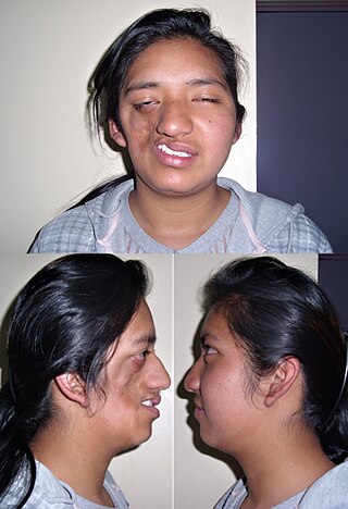
Parry–Romberg syndrome (PRS) is a rare disease characterized by progressive shrinkage and degeneration of the tissues beneath the skin, usually on only one side of the face but occasionally extending to other parts of the body. An autoimmune mechanism is suspected, and the syndrome may be a variant of localized scleroderma, but the precise cause and pathogenesis of this acquired disorder remains unknown. It has been reported in the literature as a possible consequence of sympathectomy. The syndrome has a higher prevalence in females and typically appears between 5 and 15 years of age.

Spasmodic dysphonia, also known as laryngeal dystonia, is a disorder in which the muscles that generate a person's voice go into periods of spasm. This results in breaks or interruptions in the voice, often every few sentences, which can make a person difficult to understand. The person's voice may also sound strained or they may be nearly unable to speak. Onset is often gradual and the condition is lifelong.

Synkinesis is a neurological symptom in which a voluntary muscle movement causes the simultaneous involuntary contraction of other muscles. An example might be smiling inducing an involuntary contraction of the eye muscles, causing a person to squint when smiling. Facial and extraocular muscles are affected most often; in rare cases, a person's hands might perform mirror movements.

<span class="mw-page-title-main">Avulsion injury</span> Injury where a body structure is torn off

In medicine, an avulsion is an injury in which a body structure is torn off by either trauma or surgery. The term most commonly refers to a surface trauma where all layers of the skin have been torn away, exposing the underlying structures. This is similar to an abrasion but more severe, as body parts such as an eyelid or an ear can be partially or fully detached from the body.

Migraine surgery is a surgical operation undertaken with the goal of reducing or preventing migraines. Migraine surgery most often refers to surgical decompression of one or several nerves in the head and neck which have been shown to trigger migraine symptoms in many migraine sufferers. Following the development of nerve decompression techniques for the relief of migraine pain in the year 2000, these procedures have been extensively studied and shown to be effective in appropriate candidates. The nerves that are most often addressed in migraine surgery are found outside of the skull, in the face and neck, and include the supra-orbital and supra-trochlear nerves in the forehead, the zygomaticotemporal nerve and auriculotemporal nerves in the temple region, and the greater occipital, lesser occipital, and third occipital nerves in the back of the neck. Nerve impingement in the nasal cavity has additionally been shown to be a trigger of migraine symptoms.

<span class="mw-page-title-main">Atypical trigeminal neuralgia</span> Medical condition

Atypical trigeminal neuralgia (ATN), or type 2 trigeminal neuralgia, is a form of trigeminal neuralgia, a disorder of the fifth cranial nerve. This form of nerve pain is difficult to diagnose, as it is rare and the symptoms overlap with several other disorders. The symptoms can occur in addition to having migraine headache, or can be mistaken for migraine alone, or dental problems such as temporomandibular joint disorder or musculoskeletal issues. ATN can have a wide range of symptoms and the pain can fluctuate in intensity from mild aching to a crushing or burning sensation, and also to the extreme pain experienced with the more common trigeminal neuralgia.

Botulinum toxin therapy of strabismus is a medical technique used sometimes in the management of strabismus, in which botulinum toxin is injected into selected extraocular muscles in order to reduce the misalignment of the eyes. The injection of the toxin to treat strabismus, reported upon in 1981, is considered to be the first ever use of botulinum toxin for therapeutic purposes. Today, the injection of botulinum toxin into the muscles that surround the eyes is one of the available options in the management of strabismus. Other options for strabismus management are vision therapy and occlusion therapy, corrective glasses and prism glasses, and strabismus surgery.

The management of strabismus may include the use of drugs or surgery to correct the strabismus. Agents used include paralytic agents such as botox used on extraocular muscles, topical autonomic nervous system agents to alter the refractive index in the eyes, and agents that act in the central nervous system to correct amblyopia.

Alan Brown Scott was an American ophthalmologist specializing in eye muscles and their disorders, such as strabismus. He is best known for his work in developing and manufacturing the drug that became known as Botox, research described as "groundbreaking" by the ASCRS.

References

  1. 1 2 3 Kong, Doo-Sik; Kwan Park (2007). "Hemifacial Spasm: A Neurological Perspective". Journal of Korean Neurosurgical Society . 5. 42 (5): 355–362. doi:10.3340/jkns.2007.42.5.355. PMC   2588188 . PMID   19096569.
  2. 1 2 3 Jannetta, PJ (1998). "Typical or atypical hemifacial spasm". Journal of Neurosurgery. 89 (2): 346–7. doi:10.3171/jns.1998.89.2.0346. PMID   9688136.
  3. Ryu, H.; Yamamoto S.; Miyamoto T. (1998). "Atypical hemifacial spasm". Acta Neurochir. 40 (11): 1173–76. doi:10.1007/s007010050233. PMID   9870064. S2CID   24709653.
  4. 1 2 3 4 Fukushima, T (1995). "Microvascular decompression for hemifacial spasm: Result in 2890 cases". Neurovascular Surgery. New York: McGraw Hill: 1133–45.
  5. "Hemifacial Spasm Information Page". National Institute for Neurological Disorders and Stroke. 2018. Retrieved January 3, 2019.
  6. 1 2 3 Jannetta, PJ; Abbasy M; Maroon JC; Ramos FM; Albin MS. (1977). "Etiology and definitive microsurgical treatment of hemifacial spasm. Operative techniques and results in 47 patients". Journal of Neurosurgery. 47 (3): 321–8. doi:10.3171/jns.1977.47.3.0321. PMID   894338.
  7. Office of Communications and Public Liaison. "NINDS Hemifacial Spasm Information Page". National Institute of Neurological Disorders and Strokes. Archived from the original on 28 December 2011. Retrieved 11 March 2012.
  8. 1 2 Gardner, J.W.; Sava, Gerard A. (1962). "Hemifacial Spasm - A reversible pathophysiologic state". Journal of Neurosurgery. 27 (3): 240–47. doi:10.3171/jns.1962.19.3.0240.
  9. 1 2 3 Eby, Joseph; Sung Tae Cha; Hrayr K. Shahinian (2002). "Fully endoscopic vascular decompression of the glossopharyngeal nerve". The Journal of Craniofacial Surgery. 13 (1): 90–95. doi:10.1097/00001665-200201000-00021. PMID   11887002. S2CID   20943041.
  10. Caces, F; Chays A; Locatelli P; Bruzzo M; Epron JP; Fiacre E; Magnan J. (1996). "Neuro-vascular decompression in hemifacial spasm: anatomical, electrophysiological and therapeutic results apropos of 100 cases". Rev Laryngol Otol Rhinol (Bord). 5. 117 (5): 347–51. PMID   9183904.
  11. 1 2 Lee SH, Rhee BA, Choi SK, Koh JS, Lim YJ; Jankovic J (2010). "Cerebellopontine angle tumors causing hemifacial spasm: types, incidence, and mechanism in nine reported cases and literature review". Acta Neurochir (Wien). 11. 150 (11): 1901–8. doi:10.1007/s00701-010-0796-1. PMID   20845049. S2CID   43823317.
  12. 1 2 Yaltho, Toby C.; Jankovic, Joseph (1 August 2011). "The Many Faces of Hemifacial Spasm: Differential Diagnosis of Unilateral Facial Spasms". Movement Disorders. 26 (9): 1582–92. doi:10.1002/mds.23692. PMID   21469208. S2CID   12473692.
  13. Zappia JJ; Wiet RJ; Chouhan A; Zhao JC, JJ; Wiet, RJ; Chouhan, A; Zhao, JC (1997). "Pitfalls in the diagnosis of hemifacial spasm". The Laryngoscope. 4. 107 (4): 461–465. doi:10.1097/00005537-199704000-00007. PMID   9111374. S2CID   12359030.{{cite journal}}: CS1 maint: multiple names: authors list (link)
  14. 1 2 3 I. T. Lorentz (April 1995). "Treatment of hemifacial spasm with botulinum toxin". Journal of Clinical Neuroscience. 2. 2 (2): 132–135. doi:10.1016/0967-5868(95)90005-5. PMID   18638798. S2CID   20818813.
  15. Alexander GE, Moses H (1982). "Carbamazepine for hemi facial spasm". Neurology. 32 (3): 286–287. doi:10.1212/wnl.32.3.286. PMID   7199640. S2CID   73383442.
  16. PiattJH, J'r, Wilkins RH., JH; Wilkins, RH (1984). "Treatment of tic douloureux and hemifacial spasm by posterior fossa exploration: therapeutic implications of various neurovascular relationships". Neurosurgery. 14 (4): 462–71. doi:10.1227/00006123-198404000-00012. PMID   6728149.{{cite journal}}: CS1 maint: multiple names: authors list (link)
  17. HanakitaJ, Kondo A., J; Kondo, A (1988). "Serious complications of microvascular decompression operations for trigeminal neuralgia and hemifacial spasm". Neurosurgery. 22 (2): 348–52. doi: 10.1227/00006123-198802000-00012 . PMID   3352885.
  18. Duarte, Gonçalo S.; Rodrigues, Filipe B.; Castelão, Mafalda; Marques, Raquel E.; Ferreira, Joaquim; Sampaio, Cristina; Moore, Austen P.; Costa, João (November 19, 2020). "Botulinum toxin type A therapy for hemifacial spasm". The Cochrane Database of Systematic Reviews. 2020 (11): CD004899. doi:10.1002/14651858.CD004899.pub3. ISSN   1469-493X. PMC   8078498 . PMID   33211908.
  19. LoeserJD, ChenJ., John D.; Chen, James (1982). "Hemifacial spasm: treatment by microsurgical facial nerve decompression". Neurosurgery. 13 (2): 141–46. doi:10.1227/00006123-198308000-00006. PMID   6888693.
  20. Tan, Eng-ing; Stephanie Fook-Chong; Sau-Ying Lum; Erle Lim (16 January 2004). "Botulinum toxin improves quality of life in hemifacial spasm: validation of a questionnaire (HFS-30)". Journal of the Neurological Sciences. 219 (1–2): 151–155. doi:10.1016/j.jns.2004.01.010. PMID   15050451. S2CID   22572909.
  21. Wang, A.; Jankovic J. (1998). "Hemifacial spasm: clinical findings and treatment". Muscle and Nerve. 12. 21 (12): 1740–7. doi:10.1002/(SICI)1097-4598(199812)21:12<1740::AID-MUS17>3.0.CO;2-V. PMID   9843077. S2CID   31006011.
  22. Tan, N.C.; Chan, L.L; Tan E.K. (2002). "Hemifacial spasm and involuntary facial movements". QJM. 8. 95 (8): 493–500. doi: 10.1093/qjmed/95.8.493 . PMID   12145388.
  23. Kemp, LW; Reich SG (June 2004). "Hemifacial spasm". Curr Treat Options Neurol. 6 (3): 175–9. doi:10.1007/s11940-004-0009-4. PMID   15043800. S2CID   29140844.
  24. Auger, RG; Whisnant JP (1990). "Hemifacial spasm in Rochester and Olmsted County, Minnesota, 1960 to 1984". Arch Neurol. 47 (11): 1233–4. doi:10.1001/archneur.1990.00530110095023. PMID   2241620.
  25. Colosimo, C; Bologna M; Lamberti S (2006). "A comparative study of primary and secondary hemifacial spasm". Arch Neurol. 6. 63 (3): 441–4. doi: 10.1001/archneur.63.3.441 . PMID   16533973.
  26. Wilkins, RH (1991). "Hemifacial spasm: a review". Surgical Neurology. 36 (4): 251–77. doi:10.1016/0090-3019(91)90087-P. PMID   1948626.
  27. Jannetta, PJ (1975). "Trigeminal neuralgia and hemifacial spasm--etiology and definitive treatment". Trans Am Neurol Assoc. 100: 89–91. PMID   1226647.

Further reading