Hexokinase

Last updated

Hexokinase
Hexokinase 3O08 structure.png
Crystal structures of hexokinase 1 from Kluyveromyces lactis . [1] [2]
Identifiers
EC no. 2.7.1.1
CAS no. 9001-51-8
Databases
IntEnz IntEnz view
BRENDA BRENDA entry
ExPASy NiceZyme view
KEGG KEGG entry
MetaCyc metabolic pathway
PRIAM profile
PDB structures RCSB PDB PDBe PDBsum
Gene Ontology AmiGO / QuickGO
Search
PMC articles
PubMed articles
NCBI proteins
hexokinase 1
1hkc.jpg
Hexokinase 1, homodimer, Human
Identifiers
Symbol HK1
NCBI gene 3098
HGNC 4922
OMIM 142600
RefSeq NM_000188
UniProt P19367
Other data
Locus Chr. 10 q22
Search for
Structures Swiss-model
Domains InterPro
hexokinase 2
Identifiers
Symbol HK2
NCBI gene 3099
HGNC 4923
OMIM 601125
RefSeq NM_000189
UniProt P52789
Other data
Locus Chr. 2 p13
Search for
Structures Swiss-model
Domains InterPro
hexokinase 3 (white cell)
Identifiers
Symbol HK3
NCBI gene 3101
HGNC 4925
OMIM 142570
RefSeq NM_002115
UniProt P52790
Other data
Locus Chr. 5 q35.2
Search for
Structures Swiss-model
Domains InterPro
Hexokinase_1
PDB 1v4t EBI.jpg
crystal structure of human glucokinase
Identifiers
SymbolHexokinase_1
Pfam PF00349
Pfam clan CL0108
InterPro IPR022672
PROSITE PDOC00370
SCOP2 1cza / SCOPe / SUPFAM
Available protein structures:
Pfam   structures / ECOD  
PDB RCSB PDB; PDBe; PDBj
PDBsum structure summary
Hexokinase_2
PDB 1bg3 EBI.jpg
rat brain hexokinase type i complex with glucose and inhibitor glucose-6-phosphate
Identifiers
SymbolHexokinase_2
Pfam PF03727
Pfam clan CL0108
InterPro IPR022673
PROSITE PDOC00370
SCOP2 1cza / SCOPe / SUPFAM
Available protein structures:
Pfam   structures / ECOD  
PDB RCSB PDB; PDBe; PDBj
PDBsum structure summary

A hexokinase is an enzyme that irreversibly phosphorylates hexoses (six-carbon sugars), forming hexose phosphate. In most organisms, glucose is the most important substrate for hexokinases, and glucose-6-phosphate is the most important product. Hexokinase possesses the ability to transfer an inorganic phosphate group from ATP to a substrate.

Contents

Hexokinases should not be confused with glucokinase, which is a specific hexokinase found in the liver. All hexokinases are capable of phosphorylating several hexoses but hexokinase IV(D) is often misleadingly called glucokinase, though it is no more specific for glucose than the other mammalian isoenzymes. [3]

Variation

Genes that encode hexokinase have been discovered in every domain of life, and exist among a variety of species that range from bacteria, yeast, and plants to humans and other vertebrates. The enzymes from yeast, plants and vertebrates all show clear sequence evidence of homology, but those of bacteria may not be related. [4]

They are categorized as actin fold proteins, sharing a common ATP binding site core that is surrounded by more variable sequences which determine substrate affinities and other properties.

Several hexokinase isoenzymes that provide different functions can occur in a single species.

Reaction

The intracellular reactions mediated by hexokinases can be typified as:

Hexose-CH2OH + MgATP2
→ Hexose-CH2O-PO2
3
+ MgADP
+ H+

where hexose-CH2OH represents any of several hexoses (like glucose) that contain an accessible -CH2OH moiety. Hexokinase-glucose.png

Consequences of hexose phosphorylation

Phosphorylation of a hexose such as glucose often limits it to a number of intracellular metabolic processes, such as glycolysis or glycogen synthesis. This is because phosphorylated hexoses are charged, and thus more difficult to transport out of a cell.

In patients with essential fructosuria, metabolism of fructose by hexokinase to fructose-6-phosphate is the primary method of metabolizing dietary fructose; this pathway is not significant in normal individuals.

Size of different isoforms

Most bacterial hexokinases are approximately 50 kDa in size. Multicellular organisms including plants and animals often have more than one hexokinase isoform. Most are about 100 kDa in size and consist of two halves (N and C terminal), which share much sequence homology. This suggests an evolutionary origin by duplication and fusion of a 50kDa ancestral hexokinase similar to those of bacteria.

Types of mammalian hexokinase

There are four important mammalian hexokinase isozymes (EC 2.7.1.1) that vary in subcellular locations and kinetics with respect to different substrates and conditions, and physiological function. They were designated hexokinases A, B, C, and D on the basis of their electrophoretic mobility. [5] The alternative names hexokinases I, II, III, and IV (respectively) [6] proposed later are widely used.

Hexokinases I, II, and III

Hexokinases I, II, and III are referred to as low-Km isoenzymes because of a high affinity for glucose (below 1 mM). Hexokinases I and II follow Michaelis-Menten kinetics at physiological concentrations of substrates.[ citation needed ] All three are strongly inhibited by their product, glucose-6-phosphate. Molecular masses are around 100 kDa. Each consists of two similar 50kDa halves, but only in hexokinase II do both halves have functional active sites.

Hexokinase IV ("glucokinase")

Mammalian hexokinase IV, also referred to as glucokinase, differs from other hexokinases in kinetics and functions.

The location of the phosphorylation on a subcellular level occurs when glucokinase translocates between the cytoplasm and nucleus of liver cells. Glucokinase can only phosphorylate glucose if the concentration of this substrate is high enough; it does not follow Henri–Michaelis–Menten kinetics, and has no Km; It is half-saturated at glucose concentrations 100 times higher than those of hexokinases I, II, and III.

Hexokinase IV is monomeric, about 50kDa, displays positive cooperativity with glucose, and is not allosterically inhibited by its product, glucose-6-phosphate. [4]

Hexokinase IV is present in the liver, pancreas, hypothalamus, small intestine, and perhaps certain other neuroendocrine cells, and plays an important regulatory role in carbohydrate metabolism. In the β cells of the pancreatic islets, it serves as a glucose sensor to control insulin release, and similarly controls glucagon release in the α cells. In hepatocytes of the liver, glucokinase responds to changes of ambient glucose levels by increasing or reducing glycogen synthesis.

In glycolysis

Glucose is unique in that it can be used to produce ATP by all cells in both the presence and absence of molecular oxygen (O2). The first step in glycolysis is the phosphorylation of glucose by hexokinase.

D-Glucose Hexokinaseα-D-Glucose-6-phosphate
D-glucose wpmp.svg   Alpha-D-glucose-6-phosphate wpmp.png
ATP ADP
Biochem reaction arrow forward YYNN horiz med.svg


Compound C00031 at KEGG Pathway Database.Enzyme 2.7.1.1 at KEGG Pathway Database.Compound C00668 at KEGG Pathway Database.Reaction R01786 at KEGG Pathway Database.

By catalyzing the phosphorylation of glucose to yield glucose 6-phosphate, hexokinases maintain the downhill concentration gradient that favors the facilitated transport of glucose into cells. This reaction also initiates all physiologically relevant pathways of glucose utilization, including glycolysis and the pentose phosphate pathway. [9] The addition of a charged phosphate group at the 6-position of hexoses also ensures 'trapping' of glucose and 2-deoxyhexose glucose analogs (e.g. 2-deoxyglucose, and 2-fluoro-2-deoxyglucose) within cells, as charged hexose phosphates cannot easily cross the cell membrane.

Association with mitochondria

Hexokinases I and II can associate physically to the outer surface of the external membrane of mitochondria through specific binding to a porin, or voltage dependent anion channel. This association confers hexokinase direct access to ATP generated by mitochondria, which is one of the two substrates of hexokinase. Mitochondrial hexokinase is highly elevated in rapidly growing malignant tumor cells, with levels up to 200 times higher than normal tissues. Mitochondrially bound hexokinase has been demonstrated to be the driving force [10] for the extremely high glycolytic rates that take place aerobically in tumor cells (the so-called Warburg effect described by Otto Heinrich Warburg in 1930).

Deficiency

Hexokinase deficiency is a genetic autosomal recessive disease that causes chronic haemolytic anaemia. Chronic haemolytic anaemia is caused by a mutation in the gene that codes for hexokinase. The mutation causes a reduction of the hexokinase activity, and hence hexokinase deficiency. [11]

See also

Related Research Articles

<span class="mw-page-title-main">Glycolysis</span> Series of interconnected biochemical reactions

Glycolysis is the metabolic pathway that converts glucose into pyruvate and, in most organisms, occurs in the liquid part of cells. The free energy released in this process is used to form the high-energy molecules adenosine triphosphate (ATP) and reduced nicotinamide adenine dinucleotide (NADH). Glycolysis is a sequence of ten reactions catalyzed by enzymes.

<span class="mw-page-title-main">Kinase</span> Enzyme catalyzing transfer of phosphate groups onto specific substrates

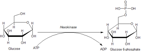
In biochemistry, a kinase is an enzyme that catalyzes the transfer of phosphate groups from high-energy, phosphate-donating molecules to specific substrates. This process is known as phosphorylation, where the high-energy ATP molecule donates a phosphate group to the substrate molecule. As a result, kinase produces a phosphorylated substrate and ADP. Conversely, it is referred to as dephosphorylation when the phosphorylated substrate donates a phosphate group and ADP gains a phosphate group. These two processes, phosphorylation and dephosphorylation, occur four times during glycolysis.

<span class="mw-page-title-main">Phosphorylation</span> Chemical process of introducing a phosphate

In biochemistry, phosphorylation is the attachment of a phosphate group to a molecule or an ion. This process and its inverse, dephosphorylation, are common in biology. Protein phosphorylation often activates many enzymes.

Gluconeogenesis (GNG) is a metabolic pathway that results in the biosynthesis of glucose from certain non-carbohydrate carbon substrates. It is a ubiquitous process, present in plants, animals, fungi, bacteria, and other microorganisms. In vertebrates, gluconeogenesis occurs mainly in the liver and, to a lesser extent, in the cortex of the kidneys. It is one of two primary mechanisms – the other being degradation of glycogen (glycogenolysis) – used by humans and many other animals to maintain blood sugar levels, avoiding low levels (hypoglycemia). In ruminants, because dietary carbohydrates tend to be metabolized by rumen organisms, gluconeogenesis occurs regardless of fasting, low-carbohydrate diets, exercise, etc. In many other animals, the process occurs during periods of fasting, starvation, low-carbohydrate diets, or intense exercise.

Carbohydrate metabolism is the whole of the biochemical processes responsible for the metabolic formation, breakdown, and interconversion of carbohydrates in living organisms.

<span class="mw-page-title-main">Phosphofructokinase 1</span> Class of enzymes

Phosphofructokinase-1 (PFK-1) is one of the most important regulatory enzymes of glycolysis. It is an allosteric enzyme made of 4 subunits and controlled by many activators and inhibitors. PFK-1 catalyzes the important "committed" step of glycolysis, the conversion of fructose 6-phosphate and ATP to fructose 1,6-bisphosphate and ADP. Glycolysis is the foundation for respiration, both anaerobic and aerobic. Because phosphofructokinase (PFK) catalyzes the ATP-dependent phosphorylation to convert fructose-6-phosphate into fructose 1,6-bisphosphate and ADP, it is one of the key regulatory steps of glycolysis. PFK is able to regulate glycolysis through allosteric inhibition, and in this way, the cell can increase or decrease the rate of glycolysis in response to the cell's energy requirements. For example, a high ratio of ATP to ADP will inhibit PFK and glycolysis. The key difference between the regulation of PFK in eukaryotes and prokaryotes is that in eukaryotes PFK is activated by fructose 2,6-bisphosphate. The purpose of fructose 2,6-bisphosphate is to supersede ATP inhibition, thus allowing eukaryotes to have greater sensitivity to regulation by hormones like glucagon and insulin.

<span class="mw-page-title-main">Pyruvate kinase</span> Class of enzymes

Pyruvate kinase is the enzyme involved in the last step of glycolysis. It catalyzes the transfer of a phosphate group from phosphoenolpyruvate (PEP) to adenosine diphosphate (ADP), yielding one molecule of pyruvate and one molecule of ATP. Pyruvate kinase was inappropriately named before it was recognized that it did not directly catalyze phosphorylation of pyruvate, which does not occur under physiological conditions. Pyruvate kinase is present in four distinct, tissue-specific isozymes in animals, each consisting of particular kinetic properties necessary to accommodate the variations in metabolic requirements of diverse tissues.

<span class="mw-page-title-main">Phosphoglucomutase</span> Metabolic enzyme

Phosphoglucomutase is an enzyme that transfers a phosphate group on an α-D-glucose monomer from the 1 to the 6 position in the forward direction or the 6 to the 1 position in the reverse direction.

Chemical specificity is the ability of binding site of a macromolecule to bind specific ligands. The fewer ligands a protein can bind, the greater its specificity.

<span class="mw-page-title-main">Glucose 6-phosphate</span> Chemical compound

Glucose 6-phosphate is a glucose sugar phosphorylated at the hydroxy group on carbon 6. This dianion is very common in cells as the majority of glucose entering a cell will become phosphorylated in this way.

<span class="mw-page-title-main">Glucokinase</span> Enzyme participating to the regulation of carbohydrate metabolism

Glucokinase is an enzyme that facilitates phosphorylation of glucose to glucose-6-phosphate. Glucokinase occurs in cells in the liver and pancreas of humans and most other vertebrates. In each of these organs it plays an important role in the regulation of carbohydrate metabolism by acting as a glucose sensor, triggering shifts in metabolism or cell function in response to rising or falling levels of glucose, such as occur after a meal or when fasting. Mutations of the gene for this enzyme can cause unusual forms of diabetes or hypoglycemia.

<span class="mw-page-title-main">Entner–Doudoroff pathway</span> Series of interconnected biochemical reactions

The Entner–Doudoroff pathway is a metabolic pathway that is most notable in Gram-negative bacteria, certain Gram-positive bacteria and archaea. Glucose is the substrate in the ED pathway and through a series of enzyme assisted chemical reactions it is catabolized into pyruvate. Entner and Doudoroff (1952) and MacGee and Doudoroff (1954) first reported the ED pathway in the bacterium Pseudomonas saccharophila. While originally thought to be just an alternative to glycolysis (EMP) and the pentose phosphate pathway (PPP), some studies now suggest that the original role of the EMP may have originally been about anabolism and repurposed over time to catabolism, meaning the ED pathway may be the older pathway. Recent studies have also shown the prevalence of the ED pathway may be more widespread than first predicted with evidence supporting the presence of the pathway in cyanobacteria, ferns, algae, mosses, and plants. Specifically, there is direct evidence that Hordeum vulgare uses the Entner–Doudoroff pathway.

<span class="mw-page-title-main">Fructose 1,6-bisphosphate</span> Chemical compound

Fructose 1,6-bisphosphate, known in older publications as Harden-Young ester, is fructose sugar phosphorylated on carbons 1 and 6. The β-D-form of this compound is common in cells. Upon entering the cell, most glucose and fructose is converted to fructose 1,6-bisphosphate.

<span class="mw-page-title-main">Fructose 2,6-bisphosphate</span> Chemical compound

Fructose 2,6-bisphosphate, abbreviated Fru-2,6-P2, is a metabolite that allosterically affects the activity of the enzymes phosphofructokinase 1 (PFK-1) and fructose 1,6-bisphosphatase (FBPase-1) to regulate glycolysis and gluconeogenesis. Fru-2,6-P2 itself is synthesized and broken down in either direction by the integrated bifunctional enzyme phosphofructokinase 2 (PFK-2/FBPase-2), which also contains a phosphatase domain and is also known as fructose-2,6-bisphosphatase. Whether the kinase and phosphatase domains of PFK-2/FBPase-2 are active or inactive depends on the phosphorylation state of the enzyme.

<span class="mw-page-title-main">Enzyme activator</span> Molecules which increase enzyme activity

Enzyme activators are molecules that bind to enzymes and increase their activity. They are the opposite of enzyme inhibitors. These molecules are often involved in the allosteric regulation of enzymes in the control of metabolism. An example of an enzyme activator working in this way is fructose 2,6-bisphosphate, which activates phosphofructokinase 1 and increases the rate of glycolysis in response to the hormone glucagon. In some cases, when a substrate binds to one catalytic subunit of an enzyme, this can trigger an increase in the substrate affinity as well as catalytic activity in the enzyme's other subunits, and thus the substrate acts as an activator.

<span class="mw-page-title-main">Inborn errors of carbohydrate metabolism</span> Medical condition

Inborn errors of carbohydrate metabolism are inborn error of metabolism that affect the catabolism and anabolism of carbohydrates.

Fructolysis refers to the metabolism of fructose from dietary sources. Though the metabolism of glucose through glycolysis uses many of the same enzymes and intermediate structures as those in fructolysis, the two sugars have very different metabolic fates in human metabolism. Unlike glucose, which is directly metabolized widely in the body, fructose is mostly metabolized in the liver in humans, where it is directed toward replenishment of liver glycogen and triglyceride synthesis. Under one percent of ingested fructose is directly converted to plasma triglyceride. 29% - 54% of fructose is converted in liver to glucose, and about a quarter of fructose is converted to lactate. 15% - 18% is converted to glycogen. Glucose and lactate are then used normally as energy to fuel cells all over the body.

<span class="mw-page-title-main">HK2</span>

Hexokinase 2 also known as HK2 is an enzyme which in humans is encoded by the HK2 gene on chromosome 2. Hexokinases phosphorylate glucose to produce glucose-6-phosphate (G6P), the first step in most glucose metabolism pathways. This gene encodes hexokinase 2, the predominant form found in skeletal muscle. It localizes to the outer membrane of mitochondria. Expression of this gene is insulin-responsive, and studies in rat suggest that it is involved in the increased rate of glycolysis seen in rapidly growing cancer cells. [provided by RefSeq, Apr 2009]

<span class="mw-page-title-main">HK3</span> Mammalian protein found in Homo sapiens

Hexokinase 3 also known as HK3 is an enzyme which in humans is encoded by the HK3 gene on chromosome 5. Hexokinases phosphorylate glucose to produce glucose-6-phosphate (G6P), the first step in most glucose metabolism pathways. This gene encodes hexokinase 3. Similar to hexokinases 1 and 2, this allosteric enzyme is inhibited by its product glucose-6-phosphate. [provided by RefSeq, Apr 2009]

<span class="mw-page-title-main">TP53-inducible glycolysis and apoptosis regulator</span> Protein-coding gene in the species Homo sapiens

The TP53-inducible glycolysis and apoptosis regulator (TIGAR) also known as fructose-2,6-bisphosphatase TIGAR is an enzyme that in humans is encoded by the C12orf5 gene.

References

  1. PDB: 3O08 ; Kuettner EB, Kettner K, Keim A, Svergun DI, Volke D (2010). "Crystal structure of dimeric KlHxk1 in crystal form I". doi:10.2210/pdb3o08/pdb.
  2. Kuettner, E. Bartholomeus; Kettner, Karina; Keim, Antje; Svergun, Dmitri I.; Volke, Daniela; Singer, David; Hoffmann, Ralf; Müller, Eva-Christina; Otto, Albrecht; Kriegel, Thomas M.; Sträter, Norbert (2010). "Crystal Structure of Hexokinase KlHxk1 of Kluyveromyces lactis". Journal of Biological Chemistry. 285 (52). Elsevier BV: 41019–41033. doi: 10.1074/jbc.m110.185850 . ISSN   0021-9258. PMC   3003401 .
  3. Cárdenas, María Luz; Rabajille, E.; Niemeyer, H. (1984). "Fructose is a good substrate for rat liver glucokinase (hexokinase D)". Biochemical Journal. 222 (2): 363–370. doi:10.1042/bj2220363. PMC   1144187 . PMID   6477520.
  4. 1 2 Cárdenas, María Luz; Cornish-Bowden, A.; Ureta, T. (1998). "Evolution and regulatory role of the hexokinases". Biochimica et Biophysica Acta. 1401 (3): 242–264. doi:10.1016/S0167-4889(97)00150-X. PMID   9540816.
  5. González, C.; Sánchez, R.; Ureta, T.; Niemeyer, H. (1964). "Multiple molecular forms of ATP:hexose 6-phosphotransferase from rat liver". Biochemical and Biophysical Research Communications. 16 (4): 347–352. doi:10.1016/0006-291X(64)90038-5. PMID   5871820.
  6. Katzen, H. M.; Sodermann, D. D.; Nitowsky, H. M. (1965). "Kinetic and electrophoretic evidence for multiple forms of glucose–ATP phosphotransferase activity from human cell cultures and rat liver". Biochemical and Biophysical Research Communications. 19 (3): 377–382. doi:10.1016/0006-291X(65)90472-9. PMID   14317406.
  7. "Hexokinase data on Uniprot". uniprot.org.
  8. Šimčíková D, Heneberg P (August 2019). "Identification of alkaline pH optimum of human glucokinase because of ATP-mediated bias correction in outcomes of enzyme assays". Scientific Reports. 9 (1): 11422. Bibcode:2019NatSR...911422S. doi:10.1038/s41598-019-47883-1. PMC   6684659 . PMID   31388064.
  9. Robey, RB; Hay, N (2006). "Mitochondrial hexokinases, novel mediators of the antiapoptotic effects of growth factors and Akt". Oncogene. 25 (34): 4683–96. doi:10.1038/sj.onc.1209595. PMID   16892082. S2CID   25230246.
  10. Bustamante E, Pedersen P (1977). "High aerobic glycolysis of rat hepatoma cells in culture: role of mitochondrial hexokinase". Proceedings of the National Academy of Sciences. 74 (9): 3735–9. Bibcode:1977PNAS...74.3735B. doi: 10.1073/pnas.74.9.3735 . PMC   431708 . PMID   198801.
  11. "Hexokinase deficiency". Enerca. Archived from the original on 8 August 2020. Retrieved 6 April 2017.