Glucose-6-phosphate isomerase | |||||||||
---|---|---|---|---|---|---|---|---|---|
Identifiers | |||||||||
EC no. | 5.3.1.9 | ||||||||
CAS no. | 9001-41-6 | ||||||||
Databases | |||||||||
IntEnz | IntEnz view | ||||||||
BRENDA | BRENDA entry | ||||||||
ExPASy | NiceZyme view | ||||||||
KEGG | KEGG entry | ||||||||
MetaCyc | metabolic pathway | ||||||||
PRIAM | profile | ||||||||
PDB structures | RCSB PDB PDBe PDBsum | ||||||||
Gene Ontology | AmiGO / QuickGO | ||||||||
|
Bacterial phosphoglucose isomerase C-terminal region | |||||||||
---|---|---|---|---|---|---|---|---|---|
Identifiers | |||||||||
Symbol | bact-PGI_C | ||||||||
Pfam | PF10432 | ||||||||
InterPro | IPR019490 | ||||||||
CDD | cd05016 | ||||||||
|
Phosphoglucose isomeras | |||||||||
---|---|---|---|---|---|---|---|---|---|
Identifiers | |||||||||
Symbol | PGI | ||||||||
Pfam | PF00342 | ||||||||
SCOP2 | 1pgi / SCOPe / SUPFAM | ||||||||
CDD | cd05015 | ||||||||
|
Glucose-6-phosphate isomerase (GPI), alternatively known as phosphoglucose isomerase/phosphoglucoisomerase (PGI) or phosphohexose isomerase (PHI), is an enzyme ( EC 5.3.1.9) that in humans is encoded by the GPI gene on chromosome 19. [4] This gene encodes a member of the glucose phosphate isomerase protein family. The encoded protein has been identified as a moonlighting protein based on its ability to perform mechanistically distinct functions. In the cytoplasm, the gene product functions as a glycolytic enzyme (glucose-6-phosphate isomerase) that interconverts glucose-6-phosphate (G6P) and fructose-6-phosphate (F6P). Extracellularly, the encoded protein (also referred to as neuroleukin) functions as a neurotrophic factor that promotes survival of skeletal motor neurons and sensory neurons, and as a lymphokine that induces immunoglobulin secretion. The encoded protein is also referred to as autocrine motility factor (AMF) based on an additional function as a tumor-secreted cytokine and angiogenic factor. Defects in this gene are the cause of nonspherocytic hemolytic anemia, and a severe enzyme deficiency can be associated with hydrops fetalis, immediate neonatal death and neurological impairment. Alternative splicing results in multiple transcript variants. [provided by RefSeq, Jan 2014] [5]
Functional GPI is a 64-kDa dimer composed of two identical monomers. [6] [7] The two monomers interact notably through the two protrusions in a hugging embrace. The active site of each monomer is formed by a cleft between the two domains and the dimer interface. [6]
GPI monomers are made of two domains, one made of two separate segments called the large domain and the other made of the segment in between called the small domain. [8] The two domains are each αβα sandwiches, with the small domain containing a five-strand β-sheet surrounded by α-helices while the large domain has a six-stranded β-sheet. [6] The large domain, located at the N-terminal, and the C-terminal of each monomer also contain "arm-like" protrusions. [8] [9] Several residues in the small domain serve to bind phosphate, while other residues, particularly His388, from the large and C-terminal domains are crucial to the sugar ring-opening step catalyzed by this enzyme. Since the isomerization activity occurs at the dimer interface, the dimer structure of this enzyme is critical to its catalytic function. [9]
It is hypothesized that serine phosphorylation of this protein induces a conformational change to its secretory form. [7]
The mechanism that GPI uses to interconvert glucose 6-phosphate and fructose 6-phosphate (aldose to ketose) consists of three major steps: opening the glucose ring, isomerizing glucose into fructose through an enediol intermediate, and closing the fructose ring. [10]
D-Glucose | Phosphoglucose isomerase | D-Fructose | |
Phosphoglucose isomerase |
α-D-Glucose 6-phosphate | Phosphoglucose isomerase | α-D-Fructose 6-phosphate | |
Phosphoglucose isomerase |
Compound C00668 at KEGG Pathway Database.Enzyme 5.3.1.9 at KEGG Pathway Database.Compound C05345 at KEGG Pathway Database.Reaction R00771 at KEGG Pathway Database.
Glucose 6-phosphate binds to GPI in its pyranose form. The ring is opened in a "push-pull" mechanism by His388, which protonates the C5 oxygen, and Lys518, which deprotonates the C1 hydroxyl group. This creates an open chain aldose. Then, the substrate is rotated about the C3-C4 bond to position it for isomerization. At this point, Glu357 deprotonates C2 to create a cis-enediolate intermediate stabilized by Arg272. To complete the isomerization, Glu357 donates its proton to C1, the C2 hydroxyl group loses its proton and the open-chain ketose fructose 6-phosphate is formed. Finally, the ring is closed by rotating the substrate about the C3-C4 bond again and deprotonating the C5 hydroxyl with Lys518. [11]
When going from fructose-6-phosphate toward glucose-6-phosphate, the result could be mannose-6-phosphate if carbon C2 is given the wrong chirality, but the enzyme does not permit that result except at a very low, non-physiological, rate. [11]
This gene belongs to the GPI family. [5] The protein encoded by this gene is a dimeric enzyme that catalyzes the reversible isomerization of G6P and F6P. [12] [13] Since the reaction is reversible, its direction is determined by G6P and F6P concentrations. [9]
glucose 6-phosphate ↔ fructose 6-phosphate
The protein has different functions inside and outside the cell. In the cytoplasm, the protein is involved in glycolysis and gluconeogenesis, as well as the pentose phosphate pathway. [9] Outside the cell, it functions as a neurotrophic factor for spinal and sensory neurons, called neuroleukin. [13] The same protein is also secreted by cancer cells, where it is called autocrine motility factor [14] and stimulates metastasis. [15] Extracellular GPI is also known to function as a maturation factor. [9] [13]
Though originally treated as separate proteins, cloning technology demonstrated that GPI is almost identical to the protein neuroleukin. [16] Neuroleukin is a neurotrophic factor for spinal and sensory neurons. It is found in large amounts in muscle, brain, heart, and kidneys. [17] Neuroleukin also acts as a lymphokine secreted by T cells stimulated by lectin. It induces immunoglobulin secretion in B cells as part of a response that activates antibody-secreting cells. [18]
Cloning experiments also revealed that GPI is identical to the protein known as autocrine motility factor (AMF). [19] AMF produced and secreted by cancer cells and stimulates cell growth and motility as a growth factor. [20] AMF is thought to play a key role in cancer metastasis by activating the MAPK/ERK or PI3K/AKT pathways. [21] [22] [23] In the PI3K/AKT pathway, AMF interacts with gp78/AMFR to regulate ER calcium release, and therefore protect against apoptosis in response to ER stress. [21]
In some archaea and bacteria glucose-6-phosphate isomerase activity occurs via a bifunctional enzyme that also exhibits phosphomannose isomerase (PMI) activity. Though not closely related to eukaryotic GPIs, the bifunctional enzyme is similar enough that the sequence includes the cluster of threonines and serines that forms the sugar phosphate-binding site in conventional GPI. The enzyme is thought to use the same catalytic mechanisms for both glucose ring-opening and isomerization for the interconversion of G6P to F6P. [24]
A deficiency of GPI is responsible for 4% of the hemolytic anemias due to glycolytic enzyme deficiencies. [12] [13] [25] [26] Several cases of GPI deficiency have recently been identified. [27]
Elevated serum GPI levels have been used as a prognostic biomarker for colorectal, breast, lung, kidney, gastrointestinal, and other cancers. [7] [13] As AMF, GPI is attributed with regulating cell migration during invasion and metastasis. [7] One study showed that the external layers of breast tumor spheroids (BTS) secrete GPI, which induces epithelial–mesenchymal transition (EMT), invasion, and metastasis in BTS. The GPI inhibitors ERI4P and 6PG were found to block metastasis of BTS but not BTS glycolysis or fibroblast viability. In addition, GPI is secreted exclusively by tumor cells and not normal cells. For these reasons, GPI inhibitors may be a safer, more targeted approach for anti-cancer therapy. [28] GPI also participates in a positive feedback loop with HER2, a major breast cancer therapeutic target, as GPI enhances HER2 expression and HER2 overexpression enhances GPI expression, and so on. As a result, GPI activity likely confers resistance in breast cancer cells against HER2-based therapies using Herceptin/Trastuzumab, and should be considered as an additional target when treating patients. [23]
Human GPI is capable of inducing arthritis in mice with varied genetic backgrounds via intradermal injection. [29] [30]
GPI is known to interact with:
Click on genes, proteins and metabolites below to link to respective articles. [§ 1]
Glycolysis is the metabolic pathway that converts glucose into pyruvate and, in most organisms, occurs in the liquid part of cells. The free energy released in this process is used to form the high-energy molecules adenosine triphosphate (ATP) and reduced nicotinamide adenine dinucleotide (NADH). Glycolysis is a sequence of ten reactions catalyzed by enzymes.
A hexokinase is an enzyme that irreversibly phosphorylates hexoses, forming hexose phosphate. In most organisms, glucose is the most important substrate for hexokinases, and glucose-6-phosphate is the most important product. Hexokinase possesses the ability to transfer an inorganic phosphate group from ATP to a substrate.
Glucose-6-phosphate dehydrogenase deficiency (G6PDD), which is the most common enzyme deficiency worldwide, is an inborn error of metabolism that predisposes to red blood cell breakdown. Most of the time, those who are affected have no symptoms. Following a specific trigger, symptoms such as yellowish skin, dark urine, shortness of breath, and feeling tired may develop. Complications can include anemia and newborn jaundice. Some people never have symptoms.
Tumor hypoxia is the situation where tumor cells have been deprived of oxygen. As a tumor grows, it rapidly outgrows its blood supply, leaving portions of the tumor with regions where the oxygen concentration is significantly lower than in healthy tissues. Hypoxic microenvironments in solid tumors are a result of available oxygen being consumed within 70 to 150 μm of tumor vasculature by rapidly proliferating tumor cells thus limiting the amount of oxygen available to diffuse further into the tumor tissue. In order to support continuous growth and proliferation in challenging hypoxic environments, cancer cells are found to alter their metabolism. Furthermore, hypoxia is known to change cell behavior and is associated with extracellular matrix remodeling and increased migratory and metastatic behavior.
Pyruvate kinase is the enzyme involved in the last step of glycolysis. It catalyzes the transfer of a phosphate group from phosphoenolpyruvate (PEP) to adenosine diphosphate (ADP), yielding one molecule of pyruvate and one molecule of ATP. Pyruvate kinase was inappropriately named before it was recognized that it did not directly catalyze phosphorylation of pyruvate, which does not occur under physiological conditions. Pyruvate kinase is present in four distinct, tissue-specific isozymes in animals, each consisting of particular kinetic properties necessary to accommodate the variations in metabolic requirements of diverse tissues.
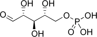
Glucose 6-phosphate is a glucose sugar phosphorylated at the hydroxy group on carbon 6. This dianion is very common in cells as the majority of glucose entering a cell will become phosphorylated in this way.
In biochemistry, isomerases are a general class of enzymes that convert a molecule from one isomer to another. Isomerases facilitate intramolecular rearrangements in which bonds are broken and formed. The general form of such a reaction is as follows:
Aldolase A, also known as fructose-bisphosphate aldolase, is an enzyme that in humans is encoded by the ALDOA gene on chromosome 16.
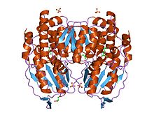
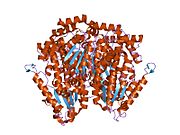
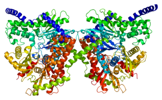
Glucose-6-phosphate dehydrogenase (G6PD or G6PDH) (EC 1.1.1.49) is a cytosolic enzyme that catalyzes the chemical reaction
Aldolase A deficiency is an autosomal recessive metabolic disorder resulting in a deficiency of the enzyme aldolase A; the enzyme is found predominantly in red blood cells and muscle tissue. The deficiency may lead to hemolytic anaemia as well as myopathy associated with exercise intolerance and rhabdomyolysis in some cases.
Ribose 5-phosphate (R5P) is both a product and an intermediate of the pentose phosphate pathway. The last step of the oxidative reactions in the pentose phosphate pathway is the production of ribulose 5-phosphate. Depending on the body's state, ribulose 5-phosphate can reversibly isomerize to ribose 5-phosphate. Ribulose 5-phosphate can alternatively undergo a series of isomerizations as well as transaldolations and transketolations that result in the production of other pentose phosphates as well as fructose 6-phosphate and glyceraldehyde 3-phosphate.
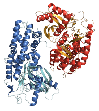
Mannose-6 phosphate isomerase (MPI), alternately phosphomannose isomerase (PMI) is an enzyme which facilitates the interconversion of fructose 6-phosphate (F6P) and mannose-6-phosphate (M6P). Mannose-6-phosphate isomerase may also enable the synthesis of GDP-mannose in eukaryotic organisms. M6P can be converted to F6P by mannose-6-phosphate isomerase and subsequently utilized in several metabolic pathways including glycolysis and capsular polysaccharide biosynthesis. PMI is monomeric and metallodependent on zinc as a cofactor ligand. PMI is inhibited by erythrose 4-phosphate, mannitol 1-phosphate, and to a lesser extent, the alpha anomer of M6P.
Hexokinase-1 (HK1) is an enzyme that in humans is encoded by the HK1 gene on chromosome 10. Hexokinases phosphorylate glucose to produce glucose-6-phosphate (G6P), the first step in most glucose metabolism pathways. This gene encodes a ubiquitous form of hexokinase which localizes to the outer membrane of mitochondria. Mutations in this gene have been associated with hemolytic anemia due to hexokinase deficiency. Alternative splicing of this gene results in five transcript variants which encode different isoforms, some of which are tissue-specific. Each isoform has a distinct N-terminus; the remainder of the protein is identical among all the isoforms. A sixth transcript variant has been described, but due to the presence of several stop codons, it is not thought to encode a protein. [provided by RefSeq, Apr 2009]
Ribose-5-phosphate isomerase (Rpi) encoded by the RPIA gene is an enzyme that catalyzes the conversion between ribose-5-phosphate (R5P) and ribulose-5-phosphate (Ru5P). It is a member of a larger class of isomerases which catalyze the interconversion of chemical isomers. It plays a vital role in biochemical metabolism in both the pentose phosphate pathway and the Calvin cycle. The systematic name of this enzyme class is D-ribose-5-phosphate aldose-ketose-isomerase.
Autocrine motility factor receptor, isoform 2 is a protein that in humans is encoded by the AMFR gene.
Triosephosphate isomerase is an enzyme that in humans is encoded by the TPI1 gene.
Inborn errors of carbohydrate metabolism are inborn error of metabolism that affect the catabolism and anabolism of carbohydrates.
Enolase Deficiency is a rare genetic disorder of glucose metabolism. Partial deficiencies have been observed in several caucasian families. The deficiency is transmitted through an autosomal dominant inheritance pattern. The gene for Enolase 1 has been localized to Chromosome 1 in humans. Enolase deficiency, like other glycolytic enzyme deficiences, usually manifests in red blood cells as they rely entirely on anaerobic glycolysis. Enolase deficiency is associated with a spherocytic phenotype and can result in hemolytic anemia, which is responsible for the clinical signs of Enolase deficiency.
Congenital hemolytic anemia (CHA) is a diverse group of rare hereditary conditions marked by decreased life expectancy and premature removal of erythrocytes from blood flow. Defects in erythrocyte membrane proteins and red cell enzyme metabolism, as well as changes at the level of erythrocyte precursors, lead to impaired bone marrow erythropoiesis. CAH is distinguished by variable anemia, chronic extravascular hemolysis, decreased erythrocyte life span, splenomegaly, jaundice, biliary lithiasis, and iron overload. Immune-mediated mechanisms may play a role in the pathogenesis of these uncommon diseases, despite the paucity of data regarding the immune system's involvement in CHAs.
Glycerol kinase deficiency (GKD) is an X-linked recessive enzyme defect that is heterozygous in nature. Three clinically distinct forms of this deficiency have been proposed, namely infantile, juvenile, and adult. National Institutes of Health and its Office of Rare Diseases Research branch classifies GKD as a rare disease, known to affect fewer than 200,000 individuals in the United States. The responsible gene lies in a region containing genes in which deletions can cause Duchenne muscular dystrophy and adrenal hypoplasia congenita. Combinations of these three genetic defects including GKD are addressed medically as Complex GKD.