Nerve growth factor

Last updated
NGF
NGF protein.png
Available structures
PDB Ortholog search: PDBe RCSB
Identifiers
Aliases NGF , Beta-HSAN5, NGFB, nerve growth factor
External IDs OMIM: 162030 MGI: 97321 HomoloGene: 1876 GeneCards: NGF
Orthologs
SpeciesHumanMouse
Entrez
Ensembl
UniProt
RefSeq (mRNA)

NM_002506

NM_001112698
NM_013609

RefSeq (protein)

NP_002497

NP_001106168
NP_038637

Location (UCSC) Chr 1: 115.29 – 115.34 Mb Chr 3: 102.38 – 102.43 Mb
PubMed search [3] [4]
Wikidata
View/Edit Human View/Edit Mouse

Nerve growth factor (NGF) is a neurotrophic factor and neuropeptide primarily involved in the regulation of growth, maintenance, proliferation, and survival of certain target neurons. It is perhaps the prototypical growth factor, in that it was one of the first to be described. Since it was first isolated by Nobel Laureates Rita Levi-Montalcini and Stanley Cohen in 1956, numerous biological processes involving NGF have been identified, two of them being the survival of pancreatic beta cells and the regulation of the immune system.

Contents

Structure

NGF is initially in a 7S, 130-kDa complex of 3 proteins – Alpha-NGF, Beta-NGF, and Gamma-NGF (2:1:2 ratio) when expressed. This form of NGF is also referred to as proNGF (NGF precursor). The gamma subunit of this complex acts as a serine protease, and cleaves the N-terminal of the beta subunit, thereby activating the protein into functional NGF.

The term nerve growth factor usually refers to the 2.5S, 26-kDa beta subunit of the protein, the only component of the 7S NGF complex that is biologically active (i.e. acting as a signaling molecule).

Function

As its name suggests, NGF is involved primarily in the growth, as well as the maintenance, proliferation, and survival of nerve cells (neurons) and is critical for the survival and maintenance of sympathetic and sensory neurons as they undergo apoptosis in its absence. [5] However, several recent studies suggest that NGF is also involved in pathways besides those regulating the life cycle of neurons.

Neuronal proliferation

NGF can drive the expression of genes such as bcl-2 by binding to the Tropomyosin receptor kinase A, which stimulates the proliferation and survival of the target neuron.

High affinity binding between proNGF, sortilin, and p75NTR can result in either survival or programmed cell death. Study results indicate that superior cervical ganglia neurons that express both p75NTR and TrkA die when treated with proNGF, [6] while NGF treatment of these same neurons results in survival and axonal growth. Survival and PCD mechanisms are mediated through adaptor protein binding to the death domain of the p75NTR cytoplasmic tail. Survival occurs when recruited cytoplasmic adaptor proteins facilitate signal transduction through tumor necrosis factor receptor members such as TRAF6, which results in the release of nuclear factor κB (NF-κB) transcription activator. [7] NF-κB regulates nuclear gene transcription to promote cell survival. Alternatively, programmed cell death occurs when TRAF6 and neurotrophin receptor interacting factor (NRIF) are both recruited to activate c-Jun N-terminal kinase (JNK); which phosphorylates c-Jun. The activated transcription factor c-Jun regulates nuclear transcription via AP-1 to increase pro-apoptotic gene transcription. [7]

Proliferation of pancreatic beta cells

There is evidence that pancreatic beta cells express both the TrkA and p75NTR receptors of NGF. It has been shown that the withdrawal of NGF induces apoptosis in pancreatic beta cells, signifying that NGF may play a critical role in the maintenance and survival of pancreatic beta cells. [8]

Regulation of the immune system

NGF plays a critical role in the regulation of both innate and acquired immunity. In the process of inflammation, NGF is released in high concentrations by mast cells, and induces axonal outgrowth in nearby nociceptive neurons. This leads to increased pain perception in areas under inflammation. In acquired immunity, NGF is produced by the Thymus as well as CD4+ T cell clones, inducing a cascade of maturation of T cells under infection. [9]

Ovulation

NGF is abundant in seminal plasma. Recent studies have found that it induces ovulation in some mammals e.g. "induced" ovulators, such as llamas. Surprisingly, research showed that these induced animals will also ovulate when semen from on-schedule or "spontaneous" ovulators, such as cattle is used. Its significance in humans is unknown. It was previously dubbed ovulation-inducing factor (OIF) in semen before it was identified as beta-NGF in 2012. [10]

Mechanism of action

NGF binds with at least two classes of receptors: the tropomyosin receptor kinase A (TrkA) and low-affinity NGF receptor (LNGFR/p75NTR). Both are associated with neurodegenerative disorders.

When NGF binds to the TrkA receptor, it drives the homodimerization of the receptor, which in turn causes the autophosphorylation of the tyrosine kinase segment. [11] The tropomyosin receptor kinase A receptor has five extracellular domains, and the fifth domain is sufficient in binding NGF. [12] Once bound, the complex undergoes endocytosis and activates the NGF transcriptional program, following two major pathways, the Ras/MAPK pathway and the PI3K/Akt pathway. [11] The binding of NGF to TrkA also leads to the activation of PI 3-kinase, ras, and PLC signaling pathways. [13] Alternatively, the p75NTR receptor can form a heterodimer with TrkA, which has higher affinity and specificity for NGF.

Studies suggest that NGF circulates throughout the entire body via the blood plasma, and is important for the overall maintenance of homeostasis. [14]

Neuron survival

Binding interaction between NGF and the TrkA receptor facilitates receptor dimerization and tyrosine residue phosphorylation of the cytoplasmic tail by adjacent Trk receptors. [15] Trk receptor phosphorylation sites operate as Shc adaptor protein docking sites, which undergo phosphorylation by the TrkA receptor [7] Once the cytoplasmic adaptor protein (Shc) is phosphorylated by the receptor cytoplasmic tail, cell survival is initiated through several intracellular pathways.

One major pathway leads to the activation of the serine/threonine kinase, Akt. This pathway begins with the Trk receptor complex-recruitment of a second adaptor protein called growth factor-receptor bound protein-2 (Grb2) along with a docking protein called Grb2-associated Binder-1 (GAB1). [7] Subsequently, phosphatidylinositol-3 kinase (PI3K) is activated, resulting in Akt kinase activation. [7] Study results have shown that blocking PI3K or Akt activity results in death of sympathetic neurons in culture, regardless of NGF presence. [16] However, if either kinase is constitutively active, neurons survive even without NGF. [16]

A second pathway contributing to cell survival occurs through activation of the mitogen-activated protein kinase (MAPK) kinase. In this pathway, recruitment of a guanine nucleotide exchange factor by the adaptor and docking proteins leads to activation of a membrane-associated G-protein known as Ras. [7] The guanine nucleotide exchange factor mediates Ras activation through the GDP-GTP exchange process. The active Ras protein phosphorylates several proteins, along with the serine/threonine kinase, Raf. [7] Raf in turn activates the MAPK cascade to facilitate ribosomal s6 kinase (RSK) activation and transcriptional regulation. [7]

Both Akt and RSK, components of the PI3K-Akt and MAPK pathways respectively, act to phosphorylate the cyclic AMP response element binding protein (CREB) transcription factor. [7] Phosphorylated CREB translocates into the nucleus and mediates increased expression of anti-apoptotic proteins, [7] thus promoting NGF-mediated cell survival. However, in the absence of NGF, the expression of pro-apoptotic proteins is increased when the activation of cell death-promoting transcription factors such as c-Jun are not suppressed by the aforementioned NGF-mediated cell survival pathways. [7]

History

Rita Levi-Montalcini and Stanley Cohen discovered NGF in the 1950s while faculty members at Washington University in St. Louis. The critical discover was done by Levi-Montalcini and Hertha Meyer at the Carlos Chagas Filho Biophysics Institute of the Federal University of Rio de Janeiro in 1952. Their publication in 1954 [17] became the definitive proof for the existence of the protein. [18] [19] Levi-Montalcini later remarked:

The tumor had given a first hint of its existence in St. Louis but it was in Rio de Janeiro that it revealed itself, and it did so in a theatrical and grand way, as if spurred by the bright atmosphere of that explosive and exhuberant manifestation of life that is the Carnival in Rio. [20]

However, its discovery, along with the discovery of other neurotrophins, was not widely recognized until 1986, when it won the Nobel Prize in Physiology or Medicine. [21] [22] [23]

Studies in 1971 determined the primary structure of NGF. This eventually led to the discovery of the NGF gene.

NGF is abundant in seminal plasma. Recent studies have found that it induces ovulation in some mammals. [24] Nerve Growth Factors (NGF) were initially discovered due to their actions during development, but NGF are now known to be involved in the function throughout the life of the animal. [25]

Interactions

Nerve growth factor has been shown to interact with Tropomyosin receptor kinase A. [26]

Clinical Use

NGF, specifically mouse (murine) nerve growth factor, has been available as a licensed medicine in China since 2003. [27] There is evidence of improved patient outcomes for several diseases of the nervous system, including acute intracerebral hemorrhage, [28] global developmental delay, [29] optic atrophy, [30] epilepsy [31] and cerebral palsy. [32] This is significant as there are few medicines which can treat injuries and diseases of the nervous system. Research and clinical use outside of China is limited despite a large body of evidence supporting its use.

See also

Related Research Articles

<span class="mw-page-title-main">Signal transduction</span> Cascade of intracellular and molecular events for transmission/amplification of signals

Signal transduction is the process by which a chemical or physical signal is transmitted through a cell as a series of molecular events. Most commonly, protein phosphorylation is catalyzed by protein kinases, ultimately resulting in a cellular response. Proteins responsible for detecting stimuli are generally termed receptors, although in some cases the term sensor is used. The changes elicited by ligand binding in a receptor give rise to a biochemical cascade, which is a chain of biochemical events known as a signaling pathway.

<span class="mw-page-title-main">Brain-derived neurotrophic factor</span> Protein found in humans

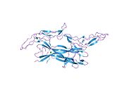
Brain-derived neurotrophic factor (BDNF), or abrineurin, is a protein that, in humans, is encoded by the BDNF gene. BDNF is a member of the neurotrophin family of growth factors, which are related to the canonical nerve growth factor (NGF), a family which also includes NT-3 and NT-4/NT-5. Neurotrophic factors are found in the brain and the periphery. BDNF was first isolated from a pig brain in 1982 by Yves-Alain Barde and Hans Thoenen.

<span class="mw-page-title-main">Neurotrophin</span> Family of proteins

Neurotrophins are a family of proteins that induce the survival, development, and function of neurons.

<span class="mw-page-title-main">Protein kinase B</span> Set of three serine threonine-specific protein kinases

Protein kinase B (PKB), also known as Akt, is the collective name of a set of three serine/threonine-specific protein kinases that play key roles in multiple cellular processes such as glucose metabolism, apoptosis, cell proliferation, transcription, and cell migration.

<span class="mw-page-title-main">Tropomyosin receptor kinase A</span> Protein-coding gene in the species Homo sapiens

Tropomyosin receptor kinase A (TrkA), also known as high affinity nerve growth factor receptor, neurotrophic tyrosine kinase receptor type 1, or TRK1-transforming tyrosine kinase protein is a protein that in humans is encoded by the NTRK1 gene.

<span class="mw-page-title-main">Tropomyosin receptor kinase B</span> Protein and coding gene in humans

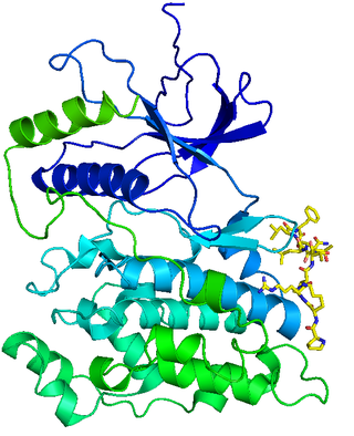
Tropomyosin receptor kinase B (TrkB), also known as tyrosine receptor kinase B, or BDNF/NT-3 growth factors receptor or neurotrophic tyrosine kinase, receptor, type 2 is a protein that in humans is encoded by the NTRK2 gene. TrkB is a receptor for brain-derived neurotrophic factor (BDNF). The standard pronunciation for this protein is "track bee".

<span class="mw-page-title-main">Low-affinity nerve growth factor receptor</span> Human protein-coding gene

The p75 neurotrophin receptor (p75NTR) was first identified in 1973 as the low-affinity nerve growth factor receptor (LNGFR) before discovery that p75NTR bound other neurotrophins equally well as nerve growth factor. p75NTR is a neurotrophic factor receptor. Neurotrophic factor receptors bind Neurotrophins including Nerve growth factor, Neurotrophin-3, Brain-derived neurotrophic factor, and Neurotrophin-4. All neurotrophins bind to p75NTR. This also includes the immature pro-neurotrophin forms. Neurotrophic factor receptors, including p75NTR, are responsible for ensuring a proper density to target ratio of developing neurons, refining broader maps in development into precise connections. p75NTR is involved in pathways that promote neuronal survival and neuronal death.

<span class="mw-page-title-main">Tropomyosin receptor kinase C</span> Protein-coding gene in the species Homo sapiens

Tropomyosin receptor kinase C (TrkC), also known as NT-3 growth factor receptor, neurotrophic tyrosine kinase receptor type 3, or TrkC tyrosine kinase is a protein that in humans is encoded by the NTRK3 gene.

Neurotrophic factors (NTFs) are a family of biomolecules – nearly all of which are peptides or small proteins – that support the growth, survival, and differentiation of both developing and mature neurons. Most NTFs exert their trophic effects on neurons by signaling through tyrosine kinases, usually a receptor tyrosine kinase. In the mature nervous system, they promote neuronal survival, induce synaptic plasticity, and modulate the formation of long-term memories. Neurotrophic factors also promote the initial growth and development of neurons in the central nervous system and peripheral nervous system, and they are capable of regrowing damaged neurons in test tubes and animal models. Some neurotrophic factors are also released by the target tissue in order to guide the growth of developing axons. Most neurotrophic factors belong to one of three families: (1) neurotrophins, (2) glial cell-line derived neurotrophic factor family ligands (GFLs), and (3) neuropoietic cytokines. Each family has its own distinct cell signaling mechanisms, although the cellular responses elicited often do overlap.

<span class="mw-page-title-main">Neurotrophin-3</span> Protein-coding gene in the species Homo sapiens

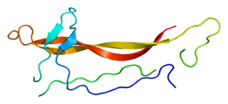
Neurotrophin-3 is a protein that in humans is encoded by the NTF3 gene.

c-Jun N-terminal kinases Chemical compounds

c-Jun N-terminal kinases (JNKs), were originally identified as kinases that bind and phosphorylate c-Jun on Ser-63 and Ser-73 within its transcriptional activation domain. They belong to the mitogen-activated protein kinase family, and are responsive to stress stimuli, such as cytokines, ultraviolet irradiation, heat shock, and osmotic shock. They also play a role in T cell differentiation and the cellular apoptosis pathway. Activation occurs through a dual phosphorylation of threonine (Thr) and tyrosine (Tyr) residues within a Thr-Pro-Tyr motif located in kinase subdomain VIII. Activation is carried out by two MAP kinase kinases, MKK4 and MKK7, and JNK can be inactivated by Ser/Thr and Tyr protein phosphatases. It has been suggested that this signaling pathway contributes to inflammatory responses in mammals and insects.

<span class="mw-page-title-main">FRS2</span> Protein-coding gene in humans

Fibroblast growth factor receptor substrate 2 is a protein that in humans is encoded by the FRS2 gene.

Trk receptors are a family of tyrosine kinases that regulates synaptic strength and plasticity in the mammalian nervous system. Trk receptors affect neuronal survival and differentiation through several signaling cascades. However, the activation of these receptors also has significant effects on functional properties of neurons.

<span class="mw-page-title-main">NCK2</span> Protein-coding gene in the species Homo sapiens

Cytoplasmic protein NCK2 is a protein that in humans is encoded by the NCK2 gene.

Neurotrophic factor receptors or neurotrophin receptors are a group of growth factor receptors which specifically bind to neurotrophins.

The Akt signaling pathway or PI3K-Akt signaling pathway is a signal transduction pathway that promotes survival and growth in response to extracellular signals. Key proteins involved are PI3K and Akt.

In cellular biology, dependence receptors are proteins that mediate programmed cell death by monitoring the absence of certain trophic factors that otherwise serve as ligands (interactors) for the dependence receptors. A trophic ligand is a molecule whose protein binding stimulates cell growth, differentiation, and/or survival. Cells depend for their survival on stimulation that is mediated by various receptors and sensors, and integrated via signaling within the cell and between cells. The withdrawal of such trophic support leads to a form of cellular suicide.

<span class="mw-page-title-main">BNN-27</span> Chemical compound

BNN-27, also known as 17α,20R-epoxypregn-5-ene-3β,21-diol, is a synthetic neurosteroid and "microneurotrophin" and analogue of the endogenous neurosteroid dehydroepiandrosterone (DHEA). It acts as a selective, high-affinity, centrally active agonist of the TrkA and p75NTR, receptors for nerve growth factor (NGF) and other neurotrophins, as well as for DHEA and DHEA sulfate (DHEA-S). BNN-27 has neuroprotective and neurogenic effects and has been suggested as a potential novel treatment for neurodegenerative diseases and brain trauma.

<span class="mw-page-title-main">Daniel Djakiew</span> Australian academic

Daniel Djakiew is a scholar, researcher, teacher, and tenured full professor in the Department of Biochemistry and Molecular & Cellular Biology, School of Medicine, Georgetown University, Washington DC.

Neurotrophin mimetics are small molecules or peptide like molecules that can modulate the action of the neurotrophin receptor. One of the main causes of neurodegeneration involves changes in the expression of neurotrophins (NTs) and/or their receptors. Indeed, these imbalances or changes in their activity, lead to neuronal damage resulting in neurological and neurodegenerative conditions. The therapeutic properties of neurotrophins attracted the focus of many researchers during the years, but the poor pharmacokinetic properties, such as reduced bioavailability and low metabolic stability, the hyperalgesia, the inability to penetrate the blood–brain barrier and the short half-lives render the large neurotrophin proteins not suitable to be implemented as drugs.

References

  1. 1 2 3 GRCh38: Ensembl release 89: ENSG00000134259 Ensembl, May 2017
  2. 1 2 3 GRCm38: Ensembl release 89: ENSMUSG00000027859 Ensembl, May 2017
  3. "Human PubMed Reference:". National Center for Biotechnology Information, U.S. National Library of Medicine.
  4. "Mouse PubMed Reference:". National Center for Biotechnology Information, U.S. National Library of Medicine.
  5. Freeman RS, Burch RL, Crowder RJ, Lomb DJ, Schoell MC, Straub JA, Xie L (2004). "NGF deprivation-induced gene expression: After ten years, where do we stand?". NGF and Related Molecules in Health and Disease. Progress in Brain Research. Vol. 146. pp. 111–26. doi:10.1016/S0079-6123(03)46008-1. ISBN   978-0-444-51472-1. PMID   14699960.
  6. Lee R, Kermani P, Teng KK, Hempstead BL (Nov 2001). "Regulation of cell survival by secreted proneurotrophins". Science. 294 (5548): 1945–48. Bibcode:2001Sci...294.1945L. doi:10.1126/science.1065057. PMID   11729324. S2CID   872149.
  7. 1 2 3 4 5 6 7 8 9 10 11 Sanes DH, Thomas AR, Harris WA (2011). "Naturally-occurring neuron death". Development of the Nervous System, Third Edition. Boston: Academic Press. pp. 171–208. ISBN   978-0-12-374539-2.
  8. Pierucci D, Cicconi S, Bonini P, Ferrelli F, Pastore D, Matteucci C, Marselli L, Marchetti P, Ris F, Halban P, Oberholzer J, Federici M, Cozzolino F, Lauro R, Borboni P, Marlier LN (Oct 2001). "NGF-withdrawal induces apoptosis in pancreatic beta cells in vitro". Diabetologia. 44 (10): 1281–95. doi: 10.1007/s001250100650 . PMID   11692177.
  9. Lambiase A, Bracci-Laudiero L, Bonini S, Bonini S, Starace G, D'Elios MM, De Carli M, Aloe L (Sep 1997). "Human CD4+ T cell clones produce and release nerve growth factor and express high-affinity nerve growth factor receptors". The Journal of Allergy and Clinical Immunology. 100 (3): 408–14. doi: 10.1016/s0091-6749(97)70256-2 . PMID   9314355.
  10. Ratto MH, Leduc YA, Valderrama XP, van Straaten KE, Delbaere LT, Pierson RA, Adams GP (Sep 2012). "The nerve of ovulation-inducing factor in semen". Proceedings of the National Academy of Sciences of the United States of America. 109 (37): 15042–47. Bibcode:2012PNAS..10915042R. doi: 10.1073/pnas.1206273109 . PMC   3443178 . PMID   22908303.
  11. 1 2 Stoleru B, Popescu A, Tache D, Neamtu O, Emami G, Tataranu L, Buteica A, Dricu A, Purcaru S (2013). "Tropomyosin-Receptor-Kinases Signaling in the Nervous System". Maedica. 8 (1): 43–48. PMC   3749761 . PMID   24023598.
  12. Wiesmann C, Ultsch MH, Bass SH, de Vos AM (September 1999). "Crystal structure of nerve growth factor in complex with the ligand-binding domain of the TrkA receptor". Nature. 401 (6749): 184–88. Bibcode:1999Natur.401..184W. doi:10.1038/43705. PMID   10490030. S2CID   4337786.
  13. Marlin MC, Li G (2015). Biogenesis and Function of the NGF/TrkA Signaling Endosome. International Review of Cell and Molecular Biology. Vol. 314. pp. 239–57. doi:10.1016/bs.ircmb.2014.10.002. ISBN   978-0-12-802283-2. PMC   4307610 . PMID   25619719.
  14. Levi-Montalcini R (2004). "The nerve growth factor and the neuroscience chess board". NGF and Related Molecules in Health and Disease. Progress in Brain Research. Vol. 146. pp. 525–27. doi:10.1016/s0079-6123(03)46033-0. ISBN   978-0-444-51472-1. PMID   14699984.
  15. Kaplan DR, Martin-Zanca D, Parada LF (Mar 1991). "Tyrosine phosphorylation and tyrosine kinase activity of the trk proto-oncogene product induced by NGF". Nature. 350 (6314): 158–60. Bibcode:1991Natur.350..158K. doi:10.1038/350158a0. PMID   1706478. S2CID   4241996.
  16. 1 2 Crowder RJ, Freeman RS (Apr 1998). "Phosphatidylinositol 3-kinase and Akt protein kinase are necessary and sufficient for the survival of nerve growth factor-dependent sympathetic neurons". The Journal of Neuroscience. 18 (8): 2933–43. doi:10.1523/JNEUROSCI.18-08-02933.1998. PMC   6792598 . PMID   9526010.
  17. Levi-Montalcini R, Meyer H, Hamburger V (1954). "In vitro experiments on the effects of mouse sarcomas 180 and 37 on the spinal and sympathetic ganglia of the chick embryo". Cancer Research. 14 (1): 49–57. ISSN   0008-5472. PMID   13126933. Archived from the original on 2024-01-11. Retrieved 2024-01-11.
  18. Levi-Montalcini R, Calissano P (1979). "The Nerve-Growth Factor". Scientific American. 240 (6): 68–77. Bibcode:1979SciAm.240f..68L. doi:10.1038/scientificamerican0679-68. ISSN   0036-8733. JSTOR   24965219. PMID   472707. Archived from the original on 2024-01-11. Retrieved 2024-01-11.
  19. Levi-Montalcini R (1998-11-16). "The saga of the nerve growth factor". NeuroReport. 9 (16): R71–83. ISSN   0959-4965. PMID   9858356. Archived from the original on 2024-01-11. Retrieved 2024-01-11.
  20. Levi-Montalcini R (1987-09-04). "The nerve growth factor 35 years later". Science. 237 (4819): 1154–1162. Bibcode:1987Sci...237.1154L. doi:10.1126/science.3306916. ISSN   0036-8075. PMID   3306916. Archived from the original on 2024-01-11. Retrieved 2024-01-11.
  21. "The 1986 Nobel Prize in Physiology or Medicine for discoveries of growth factors". Archived from the original on 2008-12-02. Retrieved 2005-11-30.
  22. Presentation Speech by Professor Kerstin Hall The Nobel Prize in Physiology or Medicine 1986 Archived 2008-10-13 at the Wayback Machine
  23. "Rita Levi-Montalcini – Nobel Lecture". Archived from the original on 2008-08-28. Retrieved 2005-11-30.
  24. "Ovulation spurred by newfound semen ingredient". 20 August 2012. Archived from the original on 2021-10-28. Retrieved 2021-10-28.
  25. Adelman, George. Encyclopedia of Neuroscience . Boston: Birkhhaeuser, 1987. Print.[ ISBN missing ][ page needed ]
  26. Nykjaer A, Lee R, Teng KK, Jansen P, Madsen P, Nielsen MS, Jacobsen C, Kliemannel M, Schwarz E, Willnow TE, Hempstead BL, Petersen CM (Feb 2004). "Sortilin is essential for proNGF-induced neuronal cell death". Nature. 427 (6977): 843–48. Bibcode:2004Natur.427..843N. doi:10.1038/nature02319. PMID   14985763. S2CID   4343450.
  27. Zhao M, Li Xy, Xu Cy, Zou Lp (2015). "Effcacy and safety of nerve growth factor for the treatment of neurological diseases: a meta-analysis of 64 randomized controlled trials involving 6,297 patients". Neural Regeneration Research. 10 (5): 819–828. doi: 10.4103/1673-5374.156989 . ISSN   1673-5374. PMC   4468778 . PMID   26109961.
  28. An S, Jia Y, Tian Y, Sun J, Wei Y, Yue S, Lin L, Wei Y, Li Y, Lei P, Zhang J, Jiang R (November 2020). "Mouse nerve growth factor promotes neurological recovery in patients with acute intracerebral hemorrhage: A proof-of-concept study". Journal of the Neurological Sciences. 418: 117069. doi:10.1016/j.jns.2020.117069. PMID   32798840. S2CID   220844712.
  29. Yuan JJ, Wu D, Wang WW, Duan J, Xu XY, Tang JL (2021). "鼠神经生长因子治疗全面性发育迟缓的前瞻性随机对照研究". 中国当代儿科杂志 (in Chinese). 23 (8): 786–790. doi:10.7499/j.issn.1008-8830.2106042 (inactive 2024-04-26). ISSN   1008-8830. PMC   8428910 . PMID   34511166. Archived from the original on 2023-01-20. Retrieved 2024-02-23.{{cite journal}}: CS1 maint: DOI inactive as of April 2024 (link)
  30. Ping L (2022). "鼠神经生长因子在视神经萎缩临床治疗中的应用效果". 黑龙江医学. 46 (10): 1164–1166+1170. ISSN   1004-5775.
  31. Li YF, Jinghou J (2023). "鼠神经生长因子联合左乙拉西坦对局灶性发作型癫痫患儿疾病发作情况及神经细胞因子水平的影响". 临床医学研究与实践. 8 (23): 65–68. ISSN   2096-1413.
  32. 赵会玲, 冯欢欢, 李晓捷, 庞伟 (2019-08-20). "注射用鼠神经生长因子联合康复训练治疗脑性瘫痪患儿的临床疗效". 中华实用儿科临床杂志 (in Chinese). 34 (16): 1237–1240. doi:10.3760/cma.j.issn.2095-428X.2019.16.011 (inactive 2024-04-26). ISSN   2095-428X.{{cite journal}}: CS1 maint: DOI inactive as of April 2024 (link)