Human Connectome Project

Last updated

The Human Connectome Project (HCP) is a five-year project sponsored by sixteen components of the National Institutes of Health, split between two consortia of research institutions. The project was launched in July 2009 [1] as the first of three Grand Challenges of the NIH's Blueprint for Neuroscience Research. [2] On September 15, 2010, the NIH announced that it would award two grants: $30 million over five years to a consortium led by Washington University in St. Louis and the University of Minnesota, with strong contributions from University of Oxford (FMRIB) and $8.5 million over three years to a consortium led by Harvard University, Massachusetts General Hospital and the University of California Los Angeles. [3]

Contents

The goal of the Human Connectome Project is to build a "network map" (connectome) that will shed light on the anatomical and functional connectivity within the healthy human brain, as well as to produce a body of data that will facilitate research into brain disorders such as dyslexia, autism, Alzheimer's disease, and schizophrenia. [4] [5]

WU-Minn-Oxford consortium

The WU-Minn-Oxford consortium developed improved MRI instrumentation, image acquisition and image analysis methods for mapping the connectivity in the human brain at spatial resolutions significantly better than previously available; using these methods, WU-Minn-Oxford consortium collected a large amount of MRI and behavioral data on 1,200 healthy adults — twin pairs and their siblings from 300 families - using a special 3 Tesla MRI instrument. In addition, it scanned 184 subjects from this pool at 7 Tesla, with higher spatial resolution. The data are being analyzed to show the anatomical and functional connections between parts of the brain for each individual, and will be related to behavioral test data. Comparing the connectomes and genetic data of genetically identical twins with fraternal twins will reveal the relative contributions of genes and environment in shaping brain circuitry and pinpoint relevant genetic variation. The maps will also shed light on how brain networks are organized.

Using a combination of non-invasive imaging technologies, including resting-state fMRI and task-based functional MRI, MEG and EEG, and diffusion MRI, the WU-Minn will be mapping connectomes at the macro scale — mapping large brain systems that can be divided into anatomically and functionally distinct areas, rather than mapping individual neurons.

Dozens of investigators and researchers from nine institutions have contributed to this project. Research institutions include: Washington University in St. Louis, the Center for Magnetic Resonance Research at the University of Minnesota, University of Oxford, Saint Louis University, Indiana University, D'Annunzio University of Chieti–Pescara, Ernst Strungmann Institute, Warwick University, Advanced MRI Technologies, and the University of California at Berkeley. [6]

The data that results from this research is being made publicly available in an open-source web-accessible neuroinformatics platform. [7] [8]

MGH/Harvard-UCLA consortium

The MGH/Harvard-UCLA consortium will focus on optimizing MRI technology for imaging the brain's structural connections using diffusion MRI, with a goal of increasing spatial resolution, quality, and speed. Diffusion MRI, employed in both projects, maps the brain's fibrous long-distance connections by tracking the motion of water. Water diffusion patterns in different types of cells allow the detection of different types of tissues. Using this imaging method, the long extensions of neurons, called white matter, can be seen in sharp relief. [9] [10]

The new scanner built at the MGH Martinos Center for this project is "4 to 8 times as powerful as conventional systems, enabling imaging of human neuroanatomy with greater sensitivity than was previously possible." [3] The scanner has a maximum gradient strength of 300 mT/m and a slew rate of 200 T/m/s, with b-values tested up to 20,000 s/mm^2. For comparison, a standard gradient coil is 45 mT/m. [11] [12] [13]

Behavioral testing and measurement

To understand the relationship between brain connectivity and behavior better, the Human Connectome Project will use a reliable and well-validated battery of measures that assess a wide range of human functions. The core of its battery is the tools and methods developed by the NIH Toolbox for Assessment of Neurological and Behavioral function. [14]

Research

The Human Connectome Project has grown into a large group of research teams. These teams make use of the style of brain scanning developed by the Project. [15] The studies usually include using large groups of participants, scanning many angles of participants' brains, and carefully documenting the location of the structures in each participant's brain. [16] Studies affiliated with the Human Connectome Project are currently cataloged by the Connectome Coordination Facility. The studies fall into three categories: Healthy Adult Connectomes, Lifespan Connectome Data, and Connectomes Related to Human Disease. Under each of these categories are research groups working on specific questions.

Healthy Adult Connectomes

[17] The Human Connectome Project Young Adult study made data on the brain connections of 1100 healthy young adults available to the scientific community. [18] Scientists have used data from the study to support theories about which areas of the brain communicate with one another. [19] For example, one study used data from the project to show that the amygdala, a part of the brain essential for emotional processing, is connected to the parts of the brain that receive information from the senses and plan movement. [20] Another study showed that healthy individuals who had a high tendency to experience anxious or depressed mood had fewer connections between the amygdala and a number of brain areas related to attention.

Lifespan Connectome Data

There are currently four research groups collecting data on connections in the brains of populations other than young adults. The purpose of these groups is to determine ordinary brain connectivity during infancy, childhood, adolescence, and aging. Scientists will use the data from these research groups in the same manner in which they have used data from the Human Connectome Project Young Adult study. [21]

Fourteen research groups investigate how connections in the brain change during the course of a particular disease. Four of the groups focus on Alzheimer's disease or dementia. Alzheimer's disease and dementia are diseases that begin during aging. Memory loss and cognitive impairment mark the progression of these diseases. While scientists consider Alzheimer's disease to be a disease with a specific cause, dementia actually describes symptoms which could be attributed to a number of causes. Two other research groups investigate how diseases that disrupt vision change connectivity in the brain. Another four of the research groups focus on anxiety disorders and major depressive disorder, psychological disorders that result in abnormal emotional regulation. Two more of the research groups focus on the effects of psychosis, a symptom of some psychological disorders in which an individual perceives reality differently than others do. One of the teams researches epilepsy, a disease characterized by seizures. Finally, one research team is documenting the brain connections of the Amish people, a religious and ethnic group that has high rates of some psychological disorders. [22]

Although theories have been put forth about the way brain connections change in the diseases under investigation, many of these theories have been supported by data from healthy populations. [20] For example, an analysis of the brains of healthy individuals supported the theory that individuals with anxiety disorders and depression have less connectivity between their emotional centers and the areas that govern attention. By collecting data specifically from individuals with these diseases, researchers hope to have a more certain idea of how brain connections in these individuals change over time.

Status

The project has yet to be officially declared complete.

HCP wiki - Human Connectome Project wiki

ICA-FIX - Documentation on ICA-FIX algorithm used on resting state fMRI data [23] [24] [25]

See also

Related Research Articles

Brain mapping is a set of neuroscience techniques predicated on the mapping of (biological) quantities or properties onto spatial representations of the brain resulting in maps.
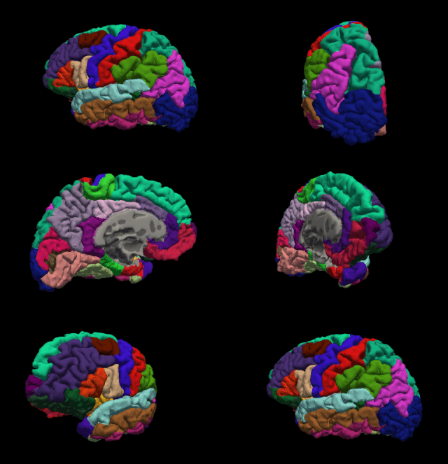
<span class="mw-page-title-main">FreeSurfer</span> Brain imaging software package

FreeSurfer is a brain imaging software package originally developed by Bruce Fischl, Anders Dale, Martin Sereno, and Doug Greve. Development and maintenance of FreeSurfer is now the primary responsibility of the Laboratory for Computational Neuroimaging at the Athinoula A. Martinos Center for Biomedical Imaging. FreeSurfer contains a set of programs with a common focus of analyzing magnetic resonance imaging (MRI) scans of brain tissue. It is an important tool in functional brain mapping and contains tools to conduct both volume based and surface based analysis. FreeSurfer includes tools for the reconstruction of topologically correct and geometrically accurate models of both the gray/white and pial surfaces, for measuring cortical thickness, surface area and folding, and for computing inter-subject registration based on the pattern of cortical folds.
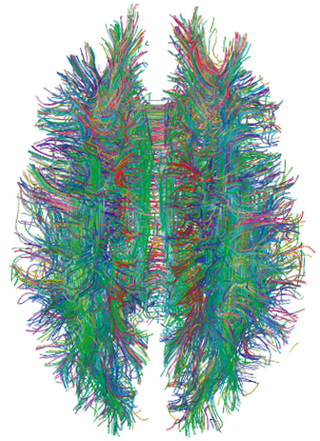
<span class="mw-page-title-main">Connectome</span> Comprehensive map of neural connections in the brain

A connectome is a comprehensive map of neural connections in the brain, and may be thought of as its "wiring diagram". An organism's nervous system is made up of neurons which communicate through synapses. A connectome is constructed by tracing the neuron in a nervous system and mapping where neurons are connected through synapses.

Connectomics is the production and study of connectomes: comprehensive maps of connections within an organism's nervous system. More generally, it can be thought of as the study of neuronal wiring diagrams with a focus on how structural connectivity, individual synapses, cellular morphology, and cellular ultrastructure contribute to the make up of a network. The nervous system is a network made of billions of connections and these connections are responsible for our thoughts, emotions, actions, memories, function and dysfunction. Therefore, the study of connectomics aims to advance our understanding of mental health and cognition by understanding how cells in the nervous system are connected and communicate. Because these structures are extremely complex, methods within this field use a high-throughput application of functional and structural neural imaging, most commonly magnetic resonance imaging (MRI), electron microscopy, and histological techniques in order to increase the speed, efficiency, and resolution of these nervous system maps. To date, tens of large scale datasets have been collected spanning the nervous system including the various areas of cortex, cerebellum, the retina, the peripheral nervous system and neuromuscular junctions.

<span class="mw-page-title-main">Default mode network</span> Large-scale brain network active when not focusing on an external task

In neuroscience, the default mode network (DMN), also known as the default network, default state network, or anatomically the medial frontoparietal network (M-FPN), is a large-scale brain network primarily composed of the dorsal medial prefrontal cortex, posterior cingulate cortex, precuneus and angular gyrus. It is best known for being active when a person is not focused on the outside world and the brain is at wakeful rest, such as during daydreaming and mind-wandering. It can also be active during detailed thoughts related to external task performance. Other times that the DMN is active include when the individual is thinking about others, thinking about themselves, remembering the past, and planning for the future.

<span class="mw-page-title-main">Magnetic resonance imaging of the brain</span>

Magnetic resonance imaging of the brain uses magnetic resonance imaging (MRI) to produce high quality two-dimensional or three-dimensional images of the brain and brainstem as well as the cerebellum without the use of ionizing radiation (X-rays) or radioactive tracers.

<span class="mw-page-title-main">Resting state fMRI</span> Type of functional magnetic resonance imaging

Resting state fMRI is a method of functional magnetic resonance imaging (fMRI) that is used in brain mapping to evaluate regional interactions that occur in a resting or task-negative state, when an explicit task is not being performed. A number of resting-state brain networks have been identified, one of which is the default mode network. These brain networks are observed through changes in blood flow in the brain which creates what is referred to as a blood-oxygen-level dependent (BOLD) signal that can be measured using fMRI.

The following outline is provided as an overview of and topical guide to brain mapping:

Connectograms are graphical representations of connectomics, the field of study dedicated to mapping and interpreting all of the white matter fiber connections in the human brain. These circular graphs based on diffusion MRI data utilize graph theory to demonstrate the white matter connections and cortical characteristics for single structures, single subjects, or populations.

Dynamic functional connectivity (DFC) refers to the observed phenomenon that functional connectivity changes over a short time. Dynamic functional connectivity is a recent expansion on traditional functional connectivity analysis which typically assumes that functional networks are static in time. DFC is related to a variety of different neurological disorders, and has been suggested to be a more accurate representation of functional brain networks. The primary tool for analyzing DFC is fMRI, but DFC has also been observed with several other mediums. DFC is a recent development within the field of functional neuroimaging whose discovery was motivated by the observation of temporal variability in the rising field of steady state connectivity research.

A brain atlas is composed of serial sections along different anatomical planes of the healthy or diseased developing or adult animal or human brain where each relevant brain structure is assigned a number of coordinates to define its outline or volume. Brain atlases are contiguous, comprehensive results of visual brain mapping and may include anatomical, genetic or functional features. A functional brain atlas is made up of regions of interest, where these regions are typically defined as spatially contiguous and functionally coherent patches of gray matter.

Randy L. Buckner is an American neuroscientist and psychologist whose research focuses on understanding how large-scale brain circuits support mental function and how dysfunction arises in illness.

<span class="mw-page-title-main">Russell Poldrack</span>

Russell "Russ" Alan Poldrack is an American psychologist and neuroscientist. He is a professor of psychology at Stanford University, associate director of Stanford Data Science, member of the Stanford Neuroscience Institute and director of the Stanford Center for Reproducible Neuroscience and the SDS Center for Open and Reproducible Science.

Brain–body interactions are patterns of neural activity in the central nervous system to coordinate the activity between the brain and body. The nervous system consists of central and peripheral nervous systems and coordinates the actions of an animal by transmitting signals to and from different parts of its body. The brain and spinal cord are interwoven with the body and interact with other organ systems through the somatic, autonomic and enteric nervous systems. Neural pathways regulate brain–body interactions and allow to sense and control its body and interact with the environment.

David C. Van Essen is an American neuroscientist specializing in neurobiology and studies the structure, function, development, connectivity and evolution of the cerebral cortex of humans and nonhuman relatives. After over two decades of teaching at the Washington University in St. Louis School of Medicine, he currently serves as an Alumni Endowed Professor of Neuroscience and maintains an active laboratory. Van Essen has held numerous positions, including Editor-in-Chief of the Journal of Neuroscience, Secretary of the Society for Neuroscience, and the President of the Society for Neuroscience from 2006 to 2007. Additionally, Van Essen has received numerous awards for his efforts in education and science, including the Krieg Cortical Discoverer Award from the Cajal Club in 2002, the Peter Raven Lifetime Achievement Award from St. Louis Academy of Science in 2007, and the Second Century Award in 2015 and the Distinguished Educator Award in 2017, both from Washington University School of Medicine.

Network neuroscience is an approach to understanding the structure and function of the human brain through an approach of network science, through the paradigm of graph theory. A network is a connection of many brain regions that interact with each other to give rise to a particular function. Network Neuroscience is a broad field that studies the brain in an integrative way by recording, analyzing, and mapping the brain in various ways. The field studies the brain at multiple scales of analysis to ultimately explain brain systems, behavior, and dysfunction of behavior in psychiatric and neurological diseases. Network neuroscience provides an important theoretical base for understanding neurobiological systems at multiple scales of analysis.

<span class="mw-page-title-main">Dimitri Van De Ville</span> Swiss-Belgian computer scientist and neuroscientist specialized in brain activity networks

Dimitri Van De Ville is a Swiss and Belgian computer scientist and neuroscientist specialized in dynamical and network aspects of brain activity. He is a professor of bioengineering at EPFL and the head of the Medical Image Processing Laboratory at EPFL's School of Engineering.

References

  1. NIH Launches the Human Connectome Project to Unravel the Brain's Connections, National Institutes of Health, 2009-07-15, retrieved 2013-02-16
  2. "The Human Connectome Project", NIH Blueprint for Neuroscience Research , National Institutes of Health , retrieved 2013-02-16
  3. 1 2 $40 million awarded to trace human brain's connections, National Institutes of Health, 2010-09-15, retrieved 2013-02-16, Souped-up scanners to reveal intricate circuitry in high resolution
  4. Ghosh, Pallab; Van Wedeen, M.D. (2013-02-16). "Inside the brain's wiring" (video). Boston: BBC News Online . Retrieved 2013-02-16.
  5. Geddes, Linda (2016). "Human brain mapped in unprecedented detail". Nature. doi:10.1038/nature.2016.20285. S2CID   89023120.
  6. "$40 Million Awarded to Trace Human Brain's Connections". NIMH. September 15, 2010. Archived from the original on January 10, 2012. Retrieved March 12, 2018.
  7. "Connectome - Homepage".
  8. "ConnectomeDB".
  9. "Sample diffusion MRI image of brain", Laboratory of Neuro Imaging , UCLA, archived from the original on 2012-03-05, retrieved 2013-02-16
  10. Fan, Qiuyun; Witzel, Thomas; Nummenmaa, Aapo; Van Dijk, Koene R. A.; Van Horn, John D.; Drews, Michelle K.; Somerville, Leah H.; Sheridan, Margaret A.; Santillana, Rosario M. (2016-01-01). "MGH-USC Human Connectome Project datasets with ultra-high b-value diffusion MRI". NeuroImage. 124 (Pt B): 1108–1114. doi:10.1016/j.neuroimage.2015.08.075. ISSN   1095-9572. PMC   4651764 . PMID   26364861.
  11. Improving SNR in high b-value diffusion imaging using Gmax = 300 mT/m human gradients, Proc. Intl. Soc. Mag. Reson. Med. 20 (2012) 2738
  12. "About the Center". 22 March 2019.
  13. Fan, Qiuyun; Nummenmaa, Aapo; Witzel, Thomas; Zanzonico, Roberta; Keil, Boris; Cauley, Stephen; Polimeni, Jonathan R.; Tisdall, Dylan; Van Dijk, Koene R. A. (2014-11-21). "Investigating the capability to resolve complex white matter structures with high b-value diffusion magnetic resonance imaging on the MGH-USC Connectom scanner". Brain Connectivity. 4 (9): 718–726. doi:10.1089/brain.2014.0305. ISSN   2158-0022. PMC   4238244 . PMID   25287963.
  14. "Components of the Human Connectome Project - Behavioral Testing - Connectome". humanconnectome.org. Retrieved 2013-03-08.
  15. Glasser, Matthew F; Smith, Stephen M; Marcus, Daniel S; Andersson, Jesper L R; Auerbach, Edward J; Behrens, Timothy E J; Coalson, Timothy S; Harms, Michael P; Jenkinson, Mark; Moeller, Steen; Robinson, Emma C; Sotiropoulos, Stamatios N; Xu, Junqian; Yacoub, Essa; Ugurbil, Kamil; Van Essen, David C (2016). "The Human Connectome Project's neuroimaging approach". Nature Neuroscience. 19 (9): 1175–87. doi:10.1038/nn.4361. PMC   6172654 . PMID   27571196.
  16. UpAndRunning. "Connectome - Homepage". www.humanconnectome.org. Retrieved 2017-11-28.
  17. UpAndRunning. "HCP Young Adult - Connectome - Publications". www.humanconnectome.org. Retrieved 2017-11-29.
  18. "Publications | Human Connectome Project". Archived from the original on 2017-05-07. Retrieved 2016-03-03.
  19. Toschi, Nicola; Duggento, Andrea; Passamonti, Luca (2017). "Functional connectivity in amygdalar-sensory/(pre)motor networks at rest: New evidence from the Human Connectome Project". European Journal of Neuroscience. 45 (9): 1224–1229. doi: 10.1111/ejn.13544 . PMID   28231395.
  20. 1 2 De Witte, Nele A. J; Mueller, Sven C (2016). "White matter integrity in brain networks relevant to anxiety and depression: Evidence from the human connectome project dataset". Brain Imaging and Behavior. 11 (6): 1604–1615. doi:10.1007/s11682-016-9642-2. hdl: 1854/LU-8163582 . PMID   27744495. S2CID   21758303.
  21. UpAndRunning. "Connectome - HCP Lifespan Studies". www.humanconnectome.org. Retrieved 2017-12-13.
  22. UpAndRunning. "Connectome - Human Connectome Studies Related To Disease". www.humanconnectome.org. Retrieved 2017-12-13.
  23. Smith, Stephen M; Beckmann, Christian F; Andersson, Jesper; Auerbach, Edward J; Bijsterbosch, Janine; Douaud, Gwenaëlle; Duff, Eugene; Feinberg, David A; Griffanti, Ludovica; Harms, Michael P; Kelly, Michael; Laumann, Timothy; Miller, Karla L; Moeller, Steen; Petersen, Steve; Power, Jonathan; Salimi-Khorshidi, Gholamreza; Snyder, Abraham Z; Vu, An T; Woolrich, Mark W; Xu, Junqian; Yacoub, Essa; Uğurbil, Kamil; Van Essen, David C; Glasser, Matthew F (2013). "Resting-state fMRI in the Human Connectome Project". NeuroImage. 80: 144–68. doi:10.1016/j.neuroimage.2013.05.039. PMC   3720828 . PMID   23702415.
  24. Griffanti, Ludovica; Douaud, Gwenaëlle; Bijsterbosch, Janine; Evangelisti, Stefania; Alfaro-Almagro, Fidel; Glasser, Matthew F; Duff, Eugene P; Fitzgibbon, Sean; Westphal, Robert; Carone, Davide; Beckmann, Christian F; Smith, Stephen M (2017). "Hand classification of fMRI ICA noise components". NeuroImage. 154: 188–205. doi:10.1016/j.neuroimage.2016.12.036. PMC   5489418 . PMID   27989777.
  25. Salimi-Khorshidi, Gholamreza; Douaud, Gwenaëlle; Beckmann, Christian F; Glasser, Matthew F; Griffanti, Ludovica; Smith, Stephen M (2014). "Automatic denoising of functional MRI data: Combining independent component analysis and hierarchical fusion of classifiers". NeuroImage. 90: 449–68. doi:10.1016/j.neuroimage.2013.11.046. PMC   4019210 . PMID   24389422.
Related Connectome Projects
Press releases
News reports