Imaging biomarker

Last updated

An imaging biomarker is a biologic feature, or biomarker detectable in an image. [1] In medicine, an imaging biomarker is a feature of an image relevant to a patient's diagnosis. For example, a number of biomarkers are frequently used to determine risk of lung cancer. First, a simple lesion in the lung detected by X-ray, CT, or MRI can lead to the suspicion of a neoplasm. The lesion itself serves as a biomarker, but the minute details of the lesion serve as biomarkers as well, and can collectively be used to assess the risk of neoplasm. Some of the imaging biomarkers used in lung nodule assessment include size, spiculation, calcification, cavitation, location within the lung, rate of growth, and rate of metabolism. Each piece of information from the image represents a probability. Spiculation increases the probability of the lesion being cancer. A slow rate of growth indicates benignity. These variables can be added to the patient's history, physical exam, laboratory tests, and pathology to reach a proposed diagnosis. Imaging biomarkers can be measured using several techniques, such as CT, electroencephalography, magnetoencephalography, and MRI.

Contents

History

Imaging biomarkers are as old as the X-ray itself. A feature of a radiograph that represent some kind of pathology was first coined "Roentgen signs" after Wilhelm Röntgen, the discoverer of the X-ray. [2] As the field of medical imaging developed and expanded to include numerous imaging modalities, imaging biomarkers have grown as well, in both quantity and complexity as finally in chemical imaging.

Quantitative imaging biomarkers

A quantitative imaging biomarkers (QIB) is an objective characteristic derived from an in vivo image measured on a ratio or interval scale as indicators of normal biological processes, pathogenic processes or a response to a therapeutic intervention. [3] advantage of QIB's over qualitative imaging biomarkers is that they are better suited to be used for follow-up of patients or in clinical trials. Examples of a frequently used QIB are the RECIST criteria, measuring the evolution in tumor size to assess treatment response for patients with cancer, the Nuchal scan used for prenatal screening, or the assessment of lesion load and brain atrophy for patients with multiple sclerosis.

Use in clinical trials

Clinical trials are known to be one of the most valuable sources of data in evidence-based medicine. For a pharmaceutical, device, or procedure to be approved for regular use in the U.S., it must be rigorously tested in clinical trials, and demonstrate sufficient efficacy. Unfortunately clinical trials are also extremely expensive and time consuming. End-points, such as morbidity and mortality, are used as measures to compare groups within a clinical trial. The most basic endpoint used in clinical trials, mortality, requires years and sometimes decades of follow-up to sufficiently assess. Morbidity, although potentially faster to measure than mortality, can also be a very difficult endpoint to measure clinically, as it is often very subjective. These are some of the reasons why biomarkers have been increasingly used in clinical trials to detect subtle changes in physiology and pathology before they can are detected clinically. The biomarkers act as surrogate endpoints. The use of surrogate endpoints has been shown to significantly decrease the time and resources used in clinical trials. Because surrogate end-points allow researchers to assess a marker rather than the patient, it allows participants to act as their own control, and in many cases allows for easier blinding.

In addition to surrogate endpoints, imaging biomarkers can be used as predictive classifiers, to assist in selecting appropriate candidates for particular treatment. Predictive classifiers are frequently used in molecular imaging in order to ensure enzymatic response to treatment.

FDA approval of surrogate end-points

The United States Congress and the Food and Drug Administration have acknowledged the value of imaging biomarkers as evidenced by recent actions that encourage their use. The FDA Modernization Act of 1997 was instituted to improve the regulatory process for medical products. Section 112 of the Act gives explicit authority to give expedited approval for drugs that treat serious conditions as long as it has shown to have an effect on a surrogate end-point that reasonably indicates a clinical benefit. Other provisions enables monitoring of the products following market approval to ensure the efficacy of the surrogate end-points and requires the FDA to establish a program that promotes the development and use of surrogate end-points for serious diseases. Although the act does not specifically mention the use of surrogate end-points for medical devices, section 205 requires that the "least burdensome means necessary" be used in their approval. [4] The wording is much more general than the provision for pharmaceuticals, but is generally accepted that surrogate endpoints will often qualify as being the "least burdensome means".

Qualification and validation

Developing an understanding of clinical significance for specific biomarkers can be a difficult process. There are two steps of certification for a surrogate endpoint to be fully established: Qualification and Validation. For a biomarker to become qualified it must go through a somewhat formal qualification process. A request must be submitted to IPRG to qualify an imaging biomarker for a specific use. The Biomarker Qualification Review Team, recruited from nonclinical and clinical review divisions, assesses the context and available data regarding the biomarker. They also evaluate the qualification study strategy methods and results and ultimately make a decision to accept or reject. After qualification, a biomarker may have limited use as a surrogate endpoint. They may be used in phase I and II clinical trials, but can only be used in phase III trials for early futility analyses.

There are two steps to validation, probable validation and known validation. "Probable validation" requires widespread agreement in the medical or scientific community as to its efficacy. "Known validation" requires a scientific framework or body of evidence that appears to elucidate the marker’s efficacy. [5] For full validation, a biomarker must demonstrate that the treatment versus control differences are similar to the treatment versus control differences for clinical outcome. It is not sufficient to simply demonstrate that the biomarker responders survive longer than the biomarker non-responders.

Quality

The following are 3 measures of quality to determine the strength of biomarker for use in clinical trials. [6]

  1. The presence of the imaging biomarker is closely coupled or linked to the presence of the target disease or condition.
  2. The detection and/or quantitative measurement of the imaging biomarker is accurate, reproducible, and feasible over time.
  3. The measured changes over time in the imaging biomarker are closely coupled or linked to the success or failure of the therapeutic effect and the true end-point sought for the medical therapy being evaluated.

Organizations

Because the project of compiling a library of validated biomarkers requires an enormous amount of resources, the FDA has encouraged the creation of consortia between public and private organization in order to facilitate the sharing of data for the qualification and validation of biomarkers.

The Biomarkers Consortium was created by the Foundation for the National Institutes of Health, National Institute of Health, Food and Drug Administration, and Pharmaceutical Research and Manufacturers of America. It is a public-private biomedical research partnership aimed to provide grants for the generation of data for clinical biomarker qualification.

The Predictive Safety Testing Consortium, was created by the Critical Path Institute and the Food and Drug Administration to develop a framework needed for data sharing between its members in order to make biomarker qualification easier. They are also working with regulatory agencies to replace the currently unstructured qualification process.

In 2001, the Radiology department at Massachusetts General Hospital, founded the MGH Center for Biomarkers in Imaging, a center dedicated to encourage the development and use of imaging biomarkers. Their initial project was to catalogue the known biomarkers in order to make them readily available to scientists, regulators, and industry representatives (now available on their website). The catalogue includes the pathology specific to the biomarkers, the investigator(s) involved in creating and using the biomarker, and the modalities used in the detection of the biomarker.

International Cancer Biomarker Consortium was created to assist in discovery of biomarkers by facilitating coordinated research and by leveraging resources. Each international team chooses a cancer site(s) for study, functions independently, and secures its own funding. The president of the organization, Leland Hartwell, is a nobel prize winner for physiology/medicine in 2001.

Uniform Protocols for Imaging in Clinical Trials (UPICT) was created by the American College of Radiology.

Imaging Response Assessment Teams was created by the National Cancer Institute and AACI to advance the role of imaging in assessment of response to therapy and to increase the application of quantitative, anatomic, functional, and molecular imaging endpoints in clinical therapeutic trials. Aims to strengthen clinical collaboration between imaging scientists and oncologic investigators.

Oncology Biomarker Qualification Initiative was created by the Food and Drug Administration and the National Cancer Institute to qualify new cancer biomarkers. Their first project involves PET imaging in non-Hodgkin lymphoma.

Related Research Articles

<span class="mw-page-title-main">Proof of concept</span> Realization of a certain method or idea in order to demonstrate its feasibility

Proof of concept, also known as proof of principle, is a realization of a certain method or idea in order to demonstrate its feasibility, or a demonstration in principle with the aim of verifying that some concept or theory has practical potential. A proof of concept is usually small and may or may not be complete.

In clinical trials, a surrogate endpoint is a measure of effect of a specific treatment that may correlate with a real clinical endpoint but does not necessarily have a guaranteed relationship. The National Institutes of Health (USA) defines surrogate endpoint as "a biomarker intended to substitute for a clinical endpoint".

Clinical endpoints or clinical outcomes are outcome measures referring to occurrence of disease, symptom, sign or laboratory abnormality constituting a target outcome in clinical research trials. The term may also refer to any disease or sign that strongly motivates withdrawal of an individual or entity from the trial, then often termed a humane (clinical) endpoint.

<span class="mw-page-title-main">Sunitinib</span> Cancer medication

Sunitinib, sold under the brand name Sutent, is an anti-cancer medication. It is a small-molecule, multi-targeted receptor tyrosine kinase (RTK) inhibitor that was approved by the FDA for the treatment of renal cell carcinoma (RCC) and imatinib-resistant gastrointestinal stromal tumor (GIST) in January 2006. Sunitinib was the first cancer drug simultaneously approved for two different indications.

In biomedical contexts, a biomarker, or biological marker, is a measurable indicator of some biological state or condition. Biomarkers are often measured and evaluated using blood, urine, or soft tissues to examine normal biological processes, pathogenic processes, or pharmacologic responses to a therapeutic intervention. Biomarkers are used in many scientific fields.

In medicine, a biomarker is a measurable indicator of the severity or presence of some disease state. It may be defined as a "cellular, biochemical or molecular alteration in cells, tissues or fluids that can be measured and evaluated to indicate normal biological processes, pathogenic processes, or pharmacological responses to a therapeutic intervention." More generally a biomarker is anything that can be used as an indicator of a particular disease state or some other physiological state of an organism. According to the WHO, the indicator may be chemical, physical, or biological in nature - and the measurement may be functional, physiological, biochemical, cellular, or molecular.

Response evaluation criteria in solid tumors (RECIST) is a set of published rules that define when tumors in cancer patients improve ("respond"), stay the same ("stabilize"), or worsen ("progress") during treatment. The criteria were published in February 2000 by an international collaboration including the European Organisation for Research and Treatment of Cancer (EORTC), National Cancer Institute of the United States, and the National Cancer Institute of Canada Clinical Trials Group. Today, the majority of clinical trials evaluating cancer treatments for objective response in solid tumors use RECIST. These criteria were developed and published in February 2000, and subsequently updated in 2009.

Leronlimab is a humanized monoclonal antibody targeted against the CCR5 receptor found on T lymphocytes of the human immune system. It is being investigated as a potential therapy in the treatment of COVID-19, triple negative breast cancer, and HIV infection. The United States Food and Drug Administration has designated PRO 140 for fast-track approval. In February 2008, the drug entered Phase 2 clinical trials and a phase 3 trial was begun in 2015. In February 2018, Cytodyn Inc reported that the primary endpoint had been achieved in the PRO 140 pivotal combination therapy trial in HIV infection. In 2020 CytoDyn submitted a fast-track biologics license application for treatment of CCR5-tropic HIV-1 Infection.

<span class="mw-page-title-main">Gadobutrol</span> Chemical compound

Gadobutrol (INN) (Gd-DO3A-butrol) is a gadolinium-based MRI contrast agent (GBCA).

Progression-free survival (PFS) is "the length of time during and after the treatment of a disease, such as cancer, that a patient lives with the disease but it does not get worse". In oncology, PFS usually refers to situations in which a tumor is present, as demonstrated by laboratory testing, radiologic testing, or clinically. Similarly, "disease-free survival" is the length of time after patients have received treatment and have no detectable disease.

The following outline is provided as an overview of and topical guide to clinical research:

DirectHit is a pharmacodiagnostic test used to determine the tumor sensitivity or resistance to drug regimens recommended for the treatment of breast cancer by the National Comprehensive Cancer Network. It is a noninvasive test performed on small amounts of tissue removed during the original surgery lumpectomy, mastectomy, or core biopsy. DirectHit was developed by CCC Diagnostics Inc., a biotechnology company established by former researchers from Johns Hopkins University. DirectHit was launched on 14 January 2010. Currently, it is the only available test for predicting treatment outcomes for anticancer chemotherapy drugs for breast cancer.

<span class="mw-page-title-main">Nintedanib</span> Chemical compound

Nintedanib, sold under the brand names Ofev and Vargatef, is an oral medication used for the treatment of idiopathic pulmonary fibrosis and along with other medications for some types of non-small-cell lung cancer.

<span class="mw-page-title-main">Phases of clinical research</span> Clinical trial stages using human subjects

The phases of clinical research are the stages in which scientists conduct experiments with a health intervention to obtain sufficient evidence for a process considered effective as a medical treatment. For drug development, the clinical phases start with testing for drug safety in a few human subjects, then expand to many study participants to determine if the treatment is effective. Clinical research is conducted on drug candidates, vaccine candidates, new medical devices, and new diagnostic assays.

<span class="mw-page-title-main">Cancer biomarker</span> Substance or process that is indicative of the presence of cancer in the body

A cancer biomarker refers to a substance or process that is indicative of the presence of cancer in the body. A biomarker may be a molecule secreted by a tumor or a specific response of the body to the presence of cancer. Genetic, epigenetic, proteomic, glycomic, and imaging biomarkers can be used for cancer diagnosis, prognosis, and epidemiology. Ideally, such biomarkers can be assayed in non-invasively collected biofluids like blood or serum.

The United States Food and Drug Administration (FDA) initiated the FDA Accelerated Approval Program in 1992 to allow faster approval of drugs for serious conditions that fill an unmet medical need. The faster approval relies on use of surrogate endpoints. Drug approval typically requires clinical trials with endpoints that demonstrate a clinical benefit, such as increased survival for cancer patients. Drugs with accelerated approval can initially be tested in clinical trials that use a surrogate endpoint, or something that is thought to predict clinical benefit. Surrogate endpoints typically require less time, and in the case of a cancer patient, it is much faster to measure a reduction in tumor size, for example, than overall patient survival.

In the field of medicine, radiomics is a method that extracts a large number of features from medical images using data-characterisation algorithms. These features, termed radiomic features, have the potential to uncover tumoral patterns and characteristics that fail to be appreciated by the naked eye. The hypothesis of radiomics is that the distinctive imaging features between disease forms may be useful for predicting prognosis and therapeutic response for various cancer types, thus providing valuable information for personalized therapy. Radiomics emerged from the medical fields of radiology and oncology and is the most advanced in applications within these fields. However, the technique can be applied to any medical study where a pathological process can be imaged.

<span class="mw-page-title-main">Adaptive design (medicine)</span> Concept in medicine referring to design of clinical trials

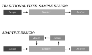
In an adaptive design of a clinical trial, the parameters and conduct of the trial for a candidate drug or vaccine may be changed based on an interim analysis. Adaptive design typically involves advanced statistics to interpret a clinical trial endpoint. This is in contrast to traditional single-arm clinical trials or randomized clinical trials (RCTs) that are static in their protocol and do not modify any parameters until the trial is completed. The adaptation process takes place at certain points in the trial, prescribed in the trial protocol. Importantly, this trial protocol is set before the trial begins with the adaptation schedule and processes specified. Adaptions may include modifications to: dosage, sample size, drug undergoing trial, patient selection criteria and/or "cocktail" mix. The PANDA provides not only a summary of different adaptive designs, but also comprehensive information on adaptive design planning, conduct, analysis and reporting.

<span class="mw-page-title-main">Piflufolastat F-18</span> Chemical compound

Piflufolastat F-18, sold under the brand name Pylarify among others, is a radioactive diagnostic agent used for positron emission tomography (PET) imaging. It is given by intravenous injection.

References

  1. Smith JJ, Sorensen AG, Thrall JH (June 2003). "Biomarkers in imaging: realizing radiology's future". Radiology. 227 (3): 633–8. doi:10.1148/radiol.2273020518. PMID   12663828.
  2. Meschan, Farrer-Meschan, Roentgen Signs in Clinical Diagnosis, W.B. Saunders Company, Philadelphia, 1956
  3. Kessler LG, Barnhart HX, Buckler AJ, Choudhury KR, Kondratovich MV, Toledano A, Guimaraes AR, Filice R, Zhang Z, Sullivan DC (February 2015). "The emerging science of quantitative imaging biomarkers terminology and definitions for scientific studies and regulatory submissions". Stat Methods Med Res. 24 (1): 9–26. doi:10.1177/0962280214537333. PMID   24919826. S2CID   21413242.
  4. Smith JJ, Shyjan AM (2000). "Defining "least burdensome means" under the Food and Drug Administration Modernization Act of 1997". Food Drug Law J. 55 (3): 435–47. PMID   11824468.
  5. Goodsaid F, Frueh F (March 2007). "Biomarker qualification pilot process at the US Food and Drug Administration". AAPS J. 9 (1): E105–8. doi:10.1208/aapsj0901010. PMC   2751298 . PMID   17408233.
  6. Schatzkin A, Gail M (2002). "The promise and peril of surrogate endpoints in cancer research". Nat Rev Cancer. 2 (1): 19–27. doi:10.1038/nrc702. PMID   11902582. S2CID   1959952.