Lateral pectoral nerve

Last updated

Lateral pectoral nerve
Nerves of the left upper extremity.gif
Nerves of the left upper extremity. (Lateral anterior thoracic visible in upper right.)
Details
From Lateral cord
Innervates Pectoralis major
Identifiers
Latin nervus pectoralis lateralis
TA98 A14.2.03.018
TA2 6419
FMA 65296
Anatomical terms of neuroanatomy

The lateral pectoral nerve (also known as the lateral anterior thoracic nerve) arises from the lateral cord of the brachial plexus, [1] [2] and through it from the C5-7. [1] [2]

Contents

It passes across the axillary artery and vein, [3] pierces the clavipectoral (coracoclavicular) fascia, and enters the deep surface of the pectoralis major to innervate it. [1] [4]

Function

The lateral pectoral nerve provides motor innervation to the pectoralis major muscle. [2] [5]

Although this nerve is described as mostly motor, it also has been considered to carry proprioceptive and nociceptive fibers. It arises either from the lateral cord or directly from the anterior divisions of the upper and middle trunks of the brachial plexus. This is unlike the medial pectoral nerve, which derives from the medial cord (or directly from the anterior division of the lower trunk). It splits into four to seven branches that pierce the clavipectoral fascia to innervate the entire pectoralis major or its superior portion.

The medial and lateral pectoral nerves form a connection, around the axillary artery, called the ansa pectoralis. The lateral pectoral nerve has been described as double, while the medial pectoral nerve has been described as single. [6]

Clinical significance

Postoperative Care

The lateral pectoral nerve is important in the pain response after breast augmentation and mastectomy, and especially in breast implant surgery, when the implant is inserted by the subpectoral route. The pectoral nerves can be anesthetized (blocked) intraoperatively by the surgeon under direct vision by three injections - one to block the medial pectoral nerve, the second to block the perforating branches of the medial pectoral nerve, and the third to block the lateral pectoral nerve. An ultrasound-guided pectoral nerve block can also be performed preventively before the operation by an anesthesiologist, experienced in regional anesthesia. It is safe and relies on ultrasound imaging to localize the pectoralis major and minor muscles, the presumed course of the pectoral nerves and the optimal spread of the local anesthetic. [7]

Nerve Block

Blockade of the lateral pectoral nerve is helpful in cases such as shoulder dislocation and other orthopedic procedures, involving the shoulder. Spasms of the pectoralis major muscle and resulting severe pain (acute or chronic) may be reduced by pectoral nerve block or neuromuscular relaxation. Decreasing the pectoral muscle tone intraoperatively by neuromuscular relaxation (paralytic agents) or by a nerve block (local anesthetic injection), can facilitate better cosmetic results during breast augmentation or post-mastectomy breast implantation. “The skin projection point of the neurovascular bundle (NVB) represents the denervation point (DP).” The NVB (thoracoacromial artery and vein, plus the lateral pectoral nerve) may be the guide for local anesthetic applications in order to achieve pectoral muscle denervation. “Routine botulinum toxin infiltration of the chest wall musculature at the time of mastectomy and immediate reconstruction… would paralyze the muscles and reduce the postoperative pain caused by muscle spasm.” [8]

Damage

The lateral pectoral nerve can be damaged, particularly during surgery, by cauterisation and avulsion. [4] Trauma to the brachial plexus can cut off innervation to the lateral pectoral nerve. [9]

See also

Related Research Articles

<span class="mw-page-title-main">Radial nerve</span> Nerve in the human body that supplies the posterior portion of the upper limb

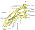
The radial nerve is a nerve in the human body that supplies the posterior portion of the upper limb. It innervates the medial and lateral heads of the triceps brachii muscle of the arm, as well as all 12 muscles in the posterior osteofascial compartment of the forearm and the associated joints and overlying skin.

<span class="mw-page-title-main">Median nerve</span> Nerve of the upper limb

The median nerve is a nerve in humans and other animals in the upper limb. It is one of the five main nerves originating from the brachial plexus.

<span class="mw-page-title-main">Axillary nerve</span> Nerve of the human body near the armpit

The axillary nerve or the circumflex nerve is a nerve of the human body, that originates from the brachial plexus at the level of the axilla (armpit) and carries nerve fibers from C5 and C6. The axillary nerve travels through the quadrangular space with the posterior circumflex humeral artery and vein to innervate the deltoid and teres minor.

<span class="mw-page-title-main">Dorsal scapular nerve</span> Branch of the brachial plexus that supplies rhomboid muscles and levator scapulae

The dorsal scapular nerve is a branch of the brachial plexus, usually derived from the ventral ramus of cervical nerve C5. It provides motor innervation to the rhomboid major muscle, rhomboid minor muscle, and levator scapulae muscle.

<span class="mw-page-title-main">Long thoracic nerve</span> Large nerve

The long thoracic nerve is a branch of the brachial plexus derived from cervical nerves C5-C7 that innervates the serratus anterior muscle.

<span class="mw-page-title-main">Deltoid muscle</span> Shoulder muscle

The deltoid muscle is the muscle forming the rounded contour of the human shoulder. It is also known as the 'common shoulder muscle', particularly in other animals such as the domestic cat. Anatomically, the deltoid muscle appears to be made up of three distinct sets of muscle fibers, namely the

  1. anterior or clavicular part
  2. posterior or scapular part
  3. intermediate or acromial part
<span class="mw-page-title-main">Levator scapulae muscle</span> Slender skeletal muscle at the back and side of the neck

The levator scapulae is a slender skeletal muscle situated at the back and side of the neck. It originates from the transverse processes of the four uppermost cervical vertebrae; it inserts onto the upper portion of the medial border of the scapula. It is innervated by the cervical nerves C3-C4, and frequently also by the dorsal scapular nerve. As the Latin name suggests, its main function is to lift the scapula.

<span class="mw-page-title-main">Pectoralis major</span> Main human chest muscle

The pectoralis major is a thick, fan-shaped or triangular convergent muscle of the human chest. It makes up the bulk of the chest muscles and lies under the breast. Beneath the pectoralis major is the pectoralis minor muscle.

<span class="mw-page-title-main">Pectoralis minor</span> Human chest muscle that protracts the shoulder

Pectoralis minor muscle is a thin, triangular muscle, situated at the upper part of the chest, beneath the pectoralis major in the human body. It arises from ribs III-V; it inserts onto the coracoid process of the scapula. It is innervated by the medial pectoral nerve. Its function is to stabilise the scapula by holding it fast in position against the chest wall.

<span class="mw-page-title-main">Cervical plexus</span> Network of nerves in the neck

The cervical plexus is a nerve plexus of the anterior rami of the first four cervical spinal nerves C1-C4. The cervical plexus provides motor innervation to some muscles of the neck, and the diaphragm; it provides sensory innervation to parts of the head, neck, and chest.

<span class="mw-page-title-main">Teres major muscle</span> Muscle of the upper limb

The teres major muscle is a muscle of the upper limb. It attaches to the scapula and the humerus and is one of the seven scapulohumeral muscles. It is a thick but somewhat flattened muscle.

<span class="mw-page-title-main">Medial cord</span>

The medial cord is the part of the brachial plexus formed by of the anterior division of the lower trunk (C8-T1). Its name comes from it being medial to the axillary artery as it passes through the axilla. The other cords of the brachial plexus are the posterior cord and lateral cord.

<span class="mw-page-title-main">Medial pectoral nerve</span>

The medial pectoral nerve is (typically) a branch of the medial cord of the brachial plexus and is derived from spinal nerve roots C8-T1. It provides motor innervation to the pectoralis minor muscle, and the lower half of the pectoralis major muscle. It runs along the inferior border of the pectoralis minor muscle.

<span class="mw-page-title-main">Thoracodorsal nerve</span> Nerve that supplies the latissimus dorsi

The thoracodorsal nerve is a nerve present in humans and other animals, also known as the middle subscapular nerve or the long subscapular nerve. It supplies the latissimus dorsi muscle.

<span class="mw-page-title-main">Anterior superior iliac spine</span> Bony projection of the iliac bone

The anterior superior iliac spine (ASIS) is a bony projection of the iliac bone, and an important landmark of surface anatomy. It refers to the anterior extremity of the iliac crest of the pelvis. It provides attachment for the inguinal ligament, and the sartorius muscle. The tensor fasciae latae muscle attaches to the lateral aspect of the superior anterior iliac spine, and also about 5 cm away at the iliac tubercle.

<span class="mw-page-title-main">Lateral cutaneous nerve of thigh</span> Nerve of the thigh

The lateral cutaneous nerve of the thigh is a cutaneous nerve of the thigh. It originates from the dorsal divisions of the second and third lumbar nerves from the lumbar plexus. It passes under the inguinal ligament to reach the thigh. It supplies sensation to the skin on the lateral part of the thigh by an anterior branch and a posterior branch.

<span class="mw-page-title-main">Lower subscapular nerve</span>

The lower subscapular nerve, also known as the inferior subscapular nerve, is the third branch of the posterior cord of the brachial plexus. It innervates the inferior portion of the subscapularis muscle and the teres major muscle.

<span class="mw-page-title-main">Bicipital aponeurosis</span> Distal end of the biceps muscle

The bicipital aponeurosis is a broad aponeurosis of the biceps brachii, which is located in the cubital fossa of the elbow. It separates superficial from deep structures in much of the fossa.

<span class="mw-page-title-main">Clavipectoral triangle</span>

The clavipectoral triangle is an anatomical region found in humans and other animals. It is bordered by the following structures:

<span class="mw-page-title-main">Brachial plexus block</span>

Brachial plexus block is a regional anesthesia technique that is sometimes employed as an alternative or as an adjunct to general anesthesia for surgery of the upper extremity. This technique involves the injection of local anesthetic agents in close proximity to the brachial plexus, temporarily blocking the sensation and ability to move the upper extremity. The subject can remain awake during the ensuing surgical procedure, or they can be sedated or even fully anesthetized if necessary.

References

PD-icon.svgThis article incorporates text in the public domain from page 933 of the 20th edition of Gray's Anatomy (1918)

  1. 1 2 3 Kahn, Elyne; Yang, Lynda J. -S. (1 January 2015), Tubbs, R. Shane; Rizk, Elias; Shoja, Mohammadali M.; Loukas, Marios (eds.), "Chapter 40 - Anatomy of the Lateral Cord and Its Branches", Nerves and Nerve Injuries, San Diego: Academic Press, pp. 547–551, doi:10.1016/b978-0-12-410390-0.00042-1, ISBN   978-0-12-410390-0 , retrieved 19 October 2020
  2. 1 2 3 Baur, Dale A.; Horan, Michael P.; Rodriguez, Juan C. (1 January 2012), Bagheri, Shahrokh C.; Bell, R. Bryan; Khan, Husain Ali (eds.), "Chapter 68 - The Pectoralis Major Myocutaneous Flap", Current Therapy In Oral and Maxillofacial Surgery, Saint Louis: W.B. Saunders, pp. 566–572, doi:10.1016/b978-1-4160-2527-6.00068-2, ISBN   978-1-4160-2527-6 , retrieved 26 October 2020
  3. Rea, Paul (1 January 2015), Rea, Paul (ed.), "Chapter 2 - Upper Limb Nerve Supply", Essential Clinically Applied Anatomy of the Peripheral Nervous System in the Limbs, Academic Press, pp. 41–100, doi:10.1016/b978-0-12-803062-2.00002-4, ISBN   978-0-12-803062-2 , retrieved 26 October 2020
  4. 1 2 Willey, Shawna C.; Feldman, Elizabeth D. (1 January 2009), Evans, Stephen R. T. (ed.), "Chapter 46 - Mastectomy", Surgical Pitfalls, Philadelphia: W.B. Saunders, pp. 475–487, doi:10.1016/b978-141602951-9.50058-x, ISBN   978-1-4160-2951-9 , retrieved 26 October 2020
  5. Wei, William Ignace; Chan, Yu-wai (1 January 2009), Wei, Fu-Chan; Mardini, Samir (eds.), "CHAPTER 17 - Pectoralis major flap", Flaps and Reconstructive Surgery, Edinburgh: W.B. Saunders, pp. 175–192, doi:10.1016/b978-0-7216-0519-7.00017-4, ISBN   978-0-7216-0519-7 , retrieved 26 October 2020
  6. Porzionato, Macchi; Stecco (2012). "Surgical Anatomy of the Pectoral Nerves and the Pectoral Musculature". Clinical Anatomy. 25 (5): 559–575. doi:10.1002/ca.21301. PMID   22125052. S2CID   22125503.
  7. Desroches, Jean; Grabs (2013). "). "Selective Ultrasound Guided Pectoral Nerve Targeting in Breast Augmentation: How to Spare the Brachial Plexus Cords?"". Clinical Anatomy. 26 (1): 49–55. doi:10.1002/ca.22117. PMID   22730005. S2CID   26711722.
  8. Titiz, Izzet; Ozel; Ozel; Toros; Marur; Yildirim; Erdogdu; Kara (19 August 2010). "Denervation Point for Neuromuscular Blockade on Lateral Pectoral Nerves: A Cadaver Study". Surgical and Radiologic Anatomy. 33 (2): 105–108. doi:10.1007/s00276-010-0712-7. PMID   20721553. S2CID   6959180.
  9. Larsen, Mikko; Bishop, Allen T.; Shin, Alexander Y. (1 January 2018), Morrey, Bernard F.; Sanchez-Sotelo, Joaquin; Morrey, Mark E. (eds.), "117 - Flaccid Dysfunction", Morrey's the Elbow and its Disorders (Fifth Edition), Philadelphia: Content Repository Only!, pp. 1078–1098, ISBN   978-0-323-34169-1 , retrieved 26 October 2020

Additional images