Livedoid vasculopathy

Last updated
Livedoid vasculopathy
Other namesLivedoid vasculitis, Livedo reticularis with summer/winter ulceration , Segmental hyalinizing vasculitis. [1]
Livedoid Vasculopathy.jpg
Livedoid Vasculopathy ulcer at the ankle.
Specialty Dermatology
Symptoms Livedoid changes, atrophie blanche, and uclers. [2]
Risk factors Antiphospholipid antibodies, thrombosis-predisposing genetic disorders, and paraproteinemias. [2]
Diagnostic method Histopathology. [2]
Differential diagnosis Chronic venous disease, peripheral vascular disease, and vasculitis. [2]
Treatment Pain management, wound care, smoking cessation, compression, antiplatelet agent, and anticoagulants. [2]
Frequency1 in 1,00,000 per year. [3]

Livedoid vasculopathy(LV) is an uncommon thrombotic dermal vasculopathy that is characterized by excruciating, recurrent ulcers on the lower limbs. [4] Livedo racemosa, a painful ulceration in the distal regions of the lower extremities, is the characteristic clinical appearance. It heals to form porcelain-white, atrophic scars, also known as Atrophie blanche. [5]

Contents

Livedoid vasculopathy has been linked to various conditions that can induce hypercoagulability, including neoplasms, autoimmune connective-tissue diseases, and inherited and acquired thrombophilias. [5]

The history, clinical findings, and histopathological analysis are combined to make the diagnosis. [5]

Prompt and suitable intervention mitigates discomfort and averts the formation of wounds and additional complications. In addition to general supportive measures, anticoagulants and antiplatelet medications can be considered the first-line treatments. [6]

Signs and symptoms

Recurrent focal non-inflammatory thrombosis of the superior superficial and medium dermal venulae, particularly on the lower extremities, bilaterally, is the initial clinical manifestation; upper extremity involvement has also been documented. [7] Livedo racemosa or, less commonly, livedo reticularis are symptoms of such thrombosis, which causes blood and pressure to build up in the dermal superficial veins. [8] The oxygen partial pressure in the skin decreases as a result of the blood flow obstruction, triggering a cutaneous response that presents as pruritus with itchy papules and erythematous-violaceous, purpuric plaques. They quickly develop into bleeding vesicles, or bullae, which, when they burst, produce painful, small ulcers about 5 mm in diameter. These ulcers eventually combine to form painful, confluent, reticulate, and geometric ulcerations. [7]

Complications

Due to the tissue exposure, the primary acute complications are pain and secondary infection. Atrophic scars, persistent hyperpigmentation, mononeuritis multiplex from vasa nervorum thrombosis, and cutaneous hemosiderosis in the lower limbs from erythrocytes oozing from the high-pressure regimen veins due to hemosiderin deposits in the skin are among the chronic complications associated with livedoid vasculopathy. [7]

Causes

Few things are known about the origins of dermal vessel thrombosis in livedoid vasculopathy, what sets off its initial episodes and relapses, and why it primarily affects the lower limbs. [4]

Risk factors

Livedoid vasculopathy has been linked to rheumatoid arthritissystemic lupus erythematosus, sclerodermapolyarteritis nodosa, mixed and undifferentiated connective tissue diseases, and Sjogren's syndrome. Individuals with systemic lupus erythematosus who also have antiphospholipid antibody syndrome are more vulnerable. [9]

Patients with hematological or solid organ cancers may also experience livedoid vasculopathy. Livedoid vasculopathy may deteriorate during pregnancy, particularly in the third trimester, though fetal compromise has not been documented. Still, a sizable fraction of cases are idiopathic. [9]

Genetics

Livedoid vasculopathy is caused by a number of inherited and acquired coagulation abnormalities, such as polymorphisms in factor V, plasminogen activator inhibitor-1 (PAI-1), prothrombin, and methylenetetrahydrofolate reductase (MTHFR). [6]

Mechanism

At this point, the pathomechanism of livedoid vasculopathy is not fully understood. [5] At first, livedoid vasculopathy was thought to be vasculitis. [10] Presently, livedoid vasculopathy is understood to be a vascular illness in which procoagulatory factors predominate, resulting in hypercoagulability. [9] Defects in endothelial dysfunction, such as reduced plasminogen activation, platelet dysfunction, or increased or restricted fibrin formation or lysis, may be the cause of the thrombotic effect. [5] As a diffusion barrier, fibrin deposition and thrombus formation cause a reduction in oxygen availability, which causes necrosis. [9] [11] Furthermore, insufficient tissue perfusion results in inadequate wound healing, creating a vicious cycle. [6] The so-called Virchow trias, hypercoagulability, stasis, and endothelial damage, also serve as risk factors for livedoid vasculopathy microvascular thrombosis. [11] [12] The manifestation of livedoid vasculopathy on the lower extremities is thought to be caused by variations in temperature and perfusion pressure, as well as a lower concentration of thrombolytic factors. [12] [13]

Diagnosis

To diagnose livedoid vasculopathy and its causes, a thorough history, dermatological examination, and laboratory work-up are necessary. [9] A skin biopsy should be performed to confirm the diagnosis of livedoid vasculopathy when it is clinically suspected. The most suitable type of biopsy is a fusiform incisional biopsy that contains subcutaneous fat. [6]

Skin biopsies' histology reveals endothelial proliferation, fibrin deposition in the vessel walls (which is frequently difficult to identify), and a high frequency of intraluminal hyaline thrombi in blood vessels, particularly in the upper and middle dermis, during the acute phase. Leukocytoclasia and a spare perivascular inflammatory infiltrate may be seen in the acute phase, but these results are not essential for the diagnosis. [14]

Following histopathological confirmation of the diagnosis, a more thorough evaluation of any potential underlying illnesses may be conducted. Naturally, the first steps in the assessment process should be a thorough history, a review of the systems, and a physical examination to look for any signs of underlying diseases. For every patient, thrombophilia laboratory testing is advised. More testing is necessary to determine whether thrombophilia is inherited or acquired, including looking into coagulating factors and their mutations. [6]

In the event that pertinent data suggestive of connective tissue diseases, such as scleroderma, rheumatoid arthritisantiphospholipid antibody syndrome, systemic lupus erythematosus, and mixed connective tissue disease, detailed laboratory research should be conducted. Furthermore, when paraproteinemia or solid organ cancers are suspected, tests for protein electrophoresisIg kappa and lambda chain levels, and immunofixation should be carried out. Again, testing for HIV and hepatitis should be done if there is a suspicion of an underlying infection. [6]

The initial step in the clinical work-up should be considering the differential diagnoses of additional common causes of atrophie blanche. Vasculitis, peripheral arterial vascular disease, and lower extremity chronic venous insufficiency are the most frequent conditions to be taken into account during the differential diagnosis process. Peripheral artery diseases are diagnosed with the help of clinical signs, abnormal arterial Doppler ultrasound findings, and the ankle-brachial index test. Cutaneous polyarteritis nodosa is another common differential diagnosis that causes similar cutaneous lesions on the legs. A proper skin biopsy can help differentiate vasculitis from livedoid vasculopathy. [6]

Treatment

Although there are numerous distinct treatment modalities for livedoid vasculopathy, no published, standardized, evidence-based therapeutic strategies exist. [5] The improvement of skin lesions, avoidance of relapses, and pain relief are the main goals of treatment for livedoid vasculopathy. [15] Since not every patient responds to a single therapy approach equally, it is necessary to evaluate or combine a number of treatment options. [6]

Treating pain related to ulcers with analgesia is crucial and frequently the patients' top priority. [16] UV light, compression, hyperbaric oxygen, and routine wound debridement are examples of local therapies for livedoid vasculopathy. [4] Hyperbaric oxygen and compression therapy have been demonstrated to enhance fibrinolysis in addition to their respective roles in reducing edema and mitigating reperfusion injury. [16] [17]

The most widely documented treatment for livedoid vasculopathy is oral anticoagulation, which directly addresses dermal vessel thrombosis. [15] The most widely used of these is rivaroxaban, which showed a significant reduction in pain after 12 weeks of therapy in an uncontrolled phase 2a trial. [18] Common substitutes are antiplatelet agents like aspirin and pentoxifylline. [4] Patients who are not responding to traditional therapies may benefit from the use of low-dose systemic thrombolytics. [19]

Epidemiology

According to estimates, the annual incidence of livedoid vasculopathy is 1:100,000, with women being affected at a ratio of 3:1. Patients may experience functional impairment for decades as the mean age of onset is in the 30s. [4]

See also

Related Research Articles

<span class="mw-page-title-main">Antiphospholipid syndrome</span> Medical condition

Antiphospholipid syndrome, or antiphospholipid antibody syndrome, is an autoimmune, hypercoagulable state caused by antiphospholipid antibodies. APS can lead to blood clots (thrombosis) in both arteries and veins, pregnancy-related complications, and other symptoms like low platelets, kidney disease, heart disease, and rash. Although the exact etiology of APS is still not clear, genetics is believed to play a key role in the development of the disease. Diagnosis is made based on symptoms and testing, but sometimes research criteria are used to aid in diagnosis. The research criteria for definite APS requires one clinical event and two positive blood test results spaced at least three months apart that detect lupus anticoagulant, anti-apolipoprotein antibodies, and/or anti-cardiolipin antibodies.

<span class="mw-page-title-main">Vasculitis</span> Medical disorders that destroy blood vessels by inflammation

Vasculitis is a group of disorders that destroy blood vessels by inflammation. Both arteries and veins are affected. Lymphangitis is sometimes considered a type of vasculitis. Vasculitis is primarily caused by leukocyte migration and resultant damage. Although both occur in vasculitis, inflammation of veins (phlebitis) or arteries (arteritis) on their own are separate entities.

<span class="mw-page-title-main">Panniculitis</span> Medical condition

Panniculitis is a group of diseases whose hallmark is inflammation of subcutaneous adipose tissue. Symptoms include tender skin nodules, and systemic signs such as weight loss and fatigue.

<span class="mw-page-title-main">Lichen planus</span> Human chronic inflammatory disease

Lichen planus (LP) is a chronic inflammatory and autoimmune disease that affects the skin, nails, hair, and mucous membranes. It is not an actual lichen, but is named for its appearance. It is characterized by polygonal, flat-topped, violaceous papules and plaques with overlying, reticulated, fine white scale, commonly affecting dorsal hands, flexural wrists and forearms, trunk, anterior lower legs and oral mucosa. The hue may be gray-brown in people with darker skin. Although there is a broad clinical range of LP manifestations, the skin and oral cavity remain as the major sites of involvement. The cause is unknown, but it is thought to be the result of an autoimmune process with an unknown initial trigger. There is no cure, but many different medications and procedures have been used in efforts to control the symptoms.

<span class="mw-page-title-main">Thromboangiitis obliterans</span> Medical condition

Thromboangiitis obliterans, also known as Buerger disease or Winiwarter-Buerger disease, is a recurring progressive inflammation and thrombosis (clotting) of small and medium arteries and veins of the hands and feet. It is strongly associated with use of tobacco products, primarily from smoking, but is also associated with smokeless tobacco.

<span class="mw-page-title-main">Polyarteritis nodosa</span> Medical condition

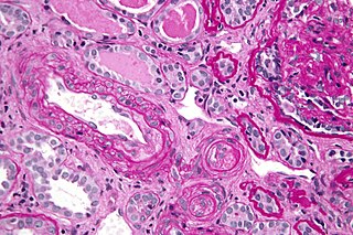
Polyarteritis nodosa (PAN) is a systemic necrotizing inflammation of blood vessels (vasculitis) affecting medium-sized muscular arteries, typically involving the arteries of the kidneys and other internal organs but generally sparing the lungs' circulation. Small aneurysms are strung like the beads of a rosary, therefore making this "rosary sign" an important diagnostic feature of the vasculitis. PAN is sometimes associated with infection by the hepatitis B or hepatitis C virus. The condition may be present in infants.

<span class="mw-page-title-main">Thrombophilia</span> Abnormality of blood coagulation

Thrombophilia is an abnormality of blood coagulation that increases the risk of thrombosis. Such abnormalities can be identified in 50% of people who have an episode of thrombosis that was not provoked by other causes. A significant proportion of the population has a detectable thrombophilic abnormality, but most of these develop thrombosis only in the presence of an additional risk factor.

<span class="mw-page-title-main">Livedo reticularis</span> Medical condition

Livedo reticularis is a common skin finding consisting of a mottled reticulated vascular pattern that appears as a lace-like purplish discoloration of the skin. The discoloration is caused by reduction in blood flow through the arterioles that supply the cutaneous capillaries, resulting in deoxygenated blood showing as blue discoloration. This can be a secondary effect of a condition that increases a person's risk of forming blood clots, including a wide array of pathological and nonpathological conditions. Examples include hyperlipidemia, microvascular hematological or anemia states, nutritional deficiencies, hyper- and autoimmune diseases, and drugs/toxins.

Catastrophic antiphospholipid syndrome (CAPS), also known as Asherson's syndrome, is a rare autoimmune disease in which widespread, intravascular clotting causes multi-organ failure. The syndrome is caused by antiphospholipid antibodies that target a group of proteins in the body that are associated with phospholipids. These antibodies activate endothelial cells, platelets, and immune cells, ultimately causing a large inflammatory immune response and widespread clotting. CAPS was first described by Ronald Asherson in 1992. The syndrome exhibits thrombotic microangiopathy, multiple organ thromboses, and in some cases tissue necrosis and is considered an extreme or catastrophic variant of the antiphospholipid syndrome.

<span class="mw-page-title-main">Sneddon's syndrome</span> Medical condition

Sneddon's syndrome is a form of arteriopathy characterized by several symptoms, including:

Anetoderma is a benign but uncommon disorder that causes localized areas of flaccid or herniated sac-like skin due to a focal reduction of dermal elastic tissue. Anetoderma is subclassified as primary anetoderma, secondary anetoderma, iatrogenic anetoderma of prematurity, congenital anetoderma, familial anetoderma, and drug-induced anetoderma.

Rheumatoid vasculitis is skin condition that is a typical feature of rheumatoid arthritis, presenting as peripheral vascular lesions that are localized purpura, cutaneous ulceration, and gangrene of the distal parts of the extremities.

<span class="mw-page-title-main">Systemic vasculitis</span> Medical condition

Necrotizing vasculitis, also called systemic necrotizing vasculitis, is a general term for the inflammation of veins and arteries that develops into necrosis and narrows the vessels.

Cryofibrinogenemia refers to a condition classified as a fibrinogen disorder in which a person's blood plasma is allowed to cool substantially, causing the (reversible) precipitation of a complex containing fibrinogen, fibrin, fibronectin, and, occasionally, small amounts of fibrin split products, albumin, immunoglobulins and other plasma proteins.

<span class="mw-page-title-main">Cutaneous small-vessel vasculitis</span> Medical condition

Cutaneous small-vessel vasculitis (CSVV), is inflammation of small blood vessels, usually accompanied by small lumps beneath the skin. The condition is also known as hypersensitivity vasculitis, cutaneous leukocytoclastic vasculitis, hypersensitivity angiitis, cutaneous leukocytoclastic angiitis, cutaneous necrotizing vasculitis and cutaneous necrotizing venulitis,

<span class="mw-page-title-main">Fungal folliculitis</span> Inflammation of hair follicles due to fungal infection

Majocchi's granuloma is a skin condition characterized by deep, pustular plaques, and is a form of tinea corporis. It is a localized form of fungal folliculitis. Lesions often have a pink and scaly central component with pustules or folliculocentric papules at the periphery. The name comes from Domenico Majocchi, who discovered the disorder in 1883. Majocchi was a professor of dermatology at the University of Parma and later the University of Bologna. The most common dermatophyte is called Trichophyton rubrum.

Blood clots are a relatively common occurrence in the general population and are seen in approximately 1-2% of the population by age 60. Typically, blood clots develop in the deep veins of the lower extremities, deep vein thrombosis (DVT) or as a blood clot in the lung, pulmonary embolism. A very small number of people who develop blood clots have a more serious and often life-threatening condition, known as thrombotic storm (TS). TS is characterized by the development of more than one blood clot in a short period of time. These clots often occur in multiple and sometimes unusual locations in the body and are often difficult to treat. TS may be associated with an existing condition or situation that predisposes a person to blood clots, such as injury, infection, or pregnancy. In many cases, a risk assessment will identify interventions that will prevent the formation of blood clots.

Vasculitic neuropathy is a peripheral neuropathic disease. In a vasculitic neuropathy there is damage to the vessels that supply blood to the nerves. It can be as part of a systemic problem or can exist as a single-organ issue only affecting the peripheral nervous system (PNS). It is diagnosed with the use of electrophysiological testing, blood tests, nerve biopsy and clinical examination. It is a serious medical condition that can cause prolonged morbidity and disability and generally requires treatment. Treatment depends on the type but it is mostly with corticosteroids or immunomodulating therapies.

Cutaneous manifestations of COVID-19 are characteristic signs or symptoms of the Coronavirus disease 2019 that occur in the skin. The American Academy of Dermatology reports that skin lesions such as morbilliform, pernio, urticaria, macular erythema, vesicular purpura, papulosquamous purpura and retiform purpura are seen in people with COVID-19. Pernio-like lesions were more common in mild disease while retiform purpura was seen only in critically ill patients. The major dermatologic patterns identified in individuals with COVID-19 are urticarial rash, confluent erythematous/morbilliform rash, papulovesicular exanthem, chilbain-like acral pattern, livedo reticularis and purpuric "vasculitic" pattern. Chilblains and Multisystem inflammatory syndrome in children are also cutaneous manifestations of COVID-19.

Retiform purpura is the result of total vascular blockage and damage to the skin's blood vessels. The skin then shows lesions, appearing due to intravascular issues where clots, proteins, or emboli block skin vessels. They can also result from direct harm to the vessel walls, as seen in conditions like vasculitis, calciphylaxis, and certain severe opportunistic infections.

References

  1. "Monarch Initiative". Monarch Initiative. Retrieved January 29, 2024.
  2. 1 2 3 4 5 "UpToDate". UpToDate. Retrieved January 29, 2024.
  3. Majmundar, Vidit D.; Baxi, Kalgi (August 8, 2023). "Livedoid Vasculopathy". StatPearls Publishing. PMID   32644463 . Retrieved January 29, 2024.
  4. 1 2 3 4 5 Eswaran, Harish; Googe, Paul; Vedak, Priyanka; Marston, William A; Moll, Stephan (2022). "Livedoid vasculopathy: A review with focus on terminology and pathogenesis". Vascular Medicine. 27 (6): 593–603. doi:10.1177/1358863X221130380. ISSN   1358-863X. PMC   9732787 . PMID   36285834.
  5. 1 2 3 4 5 6 Burg, Maria Rosa; Mitschang, Carolin; Goerge, Tobias; Schneider, Stefan Werner (October 3, 2022). "Livedoid vasculopathy – A diagnostic and therapeutic challenge". Frontiers in Medicine. Frontiers Media SA. 9. doi: 10.3389/fmed.2022.1012178 . ISSN   2296-858X. PMC   9574051 . PMID   36262273.
  6. 1 2 3 4 5 6 7 8 Bilgic, Asli; Ozcobanoglu, Salih; Bozca, Burcin Cansu; Alpsoy, Erkan (2021). "Livedoid vasculopathy: A multidisciplinary clinical approach to diagnosis and management". International Journal of Women's Dermatology. Ovid Technologies (Wolters Kluwer Health). 7 (5): 588–599. doi:10.1016/j.ijwd.2021.08.013. ISSN   2352-6475. PMC   8721056 . PMID   35024414.
  7. 1 2 3 Criado, Paulo Ricardo; Rivitti, Evandro Ararigboia; Sotto, Mirian Nacagami; Valente, Neusa Yuriko Sakai; Aoki, Valeria; Carvalho, Jozelio Freire de; Vasconcellos, Cidia (2011). "Vasculopatia livedoide: uma doença cutânea intrigante". Anais Brasileiros de Dermatologia. FapUNIFESP (SciELO). 86 (5): 961–977. doi: 10.1590/s0365-05962011000500015 . ISSN   0365-0596.
  8. Ricardo Criado, Paulo; Faillace, César; Salles Magalhães, Lidia; Brito, Késia; de Carvalho, Jozélio Freire (2012). "[Livedo reticular: classification, causes and differential diagnoses]". Acta Reumatologica Portuguesa. 37 (3): 218–225. PMID   23348110.
  9. 1 2 3 4 5 Vasudevan, Biju; Neema, Shekhar; Verma, Rajesh (2016). "Livedoid vasculopathy: A review of pathogenesis and principles of management". Indian Journal of Dermatology, Venereology and Leprology. Scientific Scholar. 82 (5): 478. doi: 10.4103/0378-6323.183635 . ISSN   0378-6323. PMID   27297279.
  10. Papi, Massimo; Didona, Biagio; De Pità, Ornella; Frezzolini, Alessandra; Di Giulio, Stefano; De Matteis, Walter; Del Principe, Domenico; Cavalieri, Rino (April 1, 1998). "Livedo Vasculopathy vs Small Vessel Cutaneous Vasculitis". Archives of Dermatology. American Medical Association (AMA). 134 (4): 447–452. doi:10.1001/archderm.134.4.447. ISSN   0003-987X. PMID   9554296.
  11. 1 2 Thornsberry, Laura A.; LoSicco, Kristen I.; English, Joseph C. (2013). "The skin and hypercoagulable states". Journal of the American Academy of Dermatology. Elsevier BV. 69 (3): 450–462. doi:10.1016/j.jaad.2013.01.043. ISSN   0190-9622. PMID   23582572.
  12. 1 2 Goerge, T. (July 24, 2011). "Livedovaskulopathie". Der Hautarzt (in German). Springer Science and Business Media LLC. 62 (8): 627–636. doi:10.1007/s00105-011-2172-3. ISSN   0017-8470. PMID   21786003. S2CID   248194522.
  13. Dodman, B.; Cunliffe, W.J.; Roberts, B.E. (1973). "Observations on tissue fibrinolytic activity in patients with cutaneous vasculitis". British Journal of Dermatology. 88 (3): 231–235. doi:10.1111/j.1365-2133.1973.tb07539.x. ISSN   0007-0963. PMID   4514551. S2CID   29460421.
  14. Weishaupt, C.; Strölin, A.; Kahle, B.; Kreuter, A.; Schneider, S. W.; Gerss, J.; Eveslage, M.; Drabik, A.; Goerge, T. (May 14, 2019). "Characteristics, risk factors and treatment reality in livedoid vasculopathy – a multicentre analysis". Journal of the European Academy of Dermatology and Venereology. Wiley. 33 (9): 1784–1791. doi:10.1111/jdv.15639. ISSN   0926-9959. PMID   31009111. S2CID   128353394.
  15. 1 2 Micieli, Robert; Alavi, Afsaneh (February 1, 2018). "Treatment for Livedoid Vasculopathy". JAMA Dermatology. American Medical Association (AMA). 154 (2): 193–202. doi:10.1001/jamadermatol.2017.4374. ISSN   2168-6068. PMID   29141075. S2CID   25609541.
  16. 1 2 Alavi, Afsaneh; Hafner, Jürg; Dutz, Jan P.; Mayer, Dieter; Sibbald, R. Gary; Criado, Paulo Ricardo; Senet, Patricia; Callen, Jeffery P.; Phillips, Tania J.; Romanelli, Marco; Kirsner, Robert S. (2013). "Livedoid vasculopathy: An in-depth analysis using a modified Delphi approach". Journal of the American Academy of Dermatology. Elsevier BV. 69 (6): 1033–1042.e1. doi:10.1016/j.jaad.2013.07.019. ISSN   0190-9622. PMID   24028907.
  17. Juan, W-H.; Chan, Y-S.; Lee, J.C.; Yang, L-C.; Hong, H-S.; Yang, C-H. (August 18, 2005). "Livedoid vasculopathy: long-term follow-up results following hyperbaric oxygen therapy". British Journal of Dermatology. Oxford University Press (OUP). 154 (2): 251–255. doi:10.1111/j.1365-2133.2005.06843.x. ISSN   0007-0963. PMID   16433793. S2CID   25099027.
  18. Weishaupt, Carsten; Strölin, Anke; Kahle, Birgit; Kreuter, Alexander; Schneider, Stefan W; Gerss, Joachim; Eveslage, Maria; Drabik, Attyla; Goerge, Tobias (2016). "Anticoagulation with rivaroxaban for livedoid vasculopathy (RILIVA): a multicentre, single-arm, open-label, phase 2a, proof-of-concept trial". The Lancet Haematology. Elsevier BV. 3 (2): e72–e79. doi:10.1016/s2352-3026(15)00251-3. ISSN   2352-3026. PMID   26853646.
  19. KLEIN, KENNETH L.; PITTELKOW, MARK R. (1992). "Tissue Plasminogen Activator for Treatment of Livedoid Vasculitis". Mayo Clinic Proceedings. Elsevier BV. 67 (10): 923–933. doi:10.1016/s0025-6196(12)60922-7. ISSN   0025-6196. PMID   1434852.

Further reading