Mass spectrometry imaging

Last updated

Mass spectrometry imaging (MSI) is a technique used in mass spectrometry to visualize the spatial distribution of molecules, as biomarkers, metabolites, peptides or proteins by their molecular masses. After collecting a mass spectrum at one spot, the sample is moved to reach another region, and so on, until the entire sample is scanned. By choosing a peak in the resulting spectra that corresponds to the compound of interest, the MS data is used to map its distribution across the sample. This results in pictures of the spatially resolved distribution of a compound pixel by pixel. Each data set contains a veritable gallery of pictures because any peak in each spectrum can be spatially mapped. Despite the fact that MSI has been generally considered a qualitative method, the signal generated by this technique is proportional to the relative abundance of the analyte. Therefore, quantification is possible, when its challenges are overcome. Although widely used traditional methodologies like radiochemistry and immunohistochemistry achieve the same goal as MSI, they are limited in their abilities to analyze multiple samples at once, and can prove to be lacking if researchers do not have prior knowledge of the samples being studied. [1] Most common ionization technologies in the field of MSI are DESI imaging, MALDI imaging, secondary ion mass spectrometry imaging (SIMS imaging) and Nanoscale SIMS (NanoSIMS). [2] [3] [4]

Contents

History

More than 50 years ago, MSI was introduced using secondary ion mass spectrometry (SIMS) to study semiconductor surfaces by Castaing and Slodzian. [5] However, it was the pioneering work of Richard Caprioli and colleagues in the late 1990s, demonstrating how matrix-assisted laser desorption/ionization (MALDI) could be applied to visualize large biomolecules (as proteins and lipids) in cells and tissue to reveal the function of these molecules and how function is changed by diseases like cancer, which led to the widespread use of MSI. Nowadays, different ionization techniques have been used, including SIMS, MALDI and desorption electrospray ionization (DESI), as well as other technologies. Still, MALDI is the current dominant technology with regard to clinical and biological applications of MSI. [6]

Operation principle

The MSI is based on the spatial distribution of the sample. Therefore, the operation principle depends on the technique that is used to obtain the spatial information. The two techniques used in MSI are: microprobe and microscope. [7]

Microprobe

This technique is performed using a focused ionization beam to analyze a specific region of the sample by generating a mass spectrum. The mass spectrum is stored along with the spatial coordination where the measurement took place. Then, a new region is selected and analyzed by moving the sample or the ionization beam. These steps are repeated until the entire sample has been scanned. By coupling all individual mass spectra, a distribution map of intensities as a function of x and y locations can be plotted. As a result, reconstructed molecular images of the sample are obtained. [7]

Microscope

In this technique, a 2D position-sensitive detector is used to measure the spatial origin of the ions generated at the sample surface by the ion optics of the instruments. The resolution of the spatial information will depend on the magnification of the microscope, the quality of the ions optics and the sensitivity of the detector. A new region still needs to be scanned, but the number of positions drastically reduces. The limitation of this mode is the finite depth of vision present with all microscopes. [7]

Ion source dependence

The ionization techniques available for MSI are suited to different applications. Some of the criteria for choosing the ionization method are the sample preparation requirement and the parameters of the measurement, as resolution, mass range and sensitivity. Based on that, the most common used ionization method are MALDI, SIMS AND DESI which are described below. Still, other minor techniques used are laser ablation electrospray ionization (LAESI), laser-ablation-inductively coupled plasma (LA-ICP) and nanospray desorption electrospray ionization (nano-DESI).

SIMS and NanoSIMS imaging

Secondary ion mass spectrometry (SIMS) is used to analyze solid surfaces and thin films by sputtering the surface with a focused primary ion beam and collecting and analyzing ejected secondary ions. There are many different sources for a primary ion beam. However, the primary ion beam must contain ions that are at the higher end of the energy scale. Some common sources are: Cs+, O2+, O, Ar+ and Ga+. [8] SIMS imaging is performed in a manner similar to electron microscopy; the primary ion beam is emitted across the sample while secondary mass spectra are recorded. [9] SIMS proves to be advantageous in providing the highest image resolution but only over small area of samples. [10] More, this technique is widely regarded as one of the most sensitive forms of mass spectrometry as it can detect elements in concentrations as small as 1012-1016 atoms per cubic centimeter. [11] [note 1] [note 2]

Multiplexed ion beam imaging (MIBI) is a SIMS method that uses metal isotope labeled antibodies to label compounds in biological samples. [12]

Developments within SIMS: Some chemical modifications have been made within SIMS to increase the efficiency of the process. There are currently two separate techniques being used to help increase the overall efficiency by increasing the sensitivity of SIMS measurements: matrix-enhanced SIMS (ME-SIMS) - This has the same sample preparation as MALDI does as this simulates the chemical ionization properties of MALDI. ME-SIMS does not sample nearly as much material. However, if the analyte being tested has a low mass value then it can produce a similar looking spectra to that of a MALDI spectra. ME-SIMS has been so effective that it has been able to detect low mass chemicals at sub cellular levels that was not possible prior to the development of the ME-SIMS technique. [4] The second technique being used is called sample metallization (Meta-SIMS) - This is the process of gold or silver addition to the sample. This forms a layer of gold or silver around the sample and it is normally no more than 1-3 nm thick. Using this technique has resulted in an increase of sensitivity for larger mass samples. The addition of the metallic layer also allows for the conversion of insulating samples to conducting samples, thus charge compensation within SIMS experiments is no longer required. [13]

Subcellular (50 nm) resolution is enabled by NanoSIMS [2] allowing for absolute quantitative analysis at the organelle level.

MALDI imaging

Mouse kidney: (a) MALDI spectra from the tissue. (b) H&E stained tissue. N-glycans at m/z = 1996.7 (c) is located in the cortex and medulla while m/z = 2158.7 (d) is in the cortex, (e) An overlay image of these two masses, (f) untreated control tissue. MALDI imaging.png
Mouse kidney: (a) MALDI spectra from the tissue. (b) H&E stained tissue. N-glycans at m/z = 1996.7 (c) is located in the cortex and medulla while m/z = 2158.7 (d) is in the cortex, (e) An overlay image of these two masses, (f) untreated control tissue.

Matrix-assisted laser desorption ionization can be used as a mass spectrometry imaging technique for relatively large molecules. [4] It has recently been shown that the most effective type of matrix to use is an ionic matrix for MALDI imaging of tissue. In this version of the technique the sample, typically a thin tissue section, is moved in two dimensions while the mass spectrum is recorded. [15] Although MALDI has the benefit of being able to record the spatial distribution of larger molecules, it comes at the cost of lower resolution than the SIMS technique. The limit for the lateral resolution for most of the modern instruments using MALDI is 20 m. MALDI experiments commonly use either an Nd:YAG (355 nm) or N2 (337 nm) laser for ionization. [4]

Pharmacodynamics and toxicodynamics in tissue have been studied by MALDI imaging. [16]

DESI imaging

Desorption electrospray Ionization is a less destructive technique, which couples simplicity and rapid analysis of the sample. The sample is sprayed with an electrically charged solvent mist at an angle that causes the ionization and desorption of various molecular species. Then, two-dimensional maps of the abundance of the selected ions in the surface of the sample in relation with the spatial distribution are generated. [17] [10] This technique is applicable to solid, liquid, frozen and gaseous samples. Moreover, DESI allows analyzing a wide range of organic and biological compounds, as animal and plant tissues and cell culture samples, without complex sample preparation [6] [10] Although, this technique has the poorest resolution among other, it can create high-quality image from a large area scan, as a whole body section scanning. [10] Fn

Comparative between the ionization techniques

Comparison of typical parameters among MSI techniques [10]
Ionization SourceType of IonizationAnalytesSpatial ResolutionMass Range
SIMSIon gunHardElemental ions, small molecules, lipids<10 m0-1000 Da
MALDIUV laser beamSoftLipids, peptide, proteins20 m0-100 000 Da
DESISolvent SpraySoftSmall molecules, lipids, peptides50 m0-2000 Da

Combination of various MSI techniques and other imaging techniques

Combining various MSI techniques can be beneficial, since each particular technique has its own advantage. For example, when information regards both proteins and lipids are necessary in the same tissue section, performing DESI to analyze the lipid, followed by MALDI to obtain information about the peptide, and finalize applying a stain (haematoxylin and eosin) for medical diagnosis of the structural characteristic of the tissue. [10] On the other side of MSI with other imaging techniques, fluorescence staining with MSI and magnetic resonance imaging (MRI) with MRI can be highlighted. Fluorescence staining can give information of the appearance of some proteins present in any process inside a tissue, while MSI may give information about the molecular changes presented in that process. Combining both techniques, multimodal picture or even 3D images of the distribution of different molecules can be generated. [10] In contrast, MRI with MSI combines the continuous 3D representation of MRI image with detailed structural representation using molecular information from MSI. Even though, MSI itself can generate 3D images, the picture is just part of the reality due to the depth limitation in the analysis, while MRI provides, for example, detailed organ shape with additional anatomical information. This coupled technique can be beneficial for cancer precise diagnosis and neurosurgery. [10]

Data processing

Standard data format for mass spectrometry imaging datasets

The imzML was proposed to exchange data in a standardized XML file based on the mzML format. [18] Several imaging MS software tools support it. The advantage of this format is the flexibility to exchange data between different instruments and data analysis software. [19]

Software

There are many free software packages available for visualization and mining of imaging mass spectrometry data. Converters from Thermo Fisher format, Analyze format, GRD format and Bruker format to imzML format were developed by the Computis project. Some software modules are also available for viewing mass spectrometry images in imzML format: Biomap (Novartis, free), Datacube Explorer (AMOLF, free), [20] EasyMSI (CEA), Mirion (JLU), MSiReader (NCSU, free) [21] and SpectralAnalysis. [22]

For processing .imzML files with the free statistical and graphics language R, a collection of R scripts is available, which permits parallel-processing of large files on a local computer, a remote cluster or on the Amazon cloud. [23]

Another free statistical package for processing imzML and Analyze 7.5 data in R exists, Cardinal. [24]

SPUTNIK [25] is an R package containing various filters to remove peaks characterized by an uncorrelated spatial distribution with the sample location or spatial randomness.

Applications

A remarkable ability of MSI is to find out the localization of biomolecules in tissues, even though there are no previous information about them. This feature has made MSI a unique tool for clinical research and pharmacological research. It provides information about biomolecular changes related with diseases by tracking proteins, lipids, and cell metabolism. For example, identifying biomarkers by MSI can show detailed cancer diagnosis. In addition, low cost imaging for pharmaceuticals studies can be acquired, such as images of molecular signatures that would be indicative of treatment response for a specific drug or the effectiveness of a particular drug delivery method. [26] [27] [28]

Ion colocalization has been studied as a way to infer local interactions between biomolecules. Similarly to colocalization in microscopy imaging, correlation has been used to quantify the similarity between ion images and generate network models. [29]

Advantages, challenges and limitations

The main advantage of MSI for studying the molecules location and distribution within the tissue is that this analysis can provide either greater selectivity, more information or more accuracy than others. Moreover, this tool requires less investment of time and resources for similar results. [17] The table below shows a comparison of advantages and disadvantages of some available techniques, including MSI, correlated with drug distribution analysis. [5]

Comparison of advantages and disadvantages of techniques assessing drug distribution [5]
MethodologyQuestion answeredAdvantagesDisadvantages
AutoradiographyWhere and how much radioactivityVery high spatial resolution; reliable quantitationEx vivo; requires radio-labelled drug; does not distinguish drug from metabolites.
ImmunohistochemistryWhereShort processing time; easy interpretation; inexpensiveEx vivo; requires antibodies, which vary in sensitivity and specificity; difficulties assigning; detection threshold; lack of standard scoring system
FluorescenceWhereIn vivo possible; reasonable costNot quantitative; poor resolution; autofluorescent interference
Positron emission tomography (PET)Where, what and activityIn vivo possible; good resolution; can be coupled to CT X-ray, gamma cameraExpensive; short-lived isotopes; need cyclotron to produce isotopes
Coherent anti-Stokes

Raman scattering

microscopy (CARS)

Where and whatLabel-free; sub-cellular spatial resolutionNot quantitative; poor selectivity; high background noise
Electrochemical atomic

force microscopy (AFM)

Where and whatLabel-free imaging; high resolutionNot quantitative; poor reproducibility; high background
MSIWhere and whatMultiplex; label-free imaging; good spatial resolutionSemi-quantitative; ion-suppression effects; complex analysis

Notes

  1. by way of comparison, 1 cc of Carbon (diamond) contains about 1.8 x 10²³ atoms. 1012 to 1016 corresponds to 6 parts per trillion (ppt) to 60 parts per billion (ppb).
  2. the sensitivity varies by element (or molecule) as well as by nature of the surface being analyzed and conditions of the analysis.

Further reading

"Imaging Trace Metals in Biological Systems" pp 81–134 in "Metals, Microbes and Minerals: The Biogeochemical Side of Life" (2021) pp xiv + 341. Authors Yu, Jyao; Harankhedkar, Shefali; Nabatilan, Arielle; Fahrni, Christopher; Walter de Gruyter, Berlin. Editors Kroneck, Peter M.H. and Sosa Torres, Martha. DOI 10.1515/9783110589771-004

Related Research Articles

<span class="mw-page-title-main">Mass spectrometry</span> Analytical technique based on determining mass to charge ratio of ions

Mass spectrometry (MS) is an analytical technique that is used to measure the mass-to-charge ratio of ions. The results are presented as a mass spectrum, a plot of intensity as a function of the mass-to-charge ratio. Mass spectrometry is used in many different fields and is applied to pure samples as well as complex mixtures.

<span class="mw-page-title-main">Ion source</span> Device that creates charged atoms and molecules (ions)

An ion source is a device that creates atomic and molecular ions. Ion sources are used to form ions for mass spectrometers, optical emission spectrometers, particle accelerators, ion implanters and ion engines.

<span class="mw-page-title-main">Lipidomics</span>

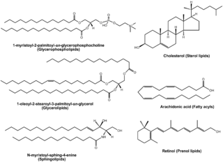
Lipidomics is the large-scale study of pathways and networks of cellular lipids in biological systems The word "lipidome" is used to describe the complete lipid profile within a cell, tissue, organism, or ecosystem and is a subset of the "metabolome" which also includes other major classes of biological molecules. Lipidomics is a relatively recent research field that has been driven by rapid advances in technologies such as mass spectrometry (MS), nuclear magnetic resonance (NMR) spectroscopy, fluorescence spectroscopy, dual polarisation interferometry and computational methods, coupled with the recognition of the role of lipids in many metabolic diseases such as obesity, atherosclerosis, stroke, hypertension and diabetes. This rapidly expanding field complements the huge progress made in genomics and proteomics, all of which constitute the family of systems biology.

<span class="mw-page-title-main">Matrix-assisted laser desorption/ionization</span> Ionization technique

In mass spectrometry, matrix-assisted laser desorption/ionization (MALDI) is an ionization technique that uses a laser energy-absorbing matrix to create ions from large molecules with minimal fragmentation. It has been applied to the analysis of biomolecules and various organic molecules, which tend to be fragile and fragment when ionized by more conventional ionization methods. It is similar in character to electrospray ionization (ESI) in that both techniques are relatively soft ways of obtaining ions of large molecules in the gas phase, though MALDI typically produces far fewer multi-charged ions.

Surface-enhanced laser desorption/ionization (SELDI) is a soft ionization method in mass spectrometry (MS) used for the analysis of protein mixtures. It is a variation of matrix-assisted laser desorption/ionization (MALDI). In MALDI, the sample is mixed with a matrix material and applied to a metal plate before irradiation by a laser, whereas in SELDI, proteins of interest in a sample become bound to a surface before MS analysis. The sample surface is a key component in the purification, desorption, and ionization of the sample. SELDI is typically used with time-of-flight (TOF) mass spectrometers and is used to detect proteins in tissue samples, blood, urine, or other clinical samples, however, SELDI technology can potentially be used in any application by simply modifying the sample surface.

Soft laser desorption (SLD) is laser desorption of large molecules that results in ionization without fragmentation. "Soft" in the context of ion formation means forming ions without breaking chemical bonds. "Hard" ionization is the formation of ions with the breaking of bonds and the formation of fragment ions.

<span class="mw-page-title-main">MALDI imaging</span> Imaging system

MALDI mass spectrometry imaging (MALDI-MSI) is the use of matrix-assisted laser desorption ionization as a mass spectrometry imaging technique in which the sample, often a thin tissue section, is moved in two dimensions while the mass spectrum is recorded. Advantages, like measuring the distribution of a large amount of analytes at one time without destroying the sample, make it a useful method in tissue-based study.

<span class="mw-page-title-main">Protein mass spectrometry</span> Application of mass spectrometry

Protein mass spectrometry refers to the application of mass spectrometry to the study of proteins. Mass spectrometry is an important method for the accurate mass determination and characterization of proteins, and a variety of methods and instrumentations have been developed for its many uses. Its applications include the identification of proteins and their post-translational modifications, the elucidation of protein complexes, their subunits and functional interactions, as well as the global measurement of proteins in proteomics. It can also be used to localize proteins to the various organelles, and determine the interactions between different proteins as well as with membrane lipids.

<span class="mw-page-title-main">Desorption electrospray ionization</span>

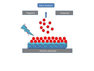
Desorption electrospray ionization (DESI) is an ambient ionization technique that can be coupled to mass spectrometry (MS) for chemical analysis of samples at atmospheric conditions. Coupled ionization sources-MS systems are popular in chemical analysis because the individual capabilities of various sources combined with different MS systems allow for chemical determinations of samples. DESI employs a fast-moving charged solvent stream, at an angle relative to the sample surface, to extract analytes from the surfaces and propel the secondary ions toward the mass analyzer. This tandem technique can be used to analyze forensics analyses, pharmaceuticals, plant tissues, fruits, intact biological tissues, enzyme-substrate complexes, metabolites and polymers. Therefore, DESI-MS may be applied in a wide variety of sectors including food and drug administration, pharmaceuticals, environmental monitoring, and biotechnology.

Sample preparation for mass spectrometry is used for the optimization of a sample for analysis in a mass spectrometer (MS). Each ionization method has certain factors that must be considered for that method to be successful, such as volume, concentration, sample phase, and composition of the analyte solution. Quite possibly the most important consideration in sample preparation is knowing what phase the sample must be in for analysis to be successful. In some cases the analyte itself must be purified before entering the ion source. In other situations, the matrix, or everything in the solution surrounding the analyte, is the most important factor to consider and adjust. Often, sample preparation itself for mass spectrometry can be avoided by coupling mass spectrometry to a chromatography method, or some other form of separation before entering the mass spectrometer. In some cases, the analyte itself must be adjusted so that analysis is possible, such as in protein mass spectrometry, where usually the protein of interest is cleaved into peptides before analysis, either by in-gel digestion or by proteolysis in solution.

<span class="mw-page-title-main">Matrix-assisted laser desorption electrospray ionization</span>

Matrix-assisted laser desorption electrospray ionization (MALDESI) was first introduced in 2006 as a novel ambient ionization technique which combines the benefits of electrospray ionization (ESI) and matrix-assisted laser desorption/ionization (MALDI). An infrared (IR) or ultraviolet (UV) laser can be utilized in MALDESI to resonantly excite an endogenous or exogenous matrix. The term 'matrix' refers to any molecule that is present in large excess and absorbs the energy of the laser, thus facilitating desorption of analyte molecules. The original MALDESI design was implemented using common organic matrices, similar to those used in MALDI, along with a UV laser. The current MALDESI source employs endogenous water or a thin layer of exogenously deposited ice as the energy-absorbing matrix where O-H symmetric and asymmetric stretching bonds are resonantly excited by a mid-IR laser.

<span class="mw-page-title-main">Desorption atmospheric pressure photoionization</span>

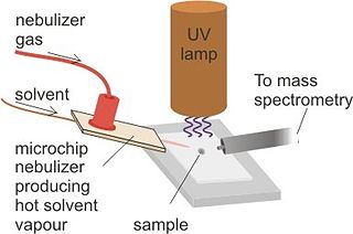
Desorption atmospheric pressure photoionization (DAPPI) is an ambient ionization technique for mass spectrometry that uses hot solvent vapor for desorption in conjunction with photoionization. Ambient Ionization techniques allow for direct analysis of samples without pretreatment. The direct analysis technique, such as DAPPI, eliminates the extraction steps seen in most nontraditional samples. DAPPI can be used to analyze bulkier samples, such as, tablets, powders, resins, plants, and tissues. The first step of this technique utilizes a jet of hot solvent vapor. The hot jet thermally desorbs the sample from a surface. The vaporized sample is then ionized by the vacuum ultraviolet light and consequently sampled into a mass spectrometer. DAPPI can detect a range of both polar and non-polar compounds, but is most sensitive when analyzing neutral or non-polar compounds. This technique also offers a selective and soft ionization for highly conjugated compounds.

<span class="mw-page-title-main">Laser ablation electrospray ionization</span>

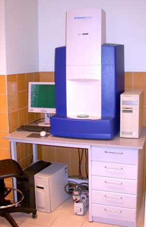
Laser ablation electrospray ionization (LAESI) is an ambient ionization method for mass spectrometry that combines laser ablation from a mid-infrared (mid-IR) laser with a secondary electrospray ionization (ESI) process. The mid-IR laser is used to generate gas phase particles which are then ionized through interactions with charged droplets from the ESI source. LAESI was developed in Professor Akos Vertes lab by Peter Nemes in 2007 and it was marketed commercially by Protea Biosciences, Inc until 2017. Fiber-LAESI for single-cell analysis approach was developed by Bindesh Shrestha in Professor Vertes lab in 2009. LAESI is a novel ionization source for mass spectrometry (MS) that has been used to perform MS imaging of plants, tissues, cell pellets, and even single cells. In addition, LAESI has been used to analyze historic documents and untreated biofluids such as urine and blood. The technique of LAESI is performed at atmospheric pressure and therefore overcomes many of the obstacles of traditional MS techniques, including extensive and invasive sample preparation steps and the use of high vacuum. Because molecules and aerosols are ionized by interacting with an electrospray plume, LAESI's ionization mechanism is similar to SESI and EESI techniques.

<span class="mw-page-title-main">Surface-assisted laser desorption/ionization</span>

Surface-assisted laser desorption/ionization (SALDI) is a soft laser desorption technique used for mass spectrometry analysis of biomolecules, polymers, and small organic molecules. In its first embodiment Koichi Tanaka used a cobalt/glycerol liquid matrix and subsequent applications included a graphite/glycerol liquid matrix as well as a solid surface of porous silicon. The porous silicon represents the first matrix-free SALDI surface analysis allowing for facile detection of intact molecular ions, these porous silicon surfaces also facilitated the analysis of small molecules at the yoctomole level. At present laser desorption/ionization methods using other inorganic matrices such as nanomaterials are often regarded as SALDI variants. As an example, silicon nanowires as well as Titania nanotube arrays (NTA) have been used as substrates to detect small molecules. SALDI is used to detect proteins and protein-protein complexes. A related method named "ambient SALDI" - which is a combination of conventional SALDI with ambient mass spectrometry incorporating the direct analysis real time (DART) ion source has also been demonstrated. SALDI is considered one of the most important techniques in MS and has many applications.

<span class="mw-page-title-main">Single-cell analysis</span> Testbg biochemical processes and reactions in an individual cell

In the field of cellular biology, single-cell analysis and subcellular analysis is the study of genomics, transcriptomics, proteomics, metabolomics and cell–cell interactions at the single cell level. The concept of single-cell analysis originated in the 1970s. Before the discovery of heterogeneity, single-cell analysis mainly referred to the analysis or manipulation of an individual cell in a bulk population of cells at a particular condition using optical or electronic microscope. To date, due to the heterogeneity seen in both eukaryotic and prokaryotic cell populations, analyzing a single cell makes it possible to discover mechanisms not seen when studying a bulk population of cells. Technologies such as fluorescence-activated cell sorting (FACS) allow the precise isolation of selected single cells from complex samples, while high throughput single cell partitioning technologies, enable the simultaneous molecular analysis of hundreds or thousands of single unsorted cells; this is particularly useful for the analysis of transcriptome variation in genotypically identical cells, allowing the definition of otherwise undetectable cell subtypes. The development of new technologies is increasing our ability to analyze the genome and transcriptome of single cells, as well as to quantify their proteome and metabolome. Mass spectrometry techniques have become important analytical tools for proteomic and metabolomic analysis of single cells. Recent advances have enabled quantifying thousands of protein across hundreds of single cells, and thus make possible new types of analysis. In situ sequencing and fluorescence in situ hybridization (FISH) do not require that cells be isolated and are increasingly being used for analysis of tissues.

iknife Medical device

An onkoknife, iKnife, or intelligent scalpel is a surgical knife that tests tissue as it contacts it during an operation and immediately gives information as to whether that tissue contains cancer cells. During a surgery this information is given continuously to the surgeon, significantly accelerating biological tissue analysis and enabling identification and removal of cancer cells. Electroknives have been in use since the 1920s and smart knife surgery is not limited only to cancer detection. In clinical studies the iKnife has shown impressive diagnostic accuracy - distinguishing benign ovarian tissue from cancerous tissue, breast tumour from normal breast tissue and recognises histological features of poor prognostic outcome in colorectal carcinoma. Furthermore, the technology behind iKnife - rapid evaporative ionisation mass spectrometry (REIMS) - can identify Candida yeasts down to species level.

<span class="mw-page-title-main">Desorption/ionization on silicon</span> Soft laser desorption method

Desorption/ionization on silicon (DIOS) is a soft laser desorption method used to generate gas-phase ions for mass spectrometry analysis. DIOS is considered the first surface-based surface-assisted laser desorption/ionization (SALDI-MS) approach. Prior approaches were accomplished using nanoparticles in a matrix of glycerol, while DIOS is a matrix-free technique in which a sample is deposited on a nanostructured surface and the sample desorbed directly from the nanostructured surface through the adsorption of laser light energy. DIOS has been used to analyze organic molecules, metabolites, biomolecules and peptides, and, ultimately, to image tissues and cells.

<span class="mw-page-title-main">Matrix-assisted ionization</span>

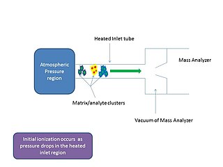
In mass spectrometry, matrix-assisted ionization is a low fragmentation (soft) ionization technique which involves the transfer of particles of the analyte and matrix sample from atmospheric pressure (AP) to the heated inlet tube connecting the AP region to the vacuum of the mass analyzer.

<span class="mw-page-title-main">Ron Heeren</span> Dutch mass spectrometry researcher

Ron M.A. Heeren is a Dutch scientist in mass spectrometry imaging. He is currently a distinguished professor at Maastricht University and the scientific director of the Multimodal Molecular Imaging Institute (M4I), where he heads the division of Imaging Mass Spectrometry.

References

  1. Monroe E, Annangudi S, Hatcher N, Gutstein H, Rubakhin S, Sweedler J (2008). "SIMS and MADLI MS Imaging of the spinal cord". Proteomics. 8 (18): 3746–3754. doi:10.1002/pmic.200800127. PMC   2706659 . PMID   18712768.
  2. 1 2 Siuzdak, Gary (September 2023). "Subcellular quantitative imaging of metabolites at the organelle level". Nature Metabolism. 5 (9): 1446–1448. doi:10.1038/s42255-023-00882-z. ISSN   2522-5812.
  3. Rohner T, Staab D, Stoeckli M (2005). "MALDI mass spectrometric imaging of biological tissue sections". Mechanisms of Ageing and Development. 126 (1): 177–185. doi:10.1016/j.mad.2004.09.032. PMID   15610777. S2CID   30982189.
  4. 1 2 3 4 McDonnell LA, Heeren RM (2007). "Imaging mass spectrometry". Mass Spectrometry Reviews. 26 (4): 606–43. Bibcode:2007MSRv...26..606M. doi:10.1002/mas.20124. hdl: 1874/26394 . PMID   17471576.
  5. 1 2 3 Cobice, D F; Goodwin, R J A; Andren, P E; Nilsson, A; Mackay, C L; Andrew, R (2015-07-01). "Future technology insight: mass spectrometry imaging as a tool in drug research and development". British Journal of Pharmacology. 172 (13): 3266–3283. doi:10.1111/bph.13135. ISSN   1476-5381. PMC   4500365 . PMID   25766375.
  6. 1 2 Addie, Ruben D.; Balluff, Benjamin; Bovée, Judith V. M. G.; Morreau, Hans; McDonnell, Liam A. (2015). "Current State and Future Challenges of Mass Spectrometry Imaging for Clinical Research". Analytical Chemistry. 87 (13): 6426–6433. doi:10.1021/acs.analchem.5b00416. PMID   25803124.
  7. 1 2 3 McDonnell, Liam A.; Heeren, Ron M.A. (2007-07-01). "Imaging mass spectrometry". Mass Spectrometry Reviews. 26 (4): 606–643. Bibcode:2007MSRv...26..606M. doi:10.1002/mas.20124. hdl: 1874/26394 . ISSN   1098-2787. PMID   17471576.
  8. Amstalden Van Hove E, Smith D, Heeren R (2010). "A concise review of mass spectrometry imaging". Journal of Chromatography A. 1217 (25): 3946–3954. doi:10.1016/j.chroma.2010.01.033. PMID   20223463.
  9. Penner-Hahn, James E. (2013). "Chapter 2. Technologies for Detecting Metals in Single Cells. Section 2.1, Secondary Ion Mass Specctrometry". In Banci, Lucia (ed.). Metallomics and the Cell. Metal Ions in Life Sciences. Vol. 12. Springer. pp. 15–40. doi:10.1007/978-94-007-5561-1_2. ISBN   978-94-007-5560-4. PMID   23595669.electronic-book ISBN   978-94-007-5561-1 ISSN   1559-0836electronic- ISSN   1868-0402
  10. 1 2 3 4 5 6 7 8 Bodzon-Kulakowska, Anna; Suder, Piotr (2016-01-01). "Imaging mass spectrometry: Instrumentation, applications, and combination with other visualization techniques". Mass Spectrometry Reviews. 35 (1): 147–169. Bibcode:2016MSRv...35..147B. doi:10.1002/mas.21468. ISSN   1098-2787. PMID   25962625.
  11. Chabala J, Soni K, Li J, Gavlirov K, Levi-Setti R (1995). "High resolution chemical imaging with scanning ion probe SIMS". International Journal of Mass Spectrometry and Ion Processes. 143: 191–212. Bibcode:1995IJMSI.143..191C. doi:10.1016/0168-1176(94)04119-R.
  12. Angelo, Michael; Bendall, Sean C; Finck, Rachel; Hale, Matthew B; Hitzman, Chuck; Borowsky, Alexander D; Levenson, Richard M; Lowe, John B; Liu, Scot D; Zhao, Shuchun; Natkunam, Yasodha; Nolan, Garry P (2014). "Multiplexed ion beam imaging of human breast tumors". Nature Medicine. 20 (4): 436–442. doi:10.1038/nm.3488. ISSN   1078-8956. PMC   4110905 . PMID   24584119.
  13. Delcorte A, Befahy S, Poleunis C, Troosters M, Bertrand P. "Improvements of metal adhesion to silicon films: a ToF-SIMS study".{{cite journal}}: Cite journal requires |journal= (help)
  14. Powers, Thomas W.; Neely, Benjamin A.; Shao, Yuan; Tang, Huiyuan; Troyer, Dean A.; Mehta, Anand S.; Haab, Brian B.; Drake, Richard R. (2014). "MALDI Imaging Mass Spectrometry Profiling of N-Glycans in Formalin-Fixed Paraffin Embedded Clinical Tissue Blocks and Tissue Microarrays". PLOS ONE. 9 (9): e106255. Bibcode:2014PLoSO...9j6255P. doi: 10.1371/journal.pone.0106255 . ISSN   1932-6203. PMC   4153616 . PMID   25184632.
  15. Chaurand P, Norris JL, Cornett DS, Mobley JA, Caprioli RM (2006). "New developments in profiling and imaging of proteins from tissue sections by MALDI mass spectrometry". J. Proteome Res. 5 (11): 2889–900. doi:10.1021/pr060346u. PMID   17081040.
  16. Patel, Ekta (1 January 2015). "MALDI-MS imaging for the study of tissue pharmacodynamics and toxicodynamics". Bioanalysis. 7 (1): 91–101. doi:10.4155/bio.14.280. PMID   25558938.
  17. 1 2 Nilsson, Anna; Goodwin, Richard J. A.; Shariatgorji, Mohammadreza; Vallianatou, Theodosia; Webborn, Peter J. H.; Andrén, Per E. (2015-02-03). "Mass Spectrometry Imaging in Drug Development". Analytical Chemistry. 87 (3): 1437–1455. doi:10.1021/ac504734s. ISSN   0003-2700. PMID   25526173.
  18. Schramm, Thorsten; Hester, Zoë; Klinkert, Ivo; Both, Jean-Pierre; Heeren, Ron M. A.; Brunelle, Alain; Laprévote, Olivier; Desbenoit, Nicolas; Robbe, Marie-France; Stoeckli, Markus; Spengler, Bernhard (2012-08-30). "imzML--a common data format for the flexible exchange and processing of mass spectrometry imaging data". Journal of Proteomics. 75 (16): 5106–5110. doi:10.1016/j.jprot.2012.07.026. ISSN   1876-7737. PMID   22842151. S2CID   25970597.
  19. A. Römpp; T. Schramm; A. Hester; I. Klinkert; J.P. Both; R.M.A. Heeren; M. Stoeckli; B. Spengler (2011). "Chapter imzML: Imaging Mass Spectrometry Markup Language: A Common Data Format for Mass Spectrometry Imaging in Data Mining in Proteomics: From Standards to Applications". Methods in Molecular Biology, Humana Press, New York. Vol. 696. pp. 205–224.
  20. Klinkert, I.; Chughtai, K.; Ellis, S. R.; Heeren, R. M. A. (2014). "Methods for Full Resolution Data Exploration and Visualization for Large 2D and 3D Mass Spectrometry Imaging Datasets". International Journal of Mass Spectrometry. 362: 40–47. Bibcode:2014IJMSp.362...40K. doi:10.1016/j.ijms.2013.12.012.
  21. Robichaud, G.; Garrard, K. P.; Barry, J. A.; Muddiman, D. C. (2013). "MSiReader: An Open-Source Interface to View and Analyze High Resolving Power MS Imaging Files on Matlab Platform". Journal of the American Society for Mass Spectrometry. 24 (5): 718–721. Bibcode:2013JASMS..24..718R. doi:10.1007/s13361-013-0607-z. PMC   3693088 . PMID   23536269.
  22. Race, A. M.; Palmer, A. D.; Dexter, A.; Steven, R. T.; Styles, I. B.; Bunch, J. (2016). "SpectralAnalysis: software for the masses" (PDF). Analytical Chemistry. 88 (19): 9451–9458. doi:10.1021/acs.analchem.6b01643. PMID   27558772.
  23. Gamboa-Becerra, Roberto; Ramírez-Chávez, Enrique; Molina-Torres, Jorge; Winkler, Robert (2015-07-01). "MSI.R scripts reveal volatile and semi-volatile features in low-temperature plasma mass spectrometry imaging (LTP-MSI) of chilli (Capsicum annuum)". Analytical and Bioanalytical Chemistry. 407 (19): 5673–5684. doi:10.1007/s00216-015-8744-9. PMID   26007697. S2CID   22813369.
  24. Bemis, Kyle D.; Harry, April; Eberlin, Livia S.; Ferreira, Christina; van de Ven, Stephanie M.; Mallick, Parag; Stolowitz, Mark; Vitek, Olga (2015-03-15). "Cardinal: an R package for statistical analysis of mass spectrometry-based imaging experiments". Bioinformatics. 31 (14): 2418–2420. doi:10.1093/bioinformatics/btv146. PMC   4495298 . PMID   25777525.
  25. Inglese, Paolo; Correia, Gonçalo; Takats, Zoltan; Nicholson, Jeremy K.; Glen, Robert C. (2018). "SPUTNIK: An R package for filtering of spatially related peaks in mass spectrometry imaging data". Bioinformatics. 35 (1): 178–180. doi:10.1093/bioinformatics/bty622. PMC   6298046 . PMID   30010780.
  26. Swales, John G.; Hamm, Gregory; Clench, Malcolm R.; Goodwin, Richard J.A. (March 2019). "Mass spectrometry imaging and its application in pharmaceutical research and development: A concise review". International Journal of Mass Spectrometry. 437: 99–112. doi:10.1016/j.ijms.2018.02.007. ISSN   1387-3806. S2CID   102892898.
  27. Addie, Ruben D.; Balluff, Benjamin; Bovée, Judith V. M. G.; Morreau, Hans; McDonnell, Liam A. (2015-07-07). "Current State and Future Challenges of Mass Spectrometry Imaging for Clinical Research". Analytical Chemistry. 87 (13): 6426–6433. doi:10.1021/acs.analchem.5b00416. ISSN   0003-2700. PMID   25803124.
  28. Aichler, Michaela; Walch, Axel (April 2015). "MALDI Imaging mass spectrometry: current frontiers and perspectives in pathology research and practice". Laboratory Investigation. 95 (4): 422–431. doi: 10.1038/labinvest.2014.156 . ISSN   1530-0307. PMID   25621874.
  29. Inglese, Paolo; Strittmatter, Nicole; Doria, Luisa; Mroz, Anna; Speller, Abigail; Poynter, Liam; Dannhorn, Andreas; Kudo, Hiromi; Mirnezami, Reza; Goldin, Robert D.; Nicholson, Jeremy K. (2018-01-09). "Network analysis of mass spectrometry imaging data from colorectal cancer identifies key metabolites common to metastatic development": 230052. doi:10.1101/230052. S2CID   90409488.{{cite journal}}: Cite journal requires |journal= (help)