Medical physics

Last updated

Medical physics [1] deals with the application of the concepts and methods of physics to the prevention, diagnosis and treatment of human diseases with a specific goal of improving human health and well-being. [2] Since 2008, medical physics has been included as a health profession according to International Standard Classification of Occupation of the International Labour Organization. [3]

Contents

Although medical physics may sometimes also be referred to as biomedical physics, medical biophysics, applied physics in medicine, physics applications in medical science, radiological physics or hospital radio-physics, a "medical physicist" is specifically a health professional [4] with specialist education and training in the concepts and techniques of applying physics in medicine and competent to practice independently in one or more of the subfields of medical physics. [5] Traditionally, medical physicists are found in the following healthcare specialties: radiation oncology (also known as radiotherapy or radiation therapy), diagnostic and interventional radiology (also known as medical imaging), nuclear medicine, and radiation protection. Medical physics of radiation therapy can involve work such as dosimetry, linac quality assurance, and brachytherapy. Medical physics of diagnostic and interventional radiology involves medical imaging techniques such as magnetic resonance imaging, ultrasound, computed tomography and x-ray. Nuclear medicine will include positron emission tomography and radionuclide therapy. However one can find Medical Physicists in many other areas such as physiological monitoring, audiology, neurology, neurophysiology, cardiology and others.

Medical physics departments may be found in institutions such as universities, hospitals, and laboratories. University departments are of two types. The first type are mainly concerned with preparing students for a career as a hospital Medical Physicist and research focuses on improving the practice of the profession. A second type (increasingly called 'biomedical physics') has a much wider scope and may include research in any applications of physics to medicine from the study of biomolecular structure to microscopy and nanomedicine.

Mission statement of medical physicists

In hospital medical physics departments, the mission statement for medical physicists as adopted by the European Federation of Organisations for Medical Physics (EFOMP) is the following: [6] [7]

"Medical Physicists will contribute to maintaining and improving the quality, safety and cost-effectiveness of healthcare services through patient-oriented activities requiring expert action, involvement or advice regarding the specification, selection, acceptance testing, commissioning, quality assurance/control and optimised clinical use of medical devices and regarding patient risks and protection from associated physical agents (e.g., x-rays, electromagnetic fields, laser light, radionuclides) including the prevention of unintended or accidental exposures; all activities will be based on current best evidence or own scientific research when the available evidence is not sufficient. The scope includes risks to volunteers in biomedical research, carers and comforters. The scope often includes risks to workers and public particularly when these impact patient risk"

The term "physical agents" refers to ionising and non-ionising electromagnetic radiations, static electric and magnetic fields, ultrasound, laser light and any other Physical Agent associated with medical e.g., x-rays in computerised tomography (CT), gamma rays/radionuclides in nuclear medicine, magnetic fields and radio-frequencies in magnetic resonance imaging (MRI), ultrasound in ultrasound imaging and Doppler measurements.

This mission includes the following 11 key activities:

  1. Scientific problem solving service: Comprehensive problem solving service involving recognition of less than optimal performance or optimised use of medical devices, identification and elimination of possible causes or misuse, and confirmation that proposed solutions have restored device performance and use to acceptable status. All activities are to be based on current best scientific evidence or own research when the available evidence is not sufficient.
  2. Dosimetry measurements: Measurement of doses had by patients, volunteers in biomedical research, carers, comforters and persons subjected to non-medical imaging exposures (e.g., for legal or employment purposes); selection, calibration and maintenance of dosimetry related instrumentation; independent checking of dose related quantities provided by dose reporting devices (including software devices); measurement of dose related quantities required as inputs to dose reporting or estimating devices (including software). Measurements to be based on current recommended techniques and protocols. Includes dosimetry of all physical agents.
  3. Patient safety/risk management (including volunteers in biomedical research, carers, comforters and persons subjected to non-medical imaging exposures. Surveillance of medical devices and evaluation of clinical protocols to ensure the ongoing protection of patients, volunteers in biomedical research, carers, comforters and persons subjected to non-medical imaging exposures from the deleterious effects of physical agents in accordance with the latest published evidence or own research when the available evidence is not sufficient. Includes the development of risk assessment protocols.
  4. Occupational and public safety/risk management (when there is an impact on medical exposure or own safety). Surveillance of medical devices and evaluation of clinical protocols with respect to protection of workers and public when impacting the exposure of patients, volunteers in biomedical research, carers, comforters and persons subjected to non-medical imaging exposures or responsibility with respect to own safety. Includes the development of risk assessment protocols in conjunction with other experts involved in occupational / public risks.
  5. Clinical medical device management: Specification, selection, acceptance testing, commissioning and quality assurance/ control of medical devices in accordance with the latest published European or International recommendations and the management and supervision of associated programmes. Testing to be based on current recommended techniques and protocols.
  6. Clinical involvement: Carrying out, participating in and supervising everyday radiation protection and quality control procedures to ensure ongoing effective and optimised use of medical radiological devices and including patient specific optimization.
  7. Development of service quality and cost-effectiveness: Leading the introduction of new medical radiological devices into clinical service, the introduction of new medical physics services and participating in the introduction/development of clinical protocols/techniques whilst giving due attention to economic issues.
  8. Expert consultancy: Provision of expert advice to outside clients (e.g., clinics with no in-house medical physics expertise).
  9. Education of healthcare professionals (including medical physics trainees: Contributing to quality healthcare professional education through knowledge transfer activities concerning the technical-scientific knowledge, skills and competences supporting the clinically effective, safe, evidence-based and economical use of medical radiological devices. Participation in the education of medical physics students and organisation of medical physics residency programmes.
  10. Health technology assessment (HTA): Taking responsibility for the physics component of health technology assessments related to medical radiological devices and /or the medical uses of radioactive substances/sources.
  11. Innovation: Developing new or modifying existing devices (including software) and protocols for the solution of hitherto unresolved clinical problems.

Medical biophysics and biomedical physics

Some education institutions house departments or programs bearing the title "medical biophysics" or "biomedical physics" or "applied physics in medicine". Generally, these fall into one of two categories: interdisciplinary departments that house biophysics, radiobiology, and medical physics under a single umbrella; [8] [9] [10] and undergraduate programs that prepare students for further study in medical physics, biophysics, or medicine. [11] [12] Most of the scientific concepts in bionanotechnology are derived from other fields. Biochemical principles that are used to understand the material properties of biological systems are central in bionanotechnology because those same principles are to be used to create new technologies. Material properties and applications studied in bionanoscience include mechanical properties (e.g. deformation, adhesion, failure), electrical/electronic (e.g. electromechanical stimulation, capacitors, energy storage/batteries), optical (e.g. absorption, luminescence, photochemistry), thermal (e.g. thermomutability, thermal management), biological (e.g. how cells interact with nanomaterials, molecular flaws/defects, biosensing, biological mechanisms such as mechanosensation), nanoscience of disease (e.g. genetic disease, cancer, organ/tissue failure), as well as computing (e.g. DNA computing) and agriculture (target delivery of pesticides, hormones and fertilizers. [13] [14] [15] [16]

Areas of specialty

The International Organization for Medical Physics (IOMP) recognizes main areas of medical physics employment and focus. [17] [18]

Medical imaging physics
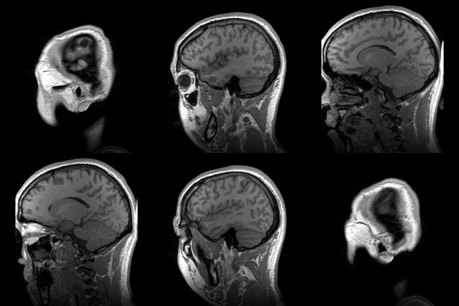
Para-sagittal MRI of the head in a patient with benign familial macrocephaly. Parasagittal MRI of human head in patient with benign familial macrocephaly prior to brain injury (ANIMATED).gif
Para-sagittal MRI of the head in a patient with benign familial macrocephaly.

Medical imaging physics is also known as diagnostic and interventional radiology physics. Clinical (both "in-house" and "consulting") physicists [19] typically deal with areas of testing, optimization, and quality assurance of diagnostic radiology physics areas such as radiographic X-rays, fluoroscopy, mammography, angiography, and computed tomography, as well as non-ionizing radiation modalities such as ultrasound, and MRI. They may also be engaged with radiation protection issues such as dosimetry (for staff and patients). In addition, many imaging physicists are often also involved with nuclear medicine systems, including single photon emission computed tomography (SPECT) and positron emission tomography (PET). Sometimes, imaging physicists may be engaged in clinical areas, but for research and teaching purposes, [20] such as quantifying intravascular ultrasound as a possible method of imaging a particular vascular object.

Radiation therapeutic physics

Radiation therapeutic physics is also known as radiotherapy physics or radiation oncologist physics. The majority of medical physicists currently working in the US, Canada, and some western countries are of this group. A radiation therapy physicist typically deals with linear accelerator (Linac) systems and kilovoltage x-ray treatment units on a daily basis, as well as other modalities such as TomoTherapy, gamma knife, Cyberknife, proton therapy, and brachytherapy. [21] [22] [23] The academic and research side of therapeutic physics may encompass fields such as boron neutron capture therapy, sealed source radiotherapy, terahertz radiation, high-intensity focused ultrasound (including lithotripsy), optical radiation lasers, ultraviolet etc. including photodynamic therapy, as well as nuclear medicine including unsealed source radiotherapy, and photomedicine, which is the use of light to treat and diagnose disease.

Nuclear medicine physics

Nuclear medicine is a branch of medicine that uses radiation to provide information about the functioning of a person's specific organs or to treat disease. The thyroid, bones, heart, liver and many other organs can be easily imaged, and disorders in their function revealed. In some cases radiation sources can be used to treat diseased organs, or tumours. Five Nobel laureates have been intimately involved with the use of radioactive tracers in medicine. Over 10,000 hospitals worldwide use radioisotopes in medicine, and about 90% of the procedures are for diagnosis. The most common radioisotope used in diagnosis is technetium-99m, with some 30 million procedures per year, accounting for 80% of all nuclear medicine procedures worldwide. [24]

Health physics

Health physics is also known as radiation safety or radiation protection. Health physics is the applied physics of radiation protection for health and health care purposes. It is the science concerned with the recognition, evaluation, and control of health hazards to permit the safe use and application of ionizing radiation. Health physics professionals promote excellence in the science and practice of radiation protection and safety.

Non-ionizing Medical Radiation Physics

Some aspects of non-ionising radiation physics may be considered under radiation protection or diagnostic imaging physics. Imaging modalities include MRI, optical imaging and ultrasound. Safety considerations include these areas and lasers

Physiological measurement

Physiological measurements have also been used to monitor and measure various physiological parameters. Many physiological measurement techniques are non-invasive and can be used in conjunction with, or as an alternative to, other invasive methods. Measurement methods include electrocardiography Many of these areas may be covered by other specialities, for example medical engineering or vascular science. [25]

Healthcare informatics and computational physics

Other closely related fields to medical physics include fields which deal with medical data, information technology and computer science for medicine.

Areas of research and academic development

ECG trace ECG principle slow.gif
ECG trace

Non-clinical physicists may or may not focus on the above areas from an academic and research point of view, but their scope of specialization may also encompass lasers and ultraviolet systems (such as photodynamic therapy), fMRI and other methods for functional imaging as well as molecular imaging, electrical impedance tomography, diffuse optical imaging, optical coherence tomography, and dual energy X-ray absorptiometry.
Legislative and advisory bodies

International

United States of America

United Kingdom

Other

Related Research Articles

<span class="mw-page-title-main">Radiography</span> Imaging technique using ionizing and non-ionizing radiation

Radiography is an imaging technique using X-rays, gamma rays, or similar ionizing radiation and non-ionizing radiation to view the internal form of an object. Applications of radiography include medical and industrial radiography. Similar techniques are used in airport security,. To create an image in conventional radiography, a beam of X-rays is produced by an X-ray generator and it is projected towards the object. A certain amount of the X-rays or other radiation are absorbed by the object, dependent on the object's density and structural composition. The X-rays that pass through the object are captured behind the object by a detector. The generation of flat two-dimensional images by this technique is called projectional radiography. In computed tomography, an X-ray source and its associated detectors rotate around the subject, which itself moves through the conical X-ray beam produced. Any given point within the subject is crossed from many directions by many different beams at different times. Information regarding the attenuation of these beams is collated and subjected to computation to generate two-dimensional images on three planes which can be further processed to produce a three-dimensional image.

<span class="mw-page-title-main">Radiology</span> Branch of Medicine

Radiology is the medical specialty that uses medical imaging to diagnose diseases and guide their treatment, within the bodies of humans and other animals. It began with radiography, but today it includes all imaging modalities, including those that use no ionizing electromagnetic radiation, as well as others that do, such as computed tomography (CT), fluoroscopy, and nuclear medicine including positron emission tomography (PET). Interventional radiology is the performance of usually minimally invasive medical procedures with the guidance of imaging technologies such as those mentioned above.

<span class="mw-page-title-main">Medical imaging</span> Technique and process of creating visual representations of the interior of a body

Medical imaging is the technique and process of imaging the interior of a body for clinical analysis and medical intervention, as well as visual representation of the function of some organs or tissues (physiology). Medical imaging seeks to reveal internal structures hidden by the skin and bones, as well as to diagnose and treat disease. Medical imaging also establishes a database of normal anatomy and physiology to make it possible to identify abnormalities. Although imaging of removed organs and tissues can be performed for medical reasons, such procedures are usually considered part of pathology instead of medical imaging.

Radiation dosimetry in the fields of health physics and radiation protection is the measurement, calculation and assessment of the ionizing radiation dose absorbed by an object, usually the human body. This applies both internally, due to ingested or inhaled radioactive substances, or externally due to irradiation by sources of radiation.

<span class="mw-page-title-main">Nuclear medicine</span> Medical specialty

Nuclear medicine or nucleology is a medical specialty involving the application of radioactive substances in the diagnosis and treatment of disease. Nuclear imaging, in a sense, is "radiology done inside out" because it records radiation emitted from within the body rather than radiation that is transmitted through the body from external sources like X-ray generators. In addition, nuclear medicine scans differ from radiology, as the emphasis is not on imaging anatomy, but on the function. For such reason, it is called a physiological imaging modality. Single photon emission computed tomography (SPECT) and positron emission tomography (PET) scans are the two most common imaging modalities in nuclear medicine.

<span class="mw-page-title-main">Health physics</span>

Health physics, also referred to as the science of radiation protection, is the profession devoted to protecting people and their environment from potential radiation hazards, while making it possible to enjoy the beneficial uses of radiation. Health physicists normally require a four-year bachelor’s degree and qualifying experience that demonstrates a professional knowledge of the theory and application of radiation protection principles and closely related sciences. Health physicists principally work at facilities where radionuclides or other sources of ionizing radiation are used or produced; these include research, industry, education, medical facilities, nuclear power, military, environmental protection, enforcement of government regulations, and decontamination and decommissioning—the combination of education and experience for health physicists depends on the specific field in which the health physicist is engaged.

A medical physicist is a health professional with specialist education and training in the concepts and techniques of applying physics in medicine and competent to practice independently in one or more of the subfields (specialties) of medical physics. A medical physicist plays a fundamental role in applying physics to medicine, but particularly in the diagnosis and treatment of cancer. The scientific and technological progress in medical physics has led to a variety of skills that must be integrated into the role of a medical physicist in order for them to perform their job. The "medical services" provided to patients undergoing diagnostic and therapeutic treatments must, therefore, be the result of different but complementary skills. In general, the medical physicist is responsible for all scientific and technical aspects of imaging, radiation treatment, and radiation safety. It is their occupational role to ensure that medical modalities offered to patients are met with the utmost quality assurance. It is the medical physicist that manage and supervise the efforts of dosimetrists, therapists and technologists in that capacity.

Articles related specifically to biomedical engineering include:

The American College of Radiology (ACR), founded in 1923, is a professional medical society representing nearly 40,000 diagnostic radiologists, radiation oncologists, interventional radiologists, nuclear medicine physicians and medical physicists.

<span class="mw-page-title-main">Walter Mauderli</span> American physicist

Walter Mauderli DSc was a pioneer in the development of the field of medical physics. He earned his doctorate from the Swiss Federal Institute of Technology under the instruction of notable physicists as Nobel Laureate physicist Wolfgang Pauli.

<span class="mw-page-title-main">Michel Ter-Pogossian</span> American medical physicist

Michel Matthew Ter-Pogossian was an American medical physicist. He was professor of radiology at the Washington University School of Medicine for over 30 years. A pioneer in nuclear medicine, he is best known for his research on the positron emission tomography (PET). He is considered one of its creators and often referred to as the "father of PET."

David A. Schauer, ScD, CHP, is executive director emeritus of the National Council on Radiation Protection and Measurements (NCRP). During his tenure a number of updated and new publications were issued by the Council.

Nuclear medicine physicians, also called nuclear radiologists or simply nucleologists, are medical specialists that use tracers, usually radiopharmaceuticals, for diagnosis and therapy. Nuclear medicine procedures are the major clinical applications of molecular imaging and molecular therapy. In the United States, nuclear medicine physicians are certified by the American Board of Nuclear Medicine and the American Osteopathic Board of Nuclear Medicine.

Wolfgang Axel Tomé is a physicist working in medicine as a researcher, inventor, and educator. He is noted for his contributions to the use of photogrammetry in high precision radiation therapy; his work on risk adaptive radiation therapy which is based on the risk level for recurrence in tumor sub-volumes using biological objective functions; and the development of hippocampal avoidant cranial radiation therapy techniques to alleviate hippocampal-dependent neurocognitive impairment following cranial irradiation.

Xie George Xu was the Edward E. Hood Chair Professor of Engineering at Rensselaer Polytechnic Institute (RPI), Troy, New York, United States, before he relocated in 2020 to China and joined the faculty of the University of Science and Technology of China.

<span class="mw-page-title-main">Madan M. Rehani</span>

Madan M. Rehani is an Indian-born medical physicist.

Radhe Mohan is a medical physicist who significantly advanced radiation treatment safety for oncology patients. He is a recipient of the ASTRO Gold Medal for outstanding contributions in the field of radiation oncology.

The European Federation of Organisations for Medical Physics (EFOMP) was founded in May 1980 in London to serve as an umbrella organisation representing the national Medical Physics societies in Europe. The office moved to Utrecht in January 2021. It is a non-profit organisation and aims to foster and coordinate the activities of its national member organisations, encourage exchange and dissemination of professional and scientific information, develop guidelines for education, training and accreditation programmes and to make recommendations on the responsibilities, organisational relationships and roles of medical physicists.

Caridad Borrás is a Spanish medical physicist. Her career started in 1964 at the Santa Creu i Sant Pau Hospital in Barcelona. From 1988 to 2000, she was Regional Advisor of the Radiological Health Program and, from 2000 to 2002, Coordinator of Essential Drugs and Technology at the Pan American Health Organization in Washington D.C.


A clinical technologist, also known as a healthcare science practitioner, is a medical professional involved in the practical delivery of medical physics and clinical engineering services. In some locations there is considerable overlap in closely related terms, for example in many countries technologist and radiographer are synonyms, while in the United Kingdom they are considered separate professions. Clinical technologists can be found in nuclear medicine, radiotherapy, radiation protection, and rehabilitation engineering departments, and they are often described by their scope of practice.

References

  1. "NYS Medical Physics". www.op.nysed.gov. Archived from the original on 2012-06-03. Retrieved 2022-02-03.
  2. "Medical Physics – International Organization for Medical Physics". 27 March 2018.
  3. Guidelines for the Certification of Clinically Qualified Medical Physicists. IAEA. Link: https://www-pub.iaea.org/MTCD/Publications/PDF/TCS-71web.pdf
  4. "IAEA Launches Guide to Promote Recognition of Medical Physicists as Health Professionals". 15 February 2021.
  5. IAEA Safety Standards: General Safety Requirements Part 3: Radiation Protection and Safety of Radiation Sources: International Basic Safety Standards. IAEA. 2014. Link: https://www-pub.iaea.org/MTCD/Publications/PDF/Pub1578_web-57265295.pdf
  6. Guibelalde E., Christofides S., Caruana C. J., Evans S. van der Putten W. (2012). Guidelines on the Medical Physics Expert' a project funded by the European Commission
  7. Caruana C.J., Christofides S., Hartmann G.H. (2014) European Federation of Organisations for Medical Physics (EFOMP) Policy Statement 12.1: Recommendations on Medical Physics Education and Training in Europe 2014 Physica Medica - European Journal of Medical Physics, 30:6, p598-603
  8. "Department of Medical Biophysics". utoronto.ca.
  9. "Medical Biophysics - Western University". uwo.ca. Archived from the original on 2013-07-03.
  10. UCLA Biomedical Physics Graduate Program
  11. "Welcome". wayne.edu. Archived from the original on 2013-08-12. Retrieved 2013-07-01.
  12. "Medical Physics". fresnostate.edu.
  13. GarciaAnoveros, J; Corey, DP (1997). "The molecules of mechanosensation". Annual Review of Neuroscience. 20: 567–94. doi:10.1146/annurev.neuro.20.1.567. PMID   9056725.
  14. Callaway DJ, Matsui T, Weiss T, Stingaciu LR, Stanley CB, Heller WT, Bu ZM (7 April 2017). "Controllable Activation of Nanoscale Dynamics in a Disordered Protein Alters Binding Kinetics". Journal of Molecular Biology. 427 (7): 987–998. doi:10.1016/j.jmb.2017.03.003. PMC   5399307 . PMID   28285124.
  15. Langer, Robert (2010). "Nanotechnology in Drug Delivery and Tissue Engineering: From Discovery to Applications". Nano Lett. 10 (9): 3223–30. Bibcode:2010NanoL..10.3223S. doi:10.1021/nl102184c. PMC   2935937 . PMID   20726522.
  16. Thangavelu, Raja Muthuramalingam; Gunasekaran, Dharanivasan; Jesse, Michael Immanuel; s.u, Mohammed Riyaz; Sundarajan, Deepan; Krishnan, Kathiravan (2018). "Nanobiotechnology approach using plant rooting hormone synthesized silver nanoparticle as "nanobullets" for the dynamic applications in horticulture – an in vitro and ex vitro study". Arabian Journal of Chemistry. 11: 48–61. doi: 10.1016/j.arabjc.2016.09.022 .
  17. "Medical Physics". International Organization for Medical Physics. Retrieved 21 October 2017.
  18. "AAPM Position Statements, Policies and Procedures - Details". aapm.org.
  19. "AAPM - What do Medical Physicists Do?". aapm.org.
  20. "COMP/OCPM - What is Medical Physics?". Archived from the original on 2013-11-13. Retrieved 2013-11-13.
  21. Hill R, Healy B, Holloway L, Kuncic Z, Thwaites D, Baldock C (2014). "Advances in kilovoltage x-ray beam dosimetry". Physics in Medicine and Biology. 59 (6): R183–231. Bibcode:2014PMB....59R.183H. doi:10.1088/0031-9155/59/6/R183. PMID   24584183. S2CID   18082594.
  22. Thwaites DI, Tuohy JB (2006). "Back to the future: the history and development of the clinical linear accelerator". Physics in Medicine and Biology. 51 (13): R343–62. Bibcode:2006PMB....51R.343T. doi:10.1088/0031-9155/51/13/R20. PMID   16790912. S2CID   7672187.
  23. Mackie, T R (2006). "The history of tomotherapy". Physics in Medicine and Biology. 51 (13): R427–53. Bibcode:2006PMB....51R.427M. doi:10.1088/0031-9155/51/13/R24. PMID   16790916. S2CID   31523227.
  24. "Radioisotopes in Medicine". World Nuclear Association. October 2017. Retrieved 21 October 2017.
  25. "Vascular science". NHS Health Careers. 25 March 2015. Retrieved 21 October 2017.