Nerve plexus

Last updated
Nerve plexus
1321 Spinal Nerve Plexuses.jpg
Superimposed image of the spinal plexuses on a human back.
Identifiers
TA2 6172
FMA 5901
Anatomical terms of neuroanatomy

A nerve plexus is a plexus (branching network) of intersecting nerves. [1] A nerve plexus is composed of afferent and efferent fibers that arise from the merging of the anterior rami of spinal nerves and blood vessels. There are five spinal nerve plexuses, except in the thoracic region, as well as other forms of autonomic plexuses, many of which are a part of the enteric nervous system. The nerves that arise from the plexuses have both sensory and motor functions. These functions include muscle contraction, the maintenance of body coordination and control, and the reaction to sensations such as heat, cold, pain, and pressure. There are several plexuses in the body, including:

Contents

Spinal plexuses

Spinal nerve Spinal nerve CAT.svg
Spinal nerve

The following table shows the nerves that arise from each spinal plexus as well as the spinal level each plexus arises from.

Spinal Plexus

Spinal Level

Nerves (superior to inferior)

Cervical Plexus

C1 – C5

· Lesser occipital

· Greater auricular

· Transverse cervical

· Ansa cervicalis

· Supraclavicular

· Phrenic

Brachial Plexus

C5 – T1

· Musculocutaneous

· Axillary

· Radial

· Median

· Ulnar

Lumbar Plexus

L1 – L4

· Iliohypogastric

· Ilioinguinal

· Genitofemoral

· Lateral femoral cutaneous

· Femoral

· Obturator

Sacral Plexus

L4, L5, S1 – S4

· Superior gluteal

· Inferior gluteal

· Posterior cutaneous

· Tibial

· Fibular (peroneal)

· Pudendal

Coccygeal Plexus

S4, S5, Co

helps contribute to pudendal

· Anococcygeal

Cervical plexus

Cervical plexus Gray804.png
Cervical plexus

The cervical plexus is formed by the ventral rami of the upper four cervical nerves and the upper part of fifth cervical ventral ramus. The network of rami is located deep to the sternocleidomastoid within the neck. The cervical plexus innervates muscles of the neck and areas of skin on the head, neck and chest. The deep branches innervate muscles, while the superficial branches supply areas of skin. A long branch (primarily of fibers of C4 and with contributions of fibers from C3 and C5; nervus phrenicus) innervates muscles of the diaphragm. The cervical plexus also communicates with the cranial nerves vagus nerve and hypoglossal nerve.

Brachial plexus

Anatomical illustration of the brachial plexus with areas of roots, trunks, divisions and cords marked. Clicking on names of branches will link to their Wikipedia entry. Brachial plexus 2.svg
Anatomical illustration of the brachial plexus with areas of roots, trunks, divisions and cords marked. Clicking on names of branches will link to their Wikipedia entry.

The brachial plexus is formed by the ventral rami of C5-C8-T1 spinal nerves, and lower and upper halves of C4 and T2 spinal nerves. The plexus extends toward the armpit. The ventral rami of C5 and C6 form upper trunk, the ventral ramus of C7 forms the middle trunk, and the ventral rami of C8 and T1 join to form the lower trunk of the brachial plexus. Under the clavicle, the trunks reorganize to form cords (fasciculi) around the axillary artery (arteria axillaris). The lateral cord (fasciculus lateralis) is formed by the upper and middle trunk, all three trunks join to form the posterior cord (fasciculus posterior), the lower trunk continues to the medial trunk (fasciculus medialis). The nerves (containing motor and sensory fibers) to the shoulder and to the upper limb emerge from the brachial plexus.

Lumbar plexus

The lumbar plexus and its branches. Lumbar plexus.svg
The lumbar plexus and its branches.

Since the lumbar plexus and sacral plexus are interconnected, they are sometimes referred to as the lumbosacral plexus. The intercostal nerves that give rami to the chest and to the upper parts of the abdominal wall efferent motor innervation and to the pleura and peritoneum afferent sensory innervation are the only ones that do not originate from a plexus. The ventral rami of L1-L5 spinal nerves with a contribution of T12 form lumbar plexus. This plexus lies within the psoas major muscle. Nervi of the plexus serve the skin and the muscles of the lower abdominal wall, the thigh and external genitals. The largest nerve of the plexus is the femoral nerve. It supplies anterior muscles of the thigh and a part of skin distal to the inguinal ligament.

Sacral plexus

Plan of sacral and coccygeal plexuses. Gray828 es.svg
Plan of sacral and coccygeal plexuses.

Ventral rami of L4-S3 with parts of L4 and S4 spinal nerves form the sacral plexus. It is located on the posterior wall of pelvic cavity (pelvis minor). Nervi of the plexus innervate the perineal region, buttocks and the lower limb. The largest nerve of the human body, the sciatic nerve, is the main branch that gives rami to the motor innervation of the muscles of the thigh, the leg, and the foot. Common peroneal nerve and its branches innervate some parts of the skin of the foot, the peroneal muscles of the leg, and the dorsal muscles of the foot.

Coccygeal plexus

Coccygeal plexus originates from the ventral rami of spinal nerves S4, S5, and Co. It is interconnected with the lower part of sacral plexus. The only nerve of the plexus is the coccygeal nerve, that serves sensory innervation of the skin in the coccygeal region.

Autonomic plexuses

Autonomic plexuses can contain both sympathetic and parasympathetic neurons.

The cardiac plexus is located near the aortic arch and the carina of the trachea.

The pulmonary plexus supplies innervation to the bronchial tree.

The celiac, or solar plexus, is located around the celiac trunk and contains the celiac ganglia. The solar plexus is the largest autonomic plexus and provides innervation to multiple abdominal and pelvic organs.

The superior mesenteric plexus includes the superior mesenteric ganglia and is located around the superior mesenteric artery. The inferior mesenteric plexus includes the inferior mesenteric ganglia and is located around the inferior mesenteric artery. Together, these plexuses innervate the intestines.

Some other plexuses include the superior and inferior hypogastric plexus, renal plexus, hepatic plexus, splenic plexus, gastric plexus, pancreatic plexus, and testicular plexus / ovarian plexus.

See also

Sources

Related Research Articles

<span class="mw-page-title-main">Peripheral nervous system</span> Part of the nervous system excluding the brain and spinal cord

The peripheral nervous system (PNS) is one of two components that make up the nervous system of bilateral animals, with the other part being the central nervous system (CNS). The PNS consists of nerves and ganglia, which lie outside the brain and the spinal cord. The main function of the PNS is to connect the CNS to the limbs and organs, essentially serving as a relay between the brain and spinal cord and the rest of the body. Unlike the CNS, the PNS is not protected by the vertebral column and skull, or by the blood–brain barrier, which leaves it exposed to toxins.

<span class="mw-page-title-main">Brachial plexus</span> Network of nerves

The brachial plexus is a network of nerves formed by the anterior rami of the lower four cervical nerves and first thoracic nerve. This plexus extends from the spinal cord, through the cervicoaxillary canal in the neck, over the first rib, and into the armpit, it supplies afferent and efferent nerve fibers to the chest, shoulder, arm, forearm, and hand.

<span class="mw-page-title-main">Spinal nerve</span> Nerve that carries signals between the spinal cord and the body

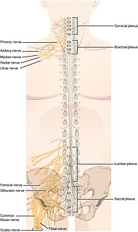
A spinal nerve is a mixed nerve, which carries motor, sensory, and autonomic signals between the spinal cord and the body. In the human body there are 31 pairs of spinal nerves, one on each side of the vertebral column. These are grouped into the corresponding cervical, thoracic, lumbar, sacral and coccygeal regions of the spine. There are eight pairs of cervical nerves, twelve pairs of thoracic nerves, five pairs of lumbar nerves, five pairs of sacral nerves, and one pair of coccygeal nerves. The spinal nerves are part of the peripheral nervous system.

<span class="mw-page-title-main">Genitofemoral nerve</span>

The genitofemoral nerve is a mixed branch of the lumbar plexus derived from anterior rami of L1-L2. It splits a genital branch and a femoral branch. It provides sensory innervation to the upper anterior thigh, as well as the skin of the anterior scrotum in males and mons pubis in females. It also provides motor innervation to the cremaster muscle.

<span class="mw-page-title-main">Cervical plexus</span>

The cervical plexus is a nerve plexus of the anterior rami of the first four cervical spinal nerves C1-C4. The cervical plexus provides motor innervation to some muscles of the neck, and the diaphragm; it provides sensory innervation to parts of the head, neck, and chest.

<span class="mw-page-title-main">Suprascapular nerve</span> Mixed nerve of the upper limb

The suprascapular nerve is a mixed nerve that branches from the upper trunk of the brachial plexus. It is derived from the ventral rami of cervical nerves C5-C6. It provides motor innervation to the supraspinatus muscle, and the infraspinatus muscle.

<span class="mw-page-title-main">Intercostal nerves</span>

The intercostal nerves are part of the somatic nervous system, and arise from the anterior rami of the thoracic spinal nerves from T1 to T11. The intercostal nerves are distributed chiefly to the thoracic pleura and abdominal peritoneum, and differ from the anterior rami of the other spinal nerves in that each pursues an independent course without plexus formation.

<span class="mw-page-title-main">Lumbar plexus</span> Web of nerves in the lower spine

The lumbar plexus is a web of nerves in the lumbar region of the body which forms part of the larger lumbosacral plexus. It is formed by the divisions of the first four lumbar nerves (L1-L4) and from contributions of the subcostal nerve (T12), which is the last thoracic nerve. Additionally, the ventral rami of the fourth lumbar nerve pass communicating branches, the lumbosacral trunk, to the sacral plexus. The nerves of the lumbar plexus pass in front of the hip joint and mainly support the anterior part of the thigh.

<span class="mw-page-title-main">Perforating cutaneous nerve</span>

The perforating cutaneous nerve is a cutaneous nerve of the sacral plexus that provides sensory innervation to the skin of the buttocks.

<span class="mw-page-title-main">Upper subscapular nerve</span> Nerve of upper limb

The upper (superior) subscapular nerve is the first branch of the posterior cord of the brachial plexus. The upper subscapular nerve contains axons from the ventral rami of the C5 and C6 cervical spinal nerves. It innervates the superior portion of the subscapularis muscle. The inferior portion of the subscapularis is innervated by the lower subscapular nerve.

<span class="mw-page-title-main">Sympathetic ganglia</span> Ganglia of the sympathetic nervous system

The sympathetic ganglia, or paravertebral ganglia are autonomic ganglia, of the sympathetic nervous system. Ganglia are 20,000 to 30,000 afferent and efferent nerve cell bodies that run along on either side of the spinal cord. Afferent nerve cell bodies bring information from the body to the brain and spinal cord, while efferent nerve cell bodies bring information from the brain and spinal cord to the rest of the body. The cell bodies create long sympathetic chains that are on either side of the spinal cord. They also form para- or pre-vertebral ganglia of gross anatomy.

<span class="mw-page-title-main">Lateral grey column</span>

The lateral grey column is one of the three grey columns of the spinal cord ; the others being the anterior and posterior grey columns. The lateral grey column is primarily involved with activity in the sympathetic division of the autonomic motor system. It projects to the side as a triangular field in the thoracic and upper lumbar regions of the postero-lateral part of the anterior grey column.

<span class="mw-page-title-main">Subclavian nerve</span>

The subclavian nerve, also known as the nerve to the subclavius, is small branch of the upper trunk of the brachial plexus. It contains axons from C5 and C6. It innervates the subclavius muscle.

<span class="mw-page-title-main">Dorsal ramus of spinal nerve</span>

The dorsal ramus of spinal nerve is the posterior division of a spinal nerve. The dorsal rami provide motor innervation to the deep muscles of the back, and sensory innervation to the skin of the posterior portion of the head, neck and back.

<span class="mw-page-title-main">Ventral ramus of spinal nerve</span>

The ventral ramus is the anterior division of a spinal nerve. The ventral rami supply the antero-lateral parts of the trunk and the limbs. They are mainly larger than the dorsal rami.

<span class="mw-page-title-main">Lumbar ganglia</span>

The lumbar ganglia are paravertebral ganglia located in the inferior portion of the sympathetic trunk. The lumbar portion of the sympathetic trunk typically has 4 lumbar ganglia. The lumbar splanchnic nerves arise from the ganglia here, and contribute sympathetic efferent fibers to the nearby plexuses. The first two lumbar ganglia have both white and gray rami communicates.

<span class="mw-page-title-main">Outline of human anatomy</span> Overview of and topical guide to human anatomy

The following outline is provided as an overview of and topical guide to human anatomy:

<span class="mw-page-title-main">Spinal cord</span> Long, tubular central nervous system structure in the vertebral column

The spinal cord is a long, thin, tubular structure made up of nervous tissue, which extends from the medulla oblongata in the brainstem to the lumbar region of the vertebral column (backbone). The backbone encloses the central canal of the spinal cord, which contains cerebrospinal fluid. The brain and spinal cord together make up the central nervous system (CNS). In humans, the spinal cord begins at the occipital bone, passing through the foramen magnum and then enters the spinal canal at the beginning of the cervical vertebrae. The spinal cord extends down to between the first and second lumbar vertebrae, where it ends. The enclosing bony vertebral column protects the relatively shorter spinal cord. It is around 45 cm (18 in) long in adult men and around 43 cm (17 in) long in adult women. The diameter of the spinal cord ranges from 13 mm in the cervical and lumbar regions to 6.4 mm in the thoracic area.

<span class="mw-page-title-main">Outline of the human nervous system</span> Overview of and topical guide to the human nervous system

The following diagram is provided as an overview of and topical guide to the human nervous system:

References

  1. Betts, J Gordon; Desaix, Peter; Johnson, Eddie; Johnson, Jody E; Korol, Oksana; Kruse, Dean; Poe, Brandon; Wise, James; Womble, Mark D; Young, Kelly A (July 16, 2023). Anatomy & Physiology. Houston: OpenStax CNX. 13.4 The Peripheral Nervous System. ISBN   978-1-947172-04-3.