Neural backpropagation

Last updated

Neural backpropagation is the phenomenon in which, after the action potential of a neuron creates a voltage spike down the axon (normal propagation), another impulse is generated from the soma and propagates towards the apical portions of the dendritic arbor or dendrites (from which much of the original input current originated). In addition to active backpropagation of the action potential, there is also passive electrotonic spread. While there is ample evidence to prove the existence of backpropagating action potentials, the function of such action potentials and the extent to which they invade the most distal dendrites remain highly controversial.

Contents

Mechanism

Methods of neural backpropagation. Left: action potential forms in axon and travels towards soma. Right: Regular action potential generates an echo that backpropagates through the dendritic tree. Mechanisms of Neural Backpropagation.jpg
Methods of neural backpropagation. Left: action potential forms in axon and travels towards soma. Right: Regular action potential generates an echo that backpropagates through the dendritic tree.

When the graded excitatory postsynaptic potentials (EPSPs) depolarize the soma to spike threshold at the axon hillock, first, the axon experiences a propagating impulse through the electrical properties of its voltage-gated sodium and voltage-gated potassium channels. An action potential occurs in the axon first as research illustrates that sodium channels at the dendrites exhibit a higher threshold than those on the membrane of the axon (Rapp et al., 1996). Moreover, the voltage-gated sodium channels on the dendritic membranes having a higher threshold helps prevent them triggering an action potential from synaptic input. Instead, only when the soma depolarizes enough from accumulating graded potentials and firing an axonal action potential will these channels be activated to propagate a signal traveling backwards (Rapp et al. 1996). Generally, EPSPs from synaptic activation are not large enough to activate the dendritic voltage-gated calcium channels (usually on the order of a couple milliamperes each) so backpropagation is typically believed to happen only when the cell is activated to fire an action potential. These sodium channels on the dendrites are abundant in certain types of neurons, especially mitral and pyramidal cells, and quickly inactivate. Initially, it was thought that an action potential could only travel down the axon in one direction (towards the axon terminal where it ultimately signaled the release of neurotransmitters). However, recent research has provided evidence for the existence of backwards-propagating action potentials (Staley 2004).

This diagram displays how the dendritic voltage spike comes after the depolarization of the axon and soma. Dendritic ap spike.gif
This diagram displays how the dendritic voltage spike comes after the depolarization of the axon and soma.

To elaborate, neural backpropagation can occur in one of two ways. First, during the initiation of an axonal action potential, the cell body, or soma, can become depolarized as well. This depolarization can spread through the cell body towards the dendritic tree where there are voltage-gated sodium channels. The depolarization of these voltage-gated sodium channels can then result in the propagation of a dendritic action potential. Such backpropagation is sometimes referred to as an echo of the forward propagating action potential (Staley 2004). It has also been shown that an action potential initiated in the axon can create a retrograde signal that travels in the opposite direction (Hausser 2000). This impulse travels up the axon eventually causing the cell body to become depolarized, thus triggering the dendritic voltage-gated calcium channels. As described in the first process, the triggering of dendritic voltage-gated calcium channels leads to the propagation of a dendritic action potential.

It is important to note that the strength of backpropagating action potentials varies greatly between different neuronal types (Hausser 2000). Some types of neuronal cells show little to no decrease in the amplitude of action potentials as they invade and travel through the dendritic tree while other neuronal cell types, such as cerebellar Purkinje neurons, exhibit very little action potential backpropagation (Stuart 1997). Additionally, there are other neuronal cell types that manifest varying degrees of amplitude decrement during backpropagation. It is thought that this is due to the fact that each neuronal cell type contains varying numbers of the voltage-gated channels required to propagate a dendritic action potential.

Regulation and inhibition

Generally, synaptic signals that are received by the dendrite are combined in the soma in order to generate an action potential that is then transmitted down the axon toward the next synaptic contact. Thus, the backpropagation of action potentials poses a threat to initiate an uncontrolled positive feedback loop between the soma and the dendrites. For example, as an action potential was triggered, its dendritic echo could enter the dendrite and potentially trigger a second action potential. If left unchecked, an endless cycle of action potentials triggered by their own echo would be created. In order to prevent such a cycle, most neurons have a relatively high density of A-type K+ channels.

A-type K+ channels belong to the superfamily of voltage-gated ion channels and are transmembrane channels that help maintain the cell's membrane potential (Cai 2007). Typically, they play a crucial role in returning the cell to its resting membrane following an action potential by allowing an inhibitory current of K+ ions to quickly flow out of the neuron. The presence of these channels in such high density in the dendrites explains their inability to initiate an action potential, even during synaptic input. Additionally, the presence of these channels provides a mechanism by which the neuron can suppress and regulate the backpropagation of action potentials through the dendrite (Vetter 2000). Pharmacological antagonists of these channels promoted the frequency of backpropagating action potentials which demonstrates their importance in keeping the cell from excessive firing (Waters et al., 2004). Results have indicated a linear increase in the density of A-type channels with increasing distance into the dendrite away from the soma. The increase in the density of A-type channels results in a dampening of the backpropagating action potential as it travels into the dendrite. Essentially, inhibition occurs because the A-type channels facilitate the outflow of K+ ions in order to maintain the membrane potential below threshold levels (Cai 2007). Such inhibition limits EPSP and protects the neuron from entering a never-ending positive-positive feedback loop between the soma and the dendrites.

History

Since the 1950s, evidence has existed that neurons in the central nervous system generate an action potential, or voltage spike, that travels both through the axon to signal the next neuron and backpropagates through the dendrites sending a retrograde signal to its presynaptic signaling neurons. This current decays significantly with travel length along the dendrites, so effects are predicted to be more significant for neurons whose synapses are near the postsynaptic cell body, with magnitude depending mainly on sodium-channel density in the dendrite. It is also dependent on the shape of the dendritic tree and, more importantly, on the rate of signal currents to the neuron. On average, a backpropagating spike loses about half its voltage after traveling nearly 500 micrometres.

Backpropagation occurs actively in the neocortex, hippocampus, substantia nigra, and spinal cord, while in the cerebellum it occurs relatively passively. This is consistent with observations that synaptic plasticity is much more apparent in areas like the hippocampus, which controls spatial memory, than the cerebellum, which controls more unconscious and vegetative functions.

The backpropagating current also causes a voltage change that increases the concentration of Ca2+ in the dendrites, an event which coincides with certain models of synaptic plasticity. This change also affects future integration of signals, leading to at least a short-term response difference between the presynaptic signals and the postsynaptic spike. [1]

Functions

While many questions have yet to be answered in regards to neural backpropagation, there exists a number of hypotheses regarding its function. Some proposed function include involvement in synaptic plasticity, involvement in dendrodendritic inhibition, boosting synaptic responses, resetting membrane potential, retrograde actions at synapses and conditional axonal output. Backpropagation is believed to help form LTP (long term potentiation) and Hebbian plasticity at hippocampal synapses. Since artificial LTP induction, using microelectrode stimulation, voltage clamp, etc. requires the postsynaptic cell to be slightly depolarized when EPSPs are elicited, backpropagation can serve as the means of depolarization of the postsynaptic cell.

Backpropagating action potentials can induce Long-term potentiation by behaving as a signal that informs the presynaptic cell that the postsynaptic cell has fired. Moreover, Spike-Time Dependent Plasticity is known as the narrow time frame for which coincidental firing of both the pre and post synaptic neurons will induce plasticity. Neural backpropagation occurs in this window to interact with NMDA receptors at the apical dendrites by assisting in the removal of voltage sensitive Mg2+ block (Waters et al., 2004). This process permits the large influx of calcium which provokes a cascade of events to cause potentiation.

Current literature also suggests that backpropagating action potentials are also responsible for the release of retrograde neurotransmitters and trophic factors which contribute to the short-term and long-term efficacy between two neurons. Since the backpropagating action potentials essentially exhibit a copy of the neurons axonal firing pattern, they help establish a synchrony between the pre and post synaptic neurons (Waters et al., 2004).

Importantly, backpropagating action potentials are necessary for the release of Brain-Derived Neurotrophic Factor (BDNF). BDNF is an essential component for inducing synaptic plasticity and development (Kuczewski N., Porcher C., Ferrand N., 2008). Moreover, backpropagating action potentials have been shown to induce BDNF-dependent phosphorylation of cyclic AMP response element-binding protein (CREB) which is known to be a major component in synaptic plasticity and memory formation (Kuczewski N., Porcher C., Lessmann V., et al. 2008).

Algorithm

While a backpropagating action potential can presumably cause changes in the weight of the presynaptic connections, there is no simple mechanism for an error signal to propagate through multiple layers of neurons, as in the computer backpropagation algorithm. However, simple linear topologies have shown that effective computation is possible through signal backpropagation in this biological sense. [2]

Related Research Articles

<span class="mw-page-title-main">Dendrite</span> Small projection on a neuron that receives signals

A dendrite or dendron is a branched protoplasmic extension of a nerve cell that propagates the electrochemical stimulation received from other neural cells to the cell body, or soma, of the neuron from which the dendrites project. Electrical stimulation is transmitted onto dendrites by upstream neurons via synapses which are located at various points throughout the dendritic tree.

<span class="mw-page-title-main">Neuron</span> Electrically excitable cell found in the nervous system of animals

Within a nervous system, a neuron, neurone, or nerve cell is an electrically excitable cell that fires electric signals called action potentials across a neural network. Neurons communicate with other cells via synapses, which are specialized connections that commonly use minute amounts of chemical neurotransmitters to pass the electric signal from the presynaptic neuron to the target cell through the synaptic gap.

<span class="mw-page-title-main">Chemical synapse</span> Biological junctions through which neurons signals can be sent

Chemical synapses are biological junctions through which neurons' signals can be sent to each other and to non-neuronal cells such as those in muscles or glands. Chemical synapses allow neurons to form circuits within the central nervous system. They are crucial to the biological computations that underlie perception and thought. They allow the nervous system to connect to and control other systems of the body.

<span class="mw-page-title-main">Action potential</span> Neuron communication by electric impulses

An action potential occurs when the membrane potential of a specific cell rapidly rises and falls. This depolarization then causes adjacent locations to similarly depolarize. Action potentials occur in several types of animal cells, called excitable cells, which include neurons, muscle cells, and in some plant cells. Certain endocrine cells such as pancreatic beta cells, and certain cells of the anterior pituitary gland are also excitable cells.

An inhibitory postsynaptic potential (IPSP) is a kind of synaptic potential that makes a postsynaptic neuron less likely to generate an action potential. The opposite of an inhibitory postsynaptic potential is an excitatory postsynaptic potential (EPSP), which is a synaptic potential that makes a postsynaptic neuron more likely to generate an action potential. IPSPs can take place at all chemical synapses, which use the secretion of neurotransmitters to create cell-to-cell signalling. EPSPs and IPSPs compete with each other at numerous synapses of a neuron. This determines whether an action potential occurring at the presynaptic terminal produces an action potential at the postsynaptic membrane. Some common neurotransmitters involved in IPSPs are GABA and glycine.

<span class="mw-page-title-main">Excitatory postsynaptic potential</span> Process causing temporary increase in postsynaptic potential

In neuroscience, an excitatory postsynaptic potential (EPSP) is a postsynaptic potential that makes the postsynaptic neuron more likely to fire an action potential. This temporary depolarization of postsynaptic membrane potential, caused by the flow of positively charged ions into the postsynaptic cell, is a result of opening ligand-gated ion channels. These are the opposite of inhibitory postsynaptic potentials (IPSPs), which usually result from the flow of negative ions into the cell or positive ions out of the cell. EPSPs can also result from a decrease in outgoing positive charges, while IPSPs are sometimes caused by an increase in positive charge outflow. The flow of ions that causes an EPSP is an excitatory postsynaptic current (EPSC).

<span class="mw-page-title-main">Excitatory synapse</span> Sort of synapse

An excitatory synapse is a synapse in which an action potential in a presynaptic neuron increases the probability of an action potential occurring in a postsynaptic cell. Neurons form networks through which nerve impulses travels, each neuron often making numerous connections with other cells of neurons. These electrical signals may be excitatory or inhibitory, and, if the total of excitatory influences exceeds that of the inhibitory influences, the neuron will generate a new action potential at its axon hillock, thus transmitting the information to yet another cell.

<span class="mw-page-title-main">Pyramidal cell</span> Projection neurons in the cerebral cortex and hippocampus

Pyramidal cells, or pyramidal neurons, are a type of multipolar neuron found in areas of the brain including the cerebral cortex, the hippocampus, and the amygdala. Pyramidal cells are the primary excitation units of the mammalian prefrontal cortex and the corticospinal tract. Pyramidal neurons are also one of two cell types where the characteristic sign, Negri bodies, are found in post-mortem rabies infection. Pyramidal neurons were first discovered and studied by Santiago Ramón y Cajal. Since then, studies on pyramidal neurons have focused on topics ranging from neuroplasticity to cognition.

<span class="mw-page-title-main">Axon hillock</span> Part of the neuronal cell soma from which the axon originates

The axon hillock is a specialized part of the cell body of a neuron that connects to the axon. It can be identified using light microscopy from its appearance and location in a neuron and from its sparse distribution of Nissl substance.

<span class="mw-page-title-main">Electrotonic potential</span>

In physiology, electrotonus refers to the passive spread of charge inside a neuron and between cardiac muscle cells or smooth muscle cells. Passive means that voltage-dependent changes in membrane conductance do not contribute. Neurons and other excitable cells produce two types of electrical potential:

<span class="mw-page-title-main">Neural circuit</span> Network or circuit of neurons

A neural circuit is a population of neurons interconnected by synapses to carry out a specific function when activated. Multiple neural circuits interconnect with one another to form large scale brain networks.

Molecular neuroscience is a branch of neuroscience that observes concepts in molecular biology applied to the nervous systems of animals. The scope of this subject covers topics such as molecular neuroanatomy, mechanisms of molecular signaling in the nervous system, the effects of genetics and epigenetics on neuronal development, and the molecular basis for neuroplasticity and neurodegenerative diseases. As with molecular biology, molecular neuroscience is a relatively new field that is considerably dynamic.

Schaffer collaterals are axon collaterals given off by CA3 pyramidal cells in the hippocampus. These collaterals project to area CA1 of the hippocampus and are an integral part of memory formation and the emotional network of the Papez circuit, and of the hippocampal trisynaptic loop. It is one of the most studied synapses in the world and named after the Hungarian anatomist-neurologist Károly Schaffer.

An apical dendrite is a dendrite that emerges from the apex of a pyramidal cell. Apical dendrites are one of two primary categories of dendrites, and they distinguish the pyramidal cells from spiny stellate cells in the cortices. Pyramidal cells are found in the prefrontal cortex, the hippocampus, the entorhinal cortex, the olfactory cortex, and other areas. Dendrite arbors formed by apical dendrites are the means by which synaptic inputs into a cell are integrated. The apical dendrites in these regions contribute significantly to memory, learning, and sensory associations by modulating the excitatory and inhibitory signals received by the pyramidal cells.

<span class="mw-page-title-main">Synapse</span> Structure connecting neurons in the nervous system

In the nervous system, a synapse is a structure that permits a neuron to pass an electrical or chemical signal to another neuron or to the target effector cell.

<span class="mw-page-title-main">Synaptic potential</span> Potential difference across the postsynaptic membrane

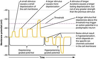
Synaptic potential refers to the potential difference across the postsynaptic membrane that results from the action of neurotransmitters at a neuronal synapse. In other words, it is the “incoming” signal that a neuron receives. There are two forms of synaptic potential: excitatory and inhibitory. The type of potential produced depends on both the postsynaptic receptor, more specifically the changes in conductance of ion channels in the post synaptic membrane, and the nature of the released neurotransmitter. Excitatory post-synaptic potentials (EPSPs) depolarize the membrane and move the potential closer to the threshold for an action potential to be generated. Inhibitory postsynaptic potentials (IPSPs) hyperpolarize the membrane and move the potential farther away from the threshold, decreasing the likelihood of an action potential occurring. The Excitatory Post Synaptic potential is most likely going to be carried out by the neurotransmitters glutamate and acetylcholine, while the Inhibitory post synaptic potential will most likely be carried out by the neurotransmitters gamma-aminobutyric acid (GABA) and glycine. In order to depolarize a neuron enough to cause an action potential, there must be enough EPSPs to both depolarize the postsynaptic membrane from its resting membrane potential to its threshold and counterbalance the concurrent IPSPs that hyperpolarize the membrane. As an example, consider a neuron with a resting membrane potential of -70 mV (millivolts) and a threshold of -50 mV. It will need to be raised 20 mV in order to pass the threshold and fire an action potential. The neuron will account for all the many incoming excitatory and inhibitory signals via summative neural integration, and if the result is an increase of 20 mV or more, an action potential will occur.

<span class="mw-page-title-main">Dendritic spike</span> Action potential generated in the dendrite of a neuron

In neurophysiology, a dendritic spike refers to an action potential generated in the dendrite of a neuron. Dendrites are branched extensions of a neuron. They receive electrical signals emitted from projecting neurons and transfer these signals to the cell body, or soma. Dendritic signaling has traditionally been viewed as a passive mode of electrical signaling. Unlike its axon counterpart which can generate signals through action potentials, dendrites were believed to only have the ability to propagate electrical signals by physical means: changes in conductance, length, cross sectional area, etc. However, the existence of dendritic spikes was proposed and demonstrated by W. Alden Spencer, Eric Kandel, Rodolfo Llinás and coworkers in the 1960s and a large body of evidence now makes it clear that dendrites are active neuronal structures. Dendrites contain voltage-gated ion channels giving them the ability to generate action potentials. Dendritic spikes have been recorded in numerous types of neurons in the brain and are thought to have great implications in neuronal communication, memory, and learning. They are one of the major factors in long-term potentiation.

<span class="mw-page-title-main">Summation (neurophysiology)</span>

Summation, which includes both spatial summation and temporal summation, is the process that determines whether or not an action potential will be generated by the combined effects of excitatory and inhibitory signals, both from multiple simultaneous inputs, and from repeated inputs. Depending on the sum total of many individual inputs, summation may or may not reach the threshold voltage to trigger an action potential.

Cellular neuroscience is a branch of neuroscience concerned with the study of neurons at a cellular level. This includes morphology and physiological properties of single neurons. Several techniques such as intracellular recording, patch-clamp, and voltage-clamp technique, pharmacology, confocal imaging, molecular biology, two photon laser scanning microscopy and Ca2+ imaging have been used to study activity at the cellular level. Cellular neuroscience examines the various types of neurons, the functions of different neurons, the influence of neurons upon each other, and how neurons work together.

<span class="mw-page-title-main">Nonsynaptic plasticity</span> Form of neuroplasticity

Nonsynaptic plasticity is a form of neuroplasticity that involves modification of ion channel function in the axon, dendrites, and cell body that results in specific changes in the integration of excitatory postsynaptic potentials and inhibitory postsynaptic potentials. Nonsynaptic plasticity is a modification of the intrinsic excitability of the neuron. It interacts with synaptic plasticity, but it is considered a separate entity from synaptic plasticity. Intrinsic modification of the electrical properties of neurons plays a role in many aspects of plasticity from homeostatic plasticity to learning and memory itself. Nonsynaptic plasticity affects synaptic integration, subthreshold propagation, spike generation, and other fundamental mechanisms of neurons at the cellular level. These individual neuronal alterations can result in changes in higher brain function, especially learning and memory. However, as an emerging field in neuroscience, much of the knowledge about nonsynaptic plasticity is uncertain and still requires further investigation to better define its role in brain function and behavior.

References

  1. Stuart, G; Spruston, N; Sakmann, B; Häusser, M (1997). "Action potential initiation and backpropagation in neurons of the mammalian CNS". Trends in Neurosciences. 20 (3): 125–31. doi:10.1016/s0166-2236(96)10075-8. PMID   9061867. S2CID   889625.
  2. Bogacz, Rafal; Malcolm W. Brown; Christophe Giraud-Carrier (2000). "Frequency-based error backpropagation in a cortical network". Proceedings of the IEEE-INNS-ENNS International Joint Conference on Neural Networks. IJCNN 2000. Neural Computing: New Challenges and Perspectives for the New Millennium (PDF). Vol. 2. pp. 211–216. CiteSeerX   10.1.1.22.8774 . doi:10.1109/IJCNN.2000.857899. ISBN   978-0-7695-0619-7. S2CID   896603. 0-7695-0619-4. Archived from the original (PDF) on June 14, 2007. Retrieved 2007-11-18.