Voltage-gated potassium channel

Last updated
Eukaryotic potassium channel
2r9r opm.png
Potassium channel, structure in a membrane-like environment. Calculated hydrocarbon boundaries of the lipid bilayer are indicated by red and blue dots.
Identifiers
SymbolIon_trans
Pfam PF00520
InterPro IPR005821
SCOP2 1bl8 / SCOPe / SUPFAM
TCDB 1.A.1
OPM superfamily 8
OPM protein 2a79
Membranome 217
Available protein structures:
Pfam   structures / ECOD  
PDB RCSB PDB; PDBe; PDBj
PDBsum structure summary
Ion channel (bacterial)
1r3j.png
Potassium channel KcsA. Calculated hydrocarbon boundaries of the lipid bilayer are indicated by red and blue dots.
Identifiers
SymbolIon_trans_2
Pfam PF07885
InterPro IPR013099
SCOP2 1bl8 / SCOPe / SUPFAM
OPM protein 1r3j
Available protein structures:
Pfam   structures / ECOD  
PDB RCSB PDB; PDBe; PDBj
PDBsum structure summary
Slow voltage-gated potassium channel (Potassium channel, voltage-dependent, beta subunit, KCNE)
Identifiers
SymbolISK_Channel
Pfam PF02060
InterPro IPR000369
TCDB 8.A.10
Membranome 218
Available protein structures:
Pfam   structures / ECOD  
PDB RCSB PDB; PDBe; PDBj
PDBsum structure summary
KCNQ voltage-gated potassium channel
Identifiers
SymbolKCNQ_channel
Pfam PF03520
InterPro IPR013821
Available protein structures:
Pfam   structures / ECOD  
PDB RCSB PDB; PDBe; PDBj
PDBsum structure summary
Kv2 voltage-gated K+ channel
Identifiers
SymbolKv2channel
Pfam PF03521
InterPro IPR003973
Available protein structures:
Pfam   structures / ECOD  
PDB RCSB PDB; PDBe; PDBj
PDBsum structure summary

Voltage-gated potassium channels (VGKCs) are transmembrane channels specific for potassium and sensitive to voltage changes in the cell's membrane potential. During action potentials, they play a crucial role in returning the depolarized cell to a resting state.

Contents

Classification

Alpha subunits

Alpha subunits form the actual conductance pore. Based on sequence homology of the hydrophobic transmembrane cores, the alpha subunits of voltage-gated potassium channels are grouped into 12 classes. These are labeled Kvα1-12. [1] The following is a list of the 40 known human voltage-gated potassium channel alpha subunits grouped first according to function and then subgrouped according to the Kv sequence homology classification scheme:

Delayed rectifier

slowly inactivating or non-inactivating

A-type potassium channel

rapidly inactivating

  • Kvα1.x - Shaker-related: Kv1.4 (KCNA4)
  • Kvα4.x - Shal-related: Kv4.1 (KCND1), Kv4.2 (KCND2), Kv4.3 (KCND3)

Outward-rectifying

Inwardly-rectifying

Passes current more easily in the inward direction (into the cell, from outside).

Slowly activating

Modifier/silencer

Unable to form functional channels as homotetramers but instead heterotetramerize with Kvα2 family members to form conductive channels.

Beta subunits

Beta subunits are auxiliary proteins that associate with alpha subunits, sometimes in a α4β4 stoichiometry. [2] These subunits do not conduct current on their own but rather modulate the activity of Kv channels. [3]

Proteins minK and MiRP1 are putative hERG beta subunits. [6]

Animal research

The voltage-gated K+ channels that provide the outward currents of action potentials have similarities to bacterial K+ channels.

These channels have been studied by X-ray diffraction, allowing determination of structural features at atomic resolution.

The function of these channels is explored by electrophysiological studies.

Genetic approaches include screening for behavioral changes in animals with mutations in K+ channel genes. Such genetic methods allowed the genetic identification of the "Shaker" K+ channel gene in Drosophila before ion channel gene sequences were well known.

Study of the altered properties of voltage-gated K+ channel proteins produced by mutated genes has helped reveal the functional roles of K+ channel protein domains and even individual amino acids within their structures.

Structure

Typically, vertebrate voltage-gated K+ channels are tetramers of four identical subunits arranged as a ring, each contributing to the wall of the trans-membrane K+ pore. Each subunit is composed of six membrane spanning hydrophobic α-helical sequences, as well as a voltage sensor in S4. The intracellular side of the membrane contains both amino and carboxy termini. [7] The high resolution crystallographic structure of the rat Kvα1.2/β2 channel has recently been solved (Protein Databank Accession Number 2A79 ), [8] and then refined in a lipid membrane-like environment ( PDB: 2r9r ).

Selectivity

Voltage-gated K+ channels are selective for K+ over other cations such as Na+. There is a selectivity filter at the narrowest part of the transmembrane pore.

Channel mutation studies have revealed the parts of the subunits that are essential for ion selectivity. They include the amino acid sequence (Thr-Val-Gly-Tyr-Gly) or (Thr-Val-Gly-Phe-Gly) typical to the selectivity filter of voltage-gated K+ channels. As K+ passes through the pore, interactions between potassium ions and water molecules are prevented and the K+ interacts with specific atomic components of the Thr-Val-Gly-[YF]-Gly sequences from the four channel subunits .

It may seem counterintuitive that a channel should allow potassium ions but not the smaller sodium ions through. However in an aqueous environment, potassium and sodium cations are solvated by water molecules. When moving through the selectivity filter of the potassium channel, the water-K+ interactions are replaced by interactions between K+ and carbonyl groups of the channel protein. The diameter of the selectivity filter is ideal for the potassium cation, but too big for the smaller sodium cation. Hence the potassium cations are well "solvated" by the protein carbonyl groups, but these same carbonyl groups are too far apart to adequately solvate the sodium cation. Hence, the passage of potassium cations through this selectivity filter is strongly favored over sodium cations.

Open and closed conformations

The structure of the mammalian voltage-gated K+ channel has been used to explain its ability to respond to the voltage across the membrane. Upon opening of the channel, conformational changes in the voltage-sensor domains (VSD) result in the transfer of 12-13 elementary charges across the membrane electric field. This charge transfer is measured as a transient capacitive current that precedes opening of the channel. Several charged residues of the VSD, in particular four arginine residues located regularly at every third position on the S4 segment, are known to move across the transmembrane field and contribute to the gating charge. The position of these arginines, known as gating arginines, are highly conserved in all voltage-gated potassium, sodium, or calcium channels. However, the extent of their movement and their displacement across the transmembrane potential has been subject to extensive debate. [9] Specific domains of the channel subunits have been identified that are responsible for voltage-sensing and converting between the open and closed conformations of the channel. There are at least two closed conformations. In the first, the channel can open if the membrane potential becomes more positive. This type of gating is mediated by a voltage-sensing domain that consists of the S4 alpha helix that contains 6–7 positive charges. Changes in membrane potential cause this alpha helix to move in the lipid bilayer. This movement in turn results in a conformational change in the adjacent S5–S6 helices that form the channel pore and cause this pore to open or close. In the second, "N-type" inactivation, voltage-gated K+ channels inactivate after opening, entering a distinctive, closed conformation. In this inactivated conformation, the channel cannot open, even if the transmembrane voltage is favorable. The amino terminal domain of the K+ channel or an auxiliary protein can mediate "N-type" inactivation. The mechanism of this type of inactivation has been described as a "ball and chain" model, where the N-terminus of the protein forms a ball that is tethered to the rest of the protein through a loop (the chain). [10] The tethered ball blocks the inner porehole, preventing ion movement through the channel. [11] [12]

Pharmacology

For blockers and activators of voltage gated potassium channels see: potassium channel blocker and potassium channel opener.

See also

Related Research Articles

<span class="mw-page-title-main">Ion channel</span> Pore-forming membrane protein

Ion channels are pore-forming membrane proteins that allow ions to pass through the channel pore. Their functions include establishing a resting membrane potential, shaping action potentials and other electrical signals by gating the flow of ions across the cell membrane, controlling the flow of ions across secretory and epithelial cells, and regulating cell volume. Ion channels are present in the membranes of all cells. Ion channels are one of the two classes of ionophoric proteins, the other being ion transporters.

<span class="mw-page-title-main">Potassium channel</span> Ion channel that selectively passes K+

Potassium channels are the most widely distributed type of ion channel found in virtually all organisms. They form potassium-selective pores that span cell membranes. Potassium channels are found in most cell types and control a wide variety of cell functions.

<span class="mw-page-title-main">Repolarization</span> Change in membrane potential

In neuroscience, repolarization refers to the change in membrane potential that returns it to a negative value just after the depolarization phase of an action potential which has changed the membrane potential to a positive value. The repolarization phase usually returns the membrane potential back to the resting membrane potential. The efflux of potassium (K+) ions results in the falling phase of an action potential. The ions pass through the selectivity filter of the K+ channel pore.

<span class="mw-page-title-main">Voltage-gated ion channel</span> Type of ion channel transmembrane protein

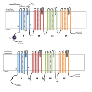
Voltage-gated ion channels are a class of transmembrane proteins that form ion channels that are activated by changes in the electrical membrane potential near the channel. The membrane potential alters the conformation of the channel proteins, regulating their opening and closing. Cell membranes are generally impermeable to ions, thus they must diffuse through the membrane through transmembrane protein channels. They have a crucial role in excitable cells such as neuronal and muscle tissues, allowing a rapid and co-ordinated depolarization in response to triggering voltage change. Found along the axon and at the synapse, voltage-gated ion channels directionally propagate electrical signals. Voltage-gated ion-channels are usually ion-specific, and channels specific to sodium (Na+), potassium (K+), calcium (Ca2+), and chloride (Cl) ions have been identified. The opening and closing of the channels are triggered by changing ion concentration, and hence charge gradient, between the sides of the cell membrane.

<span class="mw-page-title-main">KvLQT1</span> Protein-coding gene in the species Homo sapiens

Kv7.1 (KvLQT1) is a potassium channel protein whose primary subunit in humans is encoded by the KCNQ1 gene. Kv7.1 is a voltage and lipid-gated potassium channel present in the cell membranes of cardiac tissue and in inner ear neurons among other tissues. In the cardiac cells, Kv7.1 mediates the IKs (or slow delayed rectifying K+) current that contributes to the repolarization of the cell, terminating the cardiac action potential and thereby the heart's contraction. It is a member of the KCNQ family of potassium channels.

Sodium channels are integral membrane proteins that form ion channels, conducting sodium ions (Na+) through a cell's membrane. They belong to the superfamily of cation channels.

The shaker (Sh) gene, when mutated, causes a variety of atypical behaviors in the fruit fly, Drosophila melanogaster. Under ether anesthesia, the fly’s legs will shake ; even when the fly is unanaesthetized, it will exhibit aberrant movements. Sh-mutant flies have a shorter lifespan than regular flies; in their larvae, the repetitive firing of action potentials as well as prolonged exposure to neurotransmitters at neuromuscular junctions occurs.

<span class="mw-page-title-main">Kv1.1</span>

Potassium voltage-gated channel subfamily A member 1 also known as Kv1.1 is a shaker related voltage-gated potassium channel that in humans is encoded by the KCNA1 gene. Isaacs syndrome is a result of an autoimmune reaction against the Kv1.1 ion channel.

Two-pore channels (TPCs) are eukaryotic intracellular voltage-gated and ligand gated cation selective ion channels. There are two known paralogs in the human genome, TPC1s and TPC2s. In humans, TPC1s are sodium selective and TPC2s conduct sodium ions, calcium ions and possibly hydrogen ions. Plant TPC1s are non-selective channels. Expression of TPCs are found in both plant vacuoles and animal acidic organelles. These organelles consist of endosomes and lysosomes. TPCs are formed from two transmembrane non-equivalent tandem Shaker-like, pore-forming subunits, dimerized to form quasi-tetramers. Quasi-tetramers appear very similar to tetramers, but are not quite the same. Some key roles of TPCs include calcium dependent responses in muscle contraction(s), hormone secretion, fertilization, and differentiation. Disorders linked to TPCs include membrane trafficking, Parkinson's disease, Ebola, and fatty liver.

<span class="mw-page-title-main">KCNE1</span> Protein-coding gene in the species Homo sapiens

Potassium voltage-gated channel subfamily E member 1 is a protein that in humans is encoded by the KCNE1 gene.

<span class="mw-page-title-main">Cation channel superfamily</span> Family of ion channel proteins

The transmembrane cation channel superfamily was defined in InterPro and Pfam as the family of tetrameric ion channels. These include the sodium, potassium, calcium, ryanodine receptor, HCN, CNG, CatSper, and TRP channels. This large group of ion channels apparently includes families 1.A.1, 1.A.2, 1.A.3, and 1.A.4 of the TCDB transporter classification.

<span class="mw-page-title-main">Potassium channel tetramerisation domain</span>

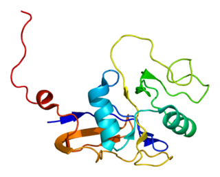
K+ channel tetramerisation domain is the N-terminal, cytoplasmic tetramerisation domain (T1) of voltage-gated K+ channels. It defines molecular determinants for subfamily-specific assembly of alpha-subunits into functional tetrameric channels. It is distantly related to the BTB/POZ domain Pfam PF00651.

<span class="mw-page-title-main">KCNB1</span> Protein-coding gene in the species Homo sapiens

Potassium voltage-gated channel, Shab-related subfamily, member 1, also known as KCNB1 or Kv2.1, is a protein that, in humans, is encoded by the KCNB1 gene.

<span class="mw-page-title-main">KCNAB2</span> Protein-coding gene in the species Homo sapiens

Voltage-gated potassium channel subunit beta-2 is a protein that in humans is encoded by the KCNAB2 gene.

<span class="mw-page-title-main">KCNE4</span> Protein-coding gene in the species Homo sapiens

Potassium voltage-gated channel subfamily E member 4, originally named MinK-related peptide 3 or MiRP3 when it was discovered, is a protein that in humans is encoded by the KCNE4 gene.

<span class="mw-page-title-main">KCNE5</span> Protein-coding gene in the species Homo sapiens

KCNE1-like also known as KCNE1L is a protein that in humans is encoded by the KCNE1L gene.

<span class="mw-page-title-main">Gating (electrophysiology)</span>

In electrophysiology, the term gating refers to the opening (activation) or closing of ion channels. This change in conformation is a response to changes in transmembrane voltage.

<span class="mw-page-title-main">KcsA potassium channel</span> Prokaryotic potassium ion channel

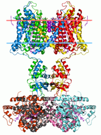
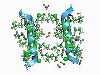
KcsA (Kchannel of streptomyces A) is a prokaryotic potassium channel from the soil bacterium Streptomyces lividans that has been studied extensively in ion channel research. The pH activated protein possesses two transmembrane segments and a highly selective pore region, responsible for the gating and shuttling of K+ ions out of the cell. The amino acid sequence found in the selectivity filter of KcsA is highly conserved among both prokaryotic and eukaryotic K+ voltage channels; as a result, research on KcsA has provided important structural and mechanistic insight on the molecular basis for K+ ion selection and conduction. As one of the most studied ion channels to this day, KcsA is a template for research on K+ channel function and its elucidated structure underlies computational modeling of channel dynamics for both prokaryotic and eukaryotic species.

<span class="mw-page-title-main">Pandinus imperator (Pi3) toxin</span>

Pi3 toxin is a purified peptide derivative of the Pandinus imperator scorpion venom. It is a potent blocker of voltage-gated potassium channel, Kv1.3 and is closely related to another peptide found in the venom, Pi2.

<span class="mw-page-title-main">Ball and chain inactivation</span> Model in neuroscience

In neuroscience, ball and chain inactivation is a model to explain the fast inactivation mechanism of voltage-gated ion channels. The process is also called hinged-lid inactivation or N-type inactivation. A voltage-gated ion channel can be in three states: open, closed, or inactivated. The inactivated state is mainly achieved through fast inactivation, by which a channel transitions rapidly from an open to an inactivated state. The model proposes that the inactivated state, which is stable and non-conducting, is caused by the physical blockage of the pore. The blockage is caused by a "ball" of amino acids connected to the main protein by a string of residues on the cytoplasmic side of the membrane. The ball enters the open channel and binds to the hydrophobic inner vestibule within the channel. This blockage causes inactivation of the channel by stopping the flow of ions. This phenomenon has mainly been studied in potassium channels and sodium channels.

References

  1. Gutman GA, Chandy KG, Grissmer S, Lazdunski M, McKinnon D, Pardo LA, Robertson GA, Rudy B, Sanguinetti MC, Stühmer W, Wang X (December 2005). "International Union of Pharmacology. LIII. Nomenclature and molecular relationships of voltage-gated potassium channels". Pharmacological Reviews. 57 (4): 473–508. doi:10.1124/pr.57.4.10. PMID   16382104. S2CID   219195192.
  2. Pongs O, Leicher T, Berger M, Roeper J, Bähring R, Wray D, Giese KP, Silva AJ, Storm JF (April 1999). "Functional and molecular aspects of voltage-gated K+ channel beta subunits". Annals of the New York Academy of Sciences. 868 (Apr 30): 344–55. Bibcode:1999NYASA.868..344P. doi:10.1111/j.1749-6632.1999.tb11296.x. PMID   10414304. S2CID   21621084.
  3. Li Y, Um SY, McDonald TV (June 2006). "Voltage-gated potassium channels: regulation by accessory subunits". The Neuroscientist. 12 (3): 199–210. doi:10.1177/1073858406287717. PMID   16684966. S2CID   24418687.
  4. Zhang M, Jiang M, Tseng GN (May 2001). "minK-related peptide 1 associates with Kv4.2 and modulates its gating function: potential role as beta subunit of cardiac transient outward channel?". Circulation Research. 88 (10): 1012–9. doi: 10.1161/hh1001.090839 . PMID   11375270.
  5. McCrossan ZA, Abbott GW (November 2004). "The MinK-related peptides". Neuropharmacology. 47 (6): 787–821. doi:10.1016/j.neuropharm.2004.06.018. PMID   15527815. S2CID   41340789.
  6. Anantharam A, Abbott GW (2005). Does hERG coassemble with a beta subunit? Evidence for roles of MinK and MiRP1. Novartis Foundation Symposia. Vol. 266. pp. 100–12, discussion 112–7, 155–8. doi:10.1002/047002142X.fmatter. ISBN   9780470021408. PMID   16050264.{{cite book}}: |journal= ignored (help)
  7. Yellen G (September 2002). "The voltage-gated potassium channels and their relatives". Nature. 419 (6902): 35–42. doi:10.1038/nature00978. PMID   12214225. S2CID   4420877.
  8. Long SB, Campbell EB, Mackinnon R (August 2005). "Crystal structure of a mammalian voltage-dependent Shaker family K+ channel". Science. 309 (5736): 897–903. Bibcode:2005Sci...309..897L. doi: 10.1126/science.1116269 . PMID   16002581. S2CID   6072007.
  9. Lee SY, Lee A, Chen J, MacKinnon R (October 2005). "Structure of the KvAP voltage-dependent K+ channel and its dependence on the lipid membrane". Proceedings of the National Academy of Sciences of the United States of America. 102 (43): 15441–6. Bibcode:2005PNAS..10215441L. doi: 10.1073/pnas.0507651102 . PMC   1253646 . PMID   16223877.
  10. Antz C, Fakler B (August 1998). "Fast Inactivation of Voltage-Gated K(+) Channels: From Cartoon to Structure" (PDF). News in Physiological Sciences. 13 (4): 177–182. doi:10.1152/physiologyonline.1998.13.4.177. PMID   11390785.
  11. Armstrong CM, Bezanilla F (April 1973). "Currents related to movement of the gating particles of the sodium channels". Nature. 242 (5398): 459–61. Bibcode:1973Natur.242..459A. doi:10.1038/242459a0. PMID   4700900. S2CID   4261606.
  12. Murrell-Lagnado RD, Aldrich RW (December 1993). "Energetics of Shaker K channels block by inactivation peptides". The Journal of General Physiology. 102 (6): 977–1003. doi:10.1085/jgp.102.6.977. PMC   2229186 . PMID   8133246.