A ligament is the fibrous connective tissue that connects bones to other bones. It is also known as articular ligament, articular larua, fibrous ligament, or true ligament. Other ligaments in the body include the:

Human teeth function to mechanically break down items of food by cutting and crushing them in preparation for swallowing and digesting. As such, they are considered part of the human digestive system. Humans have four types of teeth: incisors, canines, premolars, and molars, which each have a specific function. The incisors cut the food, the canines tear the food and the molars and premolars crush the food. The roots of teeth are embedded in the maxilla or the mandible and are covered by gums. Teeth are made of multiple tissues of varying density and hardness.

Cementum is a specialized calcified substance covering the root of a tooth. The cementum is the part of the periodontium that attaches the teeth to the alveolar bone by anchoring the periodontal ligament.

The human musculoskeletal system is an organ system that gives humans the ability to move using their muscular and skeletal systems. The musculoskeletal system provides form, support, stability, and movement to the body.

The periosteum is a membrane that covers the outer surface of all bones, except at the articular surfaces of long bones. Endosteum lines the inner surface of the medullary cavity of all long bones.
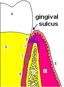
The periodontium is the specialized tissues that both surround and support the teeth, maintaining them in the maxillary and mandibular bones. The word comes from the Greek terms περί peri-, meaning "around" and -odont, meaning "tooth". Literally taken, it means that which is "around the tooth". Periodontics is the dental specialty that relates specifically to the care and maintenance of these tissues. It provides the support necessary to maintain teeth in function. It consists of four principal components, namely:
Infiltration analgesia is deposition of an analgesic drug close to the apex of a tooth so that it can diffuse to reach the nerve entering the apical foramina. It is the most routinely used in dental local treatment.
The periodontal ligament, commonly abbreviated as the PDL, is a group of specialized connective tissue fibers that essentially attach a tooth to the alveolar bone within which it sits. It inserts into root cementum on one side and onto alveolar bone on the other.

Tooth development or odontogenesis is the complex process by which teeth form from embryonic cells, grow, and erupt into the mouth. For human teeth to have a healthy oral environment, all parts of the tooth must develop during appropriate stages of fetal development. Primary (baby) teeth start to form between the sixth and eighth week of prenatal development, and permanent teeth begin to form in the twentieth week. If teeth do not start to develop at or near these times, they will not develop at all, resulting in hypodontia or anodontia.

In embryology and prenatal development, the dental papilla is a condensation of ectomesenchymal cells called odontoblasts, seen in histologic sections of a developing tooth. It lies below a cellular aggregation known as the enamel organ. The dental papilla appears after 8–10 weeks intra uteral life. The dental papilla gives rise to the dentin and pulp of a tooth.
Periodontology or periodontics is the specialty of dentistry that studies supporting structures of teeth, as well as diseases and conditions that affect them. The supporting tissues are known as the periodontium, which includes the gingiva (gums), alveolar bone, cementum, and the periodontal ligament. A periodontist is a dentist that specializes in the prevention, diagnosis and treatment of periodontal disease and in the placement of dental implants.

The dental follicle, also known as dental sac, is made up of mesenchymal cells and fibres surrounding the enamel organ and dental papilla of a developing tooth. It is a vascular fibrous sac containing the developing tooth and its odontogenic organ. The dental follicle (DF) differentiates into the periodontal ligament. In addition, it may be the precursor of other cells of the periodontium, including osteoblasts, cementoblasts and fibroblasts. They develop into the alveolar bone, the cementum with Sharpey's fibers and the periodontal ligament fibers respectively. Similar to dental papilla, the dental follicle provides nutrition to the enamel organ and dental papilla and also have an extremely rich blood supply.

The alveolar process or alveolar bone is the thickened ridge of bone that contains the tooth sockets on the jaw bones. The structures are covered by gums as part of the oral cavity.
Cementogenesis is the formation of cementum, one of the three mineralized substances of a tooth. Cementum covers the roots of teeth and serves to anchor gingival and periodontal fibers of the periodontal ligament by the fibers to the alveolar bone.
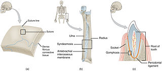
In anatomy, fibrous joints are joints connected by fibrous tissue, consisting mainly of collagen. These are fixed joints where bones are united by a layer of white fibrous tissue of varying thickness. In the skull the joints between the bones are called sutures. Such immovable joints are also referred to as synarthroses.
The gingival fibers are the connective tissue fibers that inhabit the gingival tissue adjacent to teeth and help hold the tissue firmly against the teeth. They are primarily composed of type I collagen, although type III fibers are also involved.
In dentistry, enamel matrix derivative (EMD) is an extract of porcine fetal tooth material used to biomimetically stimulate the soft and hard tissues surrounding teeth to regrow following tissue destruction.
Clinical attachment loss (CAL) is the predominant clinical manifestation and determinant of periodontal disease.
A cementicle is a small, spherical or ovoid calcified mass embedded within or attached to the cementum layer on the root surface of a tooth, or lying free within the periodontal ligament. They tend to occur in elderly individuals.
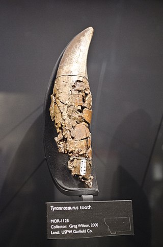
Dinosaur teeth have been studied since 1822 when Mary Ann Mantell (1795-1869) and her husband Dr Gideon Algernon Mantell (1790-1852) discovered an Iguanodon tooth in Sussex in England. Unlike mammal teeth, individual dinosaur teeth are generally not considered by paleontologists to be diagnostic to the genus or species level for unknown taxa, due morphological convergence and variability between teeth. and many historically named tooth taxa like Paronychodon and Richardoestesia are today considered nomina dubia, and are used as form taxa to refer to isolated teeth from other localities displaced considerably in time and space from the type specimens. However, it is possible to refer isolated teeth to known taxa provided that the tooth morphology is known and the teeth originate from a similar time and place.