Transdifferentiation

Last updated

Transdifferentiation, also known as lineage reprogramming, [1] is the process in which one mature somatic cell is transformed into another mature somatic cell without undergoing an intermediate pluripotent state or progenitor cell type. [2] It is a type of metaplasia, which includes all cell fate switches, including the interconversion of stem cells. Current uses of transdifferentiation include disease modeling and drug discovery and in the future may include gene therapy and regenerative medicine. [3] The term 'transdifferentiation' was originally coined by Selman and Kafatos [4] in 1974 to describe a change in cell properties as cuticle producing cells became salt-secreting cells in silk moths undergoing metamorphosis. [5]

Contents

Discovery

Davis et al. 1987 reported the first instance (sight) of transdifferentiation where a cell changed from one adult cell type to another. Forcing mouse embryonic fibroblasts to express MyoD was found to be sufficient to turn those cells into myoblasts. [6]

Natural examples

The only[ citation needed ] known instances where adult cells change directly from one lineage to another occurs in the species Turritopsis dohrnii (also known as the immortal jellyfish) and Turritopsis nutricula .

In newts, when the eye lens is removed, pigmented epithelial cells de-differentiate and then redifferentiate into the lens cells. [7] Vincenzo Colucci described this phenomenon in 1891 and Gustav Wolff described the same thing in 1894; the priority issue is examined in Holland (2021). [8]

In humans and mice, it has been demonstrated that alpha cells in the pancreas can spontaneously switch fate and transdifferentiate into beta cells. This has been demonstrated for both healthy and diabetic human and mouse pancreatic islets. [9] While it was previously believed that oesophageal cells were developed from the transdifferentiation of smooth muscle cells, that has been shown to be false. [10]

Induced and therapeutic examples

The first example of functional transdifferentiation has been provided by Ferber et al. [11] by inducing a shift in the developmental fate of cells in the liver and converting them into 'pancreatic beta-cell-like' cells. The cells induced a wide, functional and long-lasting transdifferentiation process that reduced the effects of hyperglycemia in diabetic mice. [12] Moreover, the trans-differentiated beta-like cells were found to be resistant to the autoimmune attack that characterizes type 1 diabetes. [13]

The second step was to undergo transdifferentiation in human specimens. By transducing liver cells with a single gene, Sapir et al. were able to induce human liver cells to transdifferentiate into human beta cells. [14]

This approach has been demonstrated in mice, rat, xenopus and human tissues. [15]

Schematic model of the hepatocyte-to-beta cell transdifferentiation process. Hepatocytes are obtained by liver biopsy from diabetic patient, cultured and expanded ex vivo, transduced with a PDX1 virus, transdifferentiated into functional insulin-producing beta cells, and transplanted back into the patient. [14]

Granulosa and theca cells in the ovaries of adult female mice can transdifferentiate to Sertoli and Leydig cells via induced knockout of the FOXL2 gene. [16] Similarly, Sertoli cells in the testes of adult male mice can transdifferentiate to granulosa cells via induced knockout of the DMRT1 gene. [17]

Methods

Lineage-instructive approach

In this approach, transcription factors from progenitor cells of the target cell type are transfected into a somatic cell to induce transdifferentiation. [2] There exists two different means of determining which transcription factors to use: by starting with a large pool and narrowing down factors one by one [18] or by starting with one or two and adding more. [19] One theory to explain the exact specifics is that ectopic Transcriptional factors direct the cell to an earlier progenitor state and then redirects it towards a new cell type. Rearrangement of the chromatin structure via DNA methylation or histone modification may play a role as well. [20] Here is a list of in vitro examples and in vivo examples. In vivo methods of transfecting specific mouse cells utilize the same kinds of vectors as in vitro experiments, except that the vector is injected into a specific organ. Zhou et al. (2008) injected Ngn3, Pdx1 and Mafa into the dorsal splenic lobe (pancreas) of mice to reprogram pancreatic exocrine cells into β-cells in order to ameliorate hyperglycaemia. [21]

Initial epigenetic activation phase approach

Somatic cells are first transfected with pluripotent reprogramming factors temporarily (Oct4, Sox2, Nanog, etc.) before being transfected with the desired inhibitory or activating factors. [22] Here is a list of examples in vitro.

Pharmacological agents

The DNA methylation inhibitor, 5-azacytidine is also known to promote phenotypic transdifferentiation of cardiac cells to skeletal myoblasts. [23]

In prostate cancer, treatment with androgen receptor targeted therapies induces neuroendocrine transdifferentiation in a subset of patients. [24] [25] No standard of care exists for these patients, and those diagnosed with treatment induced neuroendocrine carcinoma are typically treated palliatively. [26]

Mechanism of action

The transcription factors serve as a short term trigger to an irreversible process. The transdifferentiation liver cells observed 8 months after one single injection of pdx1. [12]

The ectopic transcription factors turn off the host repertoire of gene expression in each of the cells. However, the alternate desired repertoire is being turned on only in a subpopulation of predisposed cells. [27] Despite the massive dedifferentiation – lineage tracing approach indeed demonstrates that transdifferentiation originates in adult cells. [28]

Mogrify algorithm

Determining the unique set of cellular factors that is needed to be manipulated for each cell conversion is a long and costly process that involved much trial and error. As a result, this first step of identifying the key set of cellular factors for cell conversion is the major obstacle researchers face in the field of cell reprogramming. An international team of researchers have developed an algorithm, called Mogrify(1), that can predict the optimal set of cellular factors required to convert one human cell type to another. When tested, Mogrify was able to accurately predict the set of cellular factors required for previously published cell conversions correctly. To further validate Mogrify's predictive ability, the team conducted two novel cell conversions in the laboratory using human cells, and these were successful in both attempts solely using the predictions of Mogrify. [29] [30] [31] Mogrify has been made available online for other researchers and scientists.

Issues

Evaluation

When examining transdifferentiated cells, it is important to look for markers of the target cell type and the absence of donor cell markers which can be accomplished using green fluorescent protein or immunodetection. It is also important to examine the cell function, epigenome, transcriptome, and proteome profiles. Cells can also be evaluated based upon their ability to integrate into the corresponding tissue in vivo [18] and functionally replace its natural counterpart. In one study, transdifferentiating tail-tip fibroblasts into hepatocyte-like cells using transcription factors Gata4, Hnf1α and Foxa3, and inactivation of p19(Arf) restored hepatocyte-like liver functions in only half of the mice using survival as a means of evaluation. [32]

Transition from mouse to human cells

Generally transdifferentiation that occurs in mouse cells does not translate in effectiveness or speediness in human cells. Pang et al. found that while transcription factors Ascl1, Brn2 and Myt1l turned mouse cells into mature neurons, the same set of factors only turned human cells into immature neurons. However, the addition of NeuroD1 was able to increase efficiency and help cells reach maturity. [33]

Order of transcription factor expression

The order of expression of transcription factors can direct the fate of the cell. Iwasaki et al. (2006) showed that in hematopoietic lineages, the expression timing of Gata-2 and (C/EBPalpha) can change whether or not a lymphoid-committed progenitors can differentiate into granulocyte/monocyte progenitor, eosinophil, basophil or bipotent basophil/mast cell progenitor lineages. [34]

Immunogenicity

It has been found for induced pluripotent stem cells that when injected into mice, the immune system of the synergeic mouse rejected the teratomas forming. Part of this may be because the immune system recognized epigenetic markers of specific sequences of the injected cells. However, when embryonic stem cells were injected, the immune response was much lower. Whether or not this will occur within transdifferentiated cells remains to be researched. [3]

Method of transfection

In order to accomplish transfection, one may use integrating viral vectors such as lentiviruses or retroviruses, non-integrating vectors such as Sendai viruses or adenoviruses, microRNAs and a variety of other methods including using proteins and plasmids; [35] one example is the non-viral delivery of transcription factor-encoding plasmids with a polymeric carrier to elicit neuronal transdifferentiation of fibroblasts. [36] When foreign molecules enter cells, one must take into account the possible drawbacks and potential to cause tumorous growth. Integrating viral vectors have the chance to cause mutations when inserted into the genome. One method of going around this is to excise the viral vector once reprogramming has occurred, an example being Cre-Lox recombination [37] Non-integrating vectors have other issues concerning efficiency of reprogramming and also the removal of the vector. [38] Other methods are relatively new fields and much remains to be discovered.

Pluripotent reprogramming

See also

Related Research Articles

<span class="mw-page-title-main">Stem cell</span> Undifferentiated biological cells that can differentiate into specialized cells

In multicellular organisms, stem cells are undifferentiated or partially differentiated cells that can differentiate into various types of cells and proliferate indefinitely to produce more of the same stem cell. They are the earliest type of cell in a cell lineage. They are found in both embryonic and adult organisms, but they have slightly different properties in each. They are usually distinguished from progenitor cells, which cannot divide indefinitely, and precursor or blast cells, which are usually committed to differentiating into one cell type.

<span class="mw-page-title-main">Cellular differentiation</span> Developmental biology

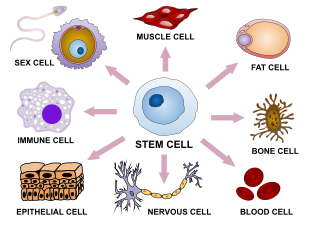
Cellular differentiation is the process in which a stem cell changes from one type to a differentiated one. Usually, the cell changes to a more specialized type. Differentiation happens multiple times during the development of a multicellular organism as it changes from a simple zygote to a complex system of tissues and cell types. Differentiation continues in adulthood as adult stem cells divide and create fully differentiated daughter cells during tissue repair and during normal cell turnover. Some differentiation occurs in response to antigen exposure. Differentiation dramatically changes a cell's size, shape, membrane potential, metabolic activity, and responsiveness to signals. These changes are largely due to highly controlled modifications in gene expression and are the study of epigenetics. With a few exceptions, cellular differentiation almost never involves a change in the DNA sequence itself. However, metabolic composition does get altered quite dramatically where stem cells are characterized by abundant metabolites with highly unsaturated structures whose levels decrease upon differentiation. Thus, different cells can have very different physical characteristics despite having the same genome.

<span class="mw-page-title-main">Embryonic stem cell</span> Pluripotent stem cell of the inner cell mass of the blastocyst

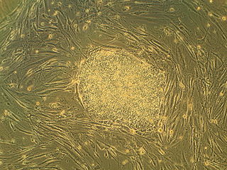
Embryonic stem cells (ESCs) are pluripotent stem cells derived from the inner cell mass of a blastocyst, an early-stage pre-implantation embryo. Human embryos reach the blastocyst stage 4–5 days post fertilization, at which time they consist of 50–150 cells. Isolating the inner cell mass (embryoblast) using immunosurgery results in destruction of the blastocyst, a process which raises ethical issues, including whether or not embryos at the pre-implantation stage have the same moral considerations as embryos in the post-implantation stage of development.

<span class="mw-page-title-main">Oct-4</span> Mammalian protein found in Homo sapiens

Oct-4, also known as POU5F1, is a protein that in humans is encoded by the POU5F1 gene. Oct-4 is a homeodomain transcription factor of the POU family. It is critically involved in the self-renewal of undifferentiated embryonic stem cells. As such, it is frequently used as a marker for undifferentiated cells. Oct-4 expression must be closely regulated; too much or too little will cause differentiation of the cells.

In biology, reprogramming refers to erasure and remodeling of epigenetic marks, such as DNA methylation, during mammalian development or in cell culture. Such control is also often associated with alternative covalent modifications of histones.

<span class="mw-page-title-main">Jun dimerization protein</span> Protein-coding gene in the species Homo sapiens

Jun dimerization protein 2 (JUNDM2) is a protein that in humans is encoded by the JDP2 gene. The Jun dimerization protein is a member of the AP-1 family of transcription factors.

<span class="mw-page-title-main">Induced pluripotent stem cell</span> Pluripotent stem cell generated directly from a somatic cell

Induced pluripotent stem cells are a type of pluripotent stem cell that can be generated directly from a somatic cell. The iPSC technology was pioneered by Shinya Yamanaka and Kazutoshi Takahashi in Kyoto, Japan, who together showed in 2006 that the introduction of four specific genes, collectively known as Yamanaka factors, encoding transcription factors could convert somatic cells into pluripotent stem cells. Shinya Yamanaka was awarded the 2012 Nobel Prize along with Sir John Gurdon "for the discovery that mature cells can be reprogrammed to become pluripotent."

<span class="mw-page-title-main">KLF4</span> Protein-coding gene in the species Homo sapiens

Kruppel-like factor 4 is a member of the KLF family of zinc finger transcription factors, which belongs to the relatively large family of SP1-like transcription factors. KLF4 is involved in the regulation of proliferation, differentiation, apoptosis and somatic cell reprogramming. Evidence also suggests that KLF4 is a tumor suppressor in certain cancers, including colorectal cancer. It has three C2H2-zinc fingers at its carboxyl terminus that are closely related to another KLF, KLF2. It has two nuclear localization sequences that signals it to localize to the nucleus. In embryonic stem cells (ESCs), KLF4 has been demonstrated to be a good indicator of stem-like capacity. It is suggested that the same is true in mesenchymal stem cells (MSCs).

<span class="mw-page-title-main">Shinya Yamanaka</span> Japanese stem cell researcher

Shinya Yamanaka is a Japanese stem cell researcher and a Nobel Prize laureate. He is the former director of Center for iPS Cell Research and Application and a professor at the Institute for Frontier Medical Sciences at Kyoto University; as a senior investigator at the UCSF-affiliated Gladstone Institutes in San Francisco, California; and as a professor of anatomy at University of California, San Francisco (UCSF). Yamanaka is also a past president of the International Society for Stem Cell Research (ISSCR).

A mesenchymal–epithelial transition (MET) is a reversible biological process that involves the transition from motile, multipolar or spindle-shaped mesenchymal cells to planar arrays of polarized cells called epithelia. MET is the reverse process of epithelial–mesenchymal transition (EMT) and it has been shown to occur in normal development, induced pluripotent stem cell reprogramming, cancer metastasis and wound healing.

<span class="mw-page-title-main">Cell potency</span> Ability of a cell to differentiate into other cell types

Cell potency is a cell's ability to differentiate into other cell types. The more cell types a cell can differentiate into, the greater its potency. Potency is also described as the gene activation potential within a cell, which like a continuum, begins with totipotency to designate a cell with the most differentiation potential, pluripotency, multipotency, oligopotency, and finally unipotency.

<span class="mw-page-title-main">FOXA2</span> Mammalian protein found in Homo sapiens

Forkhead box protein A2 (FOXA2), also known as hepatocyte nuclear factor 3-beta (HNF-3B), is a transcription factor that plays an important role during development, in mature tissues and, when dysregulated or mutated, also in cancer.

A list of examples of transdifferentiation:

A list of examples of in vivo transdifferentiation through transfection:

Induced stem cells (iSC) are stem cells derived from somatic, reproductive, pluripotent or other cell types by deliberate epigenetic reprogramming. They are classified as either totipotent (iTC), pluripotent (iPSC) or progenitor or unipotent – (iUSC) according to their developmental potential and degree of dedifferentiation. Progenitors are obtained by so-called direct reprogramming or directed differentiation and are also called induced somatic stem cells.

<span class="mw-page-title-main">GLIS1</span> Protein-coding gene

Glis1 is gene encoding a Krüppel-like protein of the same name whose locus is found on Chromosome 1p32.3. The gene is enriched in unfertilised eggs and embryos at the one cell stage and it can be used to promote direct reprogramming of somatic cells to induced pluripotent stem cells, also known as iPS cells. Glis1 is a highly promiscuous transcription factor, regulating the expression of numerous genes, either positively or negatively. In organisms, Glis1 does not appear to have any directly important functions. Mice whose Glis1 gene has been removed have no noticeable change to their phenotype.

Directed differentiation is a bioengineering methodology at the interface of stem cell biology, developmental biology and tissue engineering. It is essentially harnessing the potential of stem cells by constraining their differentiation in vitro toward a specific cell type or tissue of interest. Stem cells are by definition pluripotent, able to differentiate into several cell types such as neurons, cardiomyocytes, hepatocytes, etc. Efficient directed differentiation requires a detailed understanding of the lineage and cell fate decision, often provided by developmental biology.

<span class="mw-page-title-main">Thomas Graf (biologist)</span>

Thomas Graf is a biologist at the Centre for Genomic Regulation (CRG) in Barcelona, Spain. He is a pioneer in cell reprogramming, showing that blood cells can be transdifferentiated by transcription factors. He is also known for his early work on oncogenes carried by retroviruses and oncogene cooperation in leukemia formation.

Transflammation describes the process by which innate immune response mechanisms affect the epigenetic plasticity of a cell during nuclear reprogramming. This phenomenon is essential in dedifferentiating a somatic cell to a pluripotent cell and also in transdifferentiating a terminally differentiated cell to another terminally differentiated cell.

Inner ear regeneration is the biological process by which the hair cells and supporting cells of the ear proliferate and regrow after hair cell injury. This process depends on communication between supporting cells and the brain. Because of the volatility of the inner ear's hair cells, regeneration is crucial to the functioning of the inner ear. It is also a limited process, which contributes to the irreversibility of hearing loss in humans and other mammals.

References

  1. Orkin, S. H.; Zon, L. I. (2008). "Hematopoiesis: An Evolving Paradigm for Stem Cell Biology". Cell. 132 (4): 631–644. doi:10.1016/j.cell.2008.01.025. PMC   2628169 . PMID   18295580.
  2. 1 2 Graf, T.; Enver, T. (2009). "Forcing cells to change lineages". Nature. 462 (7273): 587–594. Bibcode:2009Natur.462..587G. doi:10.1038/nature08533. PMID   19956253. S2CID   4417323.
  3. 1 2 Pournasr, B.; Khaloughi, K.; Salekdeh, G. H.; Totonchi, M.; Shahbazi, E.; Baharvand, H. (2011). "Concise Review: Alchemy of Biology: Generating Desired Cell Types from Abundant and Accessible Cells". Stem Cells. 29 (12): 1933–1941. doi: 10.1002/stem.760 . PMID   21997905.
  4. Selman, Kelly; Kafatos, Fotis C. (1974-07-01). "Transdifferentiation in the labial gland of silk moths: is DNA required for cellular metamorphosis?". Cell Differentiation. 3 (2): 81–94. doi:10.1016/0045-6039(74)90030-X. PMID   4277742.
  5. Selman, K.; Kafatos, F. C. (2013). "Transdifferentiation in the labial gland of silk moths: Is DNA required for cellular metamorphosis?". Cell Differentiation. 3 (2): 81–94. doi:10.1016/0045-6039(74)90030-x. PMID   4277742.
  6. Davis, R. L.; Weintraub, H.; Lassar, A. B. (1987). "Expression of a single transfected cDNA converts fibroblasts to myoblasts". Cell. 51 (6): 987–1000. doi:10.1016/0092-8674(87)90585-x. PMID   3690668. S2CID   37741454.
  7. Jopling, C.; Boue, S.; Belmonte, J. C. I. (2011). "Dedifferentiation, transdifferentiation and reprogramming: Three routes to regeneration". Nature Reviews Molecular Cell Biology. 12 (2): 79–89. doi:10.1038/nrm3043. PMID   21252997. S2CID   205494805.
  8. Holland, Nicholas (2021), "Vicenzo Colucci's 1886 memoir, Intorno alla rigenerazione degli arti e della coda nei tritoni, annotated and translated into English as: Concerning regeneration of the limbs and tail in salamanders", The European Zoological Journal, 88: 837–890, doi: 10.1080/24750263.2021.1943549
  9. van der Meulen, T.; Mawla, A.M.; DiGruccio, M.R.; Adams, M.W.; Nies, V.; Dolleman, S.; Liu, S.; Ackermann, A.M.; Caceres, E.; Hunter, A.E.; Kaestner, K.H.; Donaldson, C.J.; Huising, M.O. (2017). "Virgin Beta Cells Persist throughout Life at a Neogenic Niche within Pancreatic Islets" (PDF). Cell Metabolism. 25 (4): 911–926. doi: 10.1016/j.cmet.2017.03.017 . PMC   8586897 . PMID   28380380.
  10. Rishniw, M.; Xin, H. B.; Deng, K. Y.; Kotlikoff, M. I. (2003). "Skeletal myogenesis in the mouse esophagus does not occur through transdifferentiation". Genesis. 36 (2): 81–82. doi:10.1002/gene.10198. PMID   12820168. S2CID   20010447.
  11. Ferber S, Halkin A, Cohen H, Ber I, Einav Y, Goldberg I, Barshack I, Seijffers R, Kopolovic J, Kaiser N, Karasik A (2000) Pancreatic and duodenal homeobox gene 1 induces expression of insulin genes in liver and ameliorates streptozotocin-induced hyperglycemia. http://www.nature.com/nm/journal/v6/n5/full/nm0500_568.html
  12. 1 2 Sarah Ferber, Amir Halkin, Hofit Cohen, Idit Ber, Yulia Einav, Iris Goldberg, Iris Barshack, Rhona Seijffers, Juri Kopolovic, Nurit Kaiser & Avraham Karasik (2000) - "Pancreatic and duodenal homeobox gene 1 induces expression of insulin genes in liver and ameliorates streptozotocin-induced hyperglycemia"
  13. Shternhall-Ron K et al., Ectopic PDX-1 expression in liver ameliorates type 1 diabetes, Journal of Autoimmunity (2007), doi:10.1016/j.jaut.2007.02.010. http://www.orgenesis.com/uploads/default/files/shternhall-jai-2007.pdf
  14. 1 2 Tamar Sapir, Keren Shternhall, Irit Meivar-Levy, Tamar Blumenfeld, Hamutal Cohen, Ehud Skutelsky, Smadar Eventov-Friedman, Iris Barshack, Iris Goldberg, Sarah Pri-Chen, Lya Ben-Dor, Sylvie Polak-Charcon, Avraham Karasik, Ilan Shimon, Eytan Mor, and Sarah Ferber (2005) Cell-replacement therapy for diabetes: Generating functional insulin-producing tissue from adult human liver cells
  15. Al-Hasani, K; Pfeifer, A; Courtney, M; Ben-Othman, N; Gjernes, E; Vieira, A; Druelle, N; Avolio, F; Ravassard, P; Leuckx, G; Lacas-Gervais, S; Ambrosetti, D; Benizri, E; Hecksher-Sorensen, J; Gounon, P; Ferrer, J; Gradwohl, G; Heimberg, H; Mansouri, A; Collombat, P (2013). "Adult Duct-Lining Cells Can Reprogram into β-like Cells Able to Counter Repeated Cycles of Toxin-Induced Diabetes". Dev. Cell. 26 (1): 86–100. doi: 10.1016/j.devcel.2013.05.018 . hdl: 11858/00-001M-0000-0014-3C2F-6 . PMID   23810513.
  16. Uhlenhaut NH, Jakob S, Anlag K, Eisenberger T, Sekido R, Kress J, et al. (December 2009). "Somatic sex reprogramming of adult ovaries to testes by FOXL2 ablation". Cell. 139 (6): 1130–42. doi: 10.1016/j.cell.2009.11.021 . PMID   20005806.
  17. Matson CK, Murphy MW, Sarver AL, Griswold MD, Bardwell VJ, Zarkower D (July 2011). "DMRT1 prevents female reprogramming in the postnatal mammalian testis". Nature. 476 (7358): 101–4. doi:10.1038/nature10239. PMC   3150961 . PMID   21775990.
  18. 1 2 Ieda, M.; Fu, J. D.; Delgado-Olguin, P.; Vedantham, V.; Hayashi, Y.; Bruneau, B. G.; Srivastava, D. (2010). "Direct Reprogramming of Fibroblasts into Functional Cardiomyocytes by Defined Factors". Cell. 142 (3): 375–386. doi:10.1016/j.cell.2010.07.002. PMC   2919844 . PMID   20691899.
  19. Vierbuchen, T.; Ostermeier, A.; Pang, Z. P.; Kokubu, Y.; Südhof, T. C.; Wernig, M. (2010). "Direct conversion of fibroblasts to functional neurons by defined factors". Nature. 463 (7284): 1035–1041. Bibcode:2010Natur.463.1035V. doi:10.1038/nature08797. PMC   2829121 . PMID   20107439.
  20. Ang, Y. S.; Gaspar-Maia, A.; Lemischka, I. R.; Bernstein, E. (2011). "Stem cells and reprogramming: Breaking the epigenetic barrier?". Trends in Pharmacological Sciences. 32 (7): 394–401. doi:10.1016/j.tips.2011.03.002. PMC   3128683 . PMID   21621281.
  21. Zhou, Q.; Brown, J.; Kanarek, A.; Rajagopal, J.; Melton, D. A. (2008). "In vivo reprogramming of adult pancreatic exocrine cells to β-cells". Nature. 455 (7213): 627–632. Bibcode:2008Natur.455..627Z. doi:10.1038/nature07314. PMC   9011918 . PMID   18754011. S2CID   205214877.
  22. Efe, J. A.; Hilcove, S.; Kim, J.; Zhou, H.; Ouyang, K.; Wang, G.; Chen, J.; Ding, S. (2011). "Conversion of mouse fibroblasts into cardiomyocytes using a direct reprogramming strategy". Nature Cell Biology. 13 (3): 215–222. doi:10.1038/ncb2164. PMID   21278734. S2CID   5310172.
  23. kaur, keerat; yang, jinpu; eisenberg, carol; eisenberg, leonard (2014). "5-azacytidine promotes the transdifferentiation of cardiac cells to skeletal myocytes". Cellular Reprogramming. 16 (5): 324–330. doi:10.1089/cell.2014.0021. PMID   25090621.
  24. Usmani, S; Orevi, M; Stefanelli, A; Zaniboni, A; Gofrit, ON; Bnà, C; Illuminati, S; Lojacono, G; Noventa, S; Savelli, G (June 2019). "Neuroendocrine differentiation in castration resistant prostate cancer. Nuclear medicine radiopharmaceuticals and imaging techniques: A narrative review". Critical Reviews in Oncology/Hematology. 138: 29–37. doi:10.1016/j.critrevonc.2019.03.005. PMID   31092382. S2CID   131934021.
  25. Davies, AH; Beltran, H; Amina Zoubeidi (May 2018). "Cellular plasticity and the neuroendocrine phenotype in prostate cancer". Nature Reviews. Urology. 15 (5): 271–286. doi:10.1038/nrurol.2018.22. PMID   29460922. S2CID   4732323.
  26. Aggarwal, R; Zhang, T; Small, EJ; Armstrong, AJ (May 2014). "Neuroendocrine prostate cancer: subtypes, biology, and clinical outcomes". Journal of the National Comprehensive Cancer Network. 12 (5): 719–26. doi:10.6004/jnccn.2014.0073. PMID   24812138.
  27. Meivar-Levy, I.; Sapir, T.; Gefen-Halevi, S.; Aviv, V.; Barshack, I.; Onaca, N.; Mor, E.; Ferber, S. (2007). "Pancreatic and duodenal homeobox gene 1 induces hepatic dedifferentiation by suppressing the expression of CCAAT/enhancer-binding protein β". Hepatology. 46 (3): 898–905. doi: 10.1002/hep.21766 . PMID   17705277.
  28. Mauda-Havakuk, M.; Litichever, N.; Chernichovski, E.; Nakar, O.; Winkler, E.; Mazkereth, R.; Orenstein, A.; Bar-Meir, E.; Ravassard, P.; Meivar-Levy, I.; Ferber, S. (2011). Linden, Rafael (ed.). "Ectopic PDX-1 Expression Directly Reprograms Human Keratinocytes along Pancreatic Insulin-Producing Cells Fate". PLOS ONE. 6 (10): e26298. Bibcode:2011PLoSO...626298M. doi: 10.1371/journal.pone.0026298 . PMC   3196540 . PMID   22028850.
  29. Mapping out cell conversion
  30. Owen, Rackham; Gough, Julian (2016). "A predictive computational framework for direct reprogramming between human cell types". Nature Genetics. 48 (3): 331–335. doi:10.1038/ng.3487. hdl: 1983/e6490a78-f3e8-4253-acc4-7ee181c79168 . PMID   26780608. S2CID   217524918.
  31. Jane Byrne (Jul 2021). Mogrify looks to transform cell therapy development. BIOPHARMA-REPORTER.COM
  32. Huang, P.; He, Z.; Ji, S.; Sun, H.; Xiang, D.; Liu, C.; Hu, Y.; Wang, X.; Hui, L. (2011). "Induction of functional hepatocyte-like cells from mouse fibroblasts by defined factors". Nature. 475 (7356): 386–389. doi:10.1038/nature10116. PMID   21562492. S2CID   1115749.
  33. Pang, Z. P.; Yang, N.; Vierbuchen, T.; Ostermeier, A.; Fuentes, D. R.; Yang, T. Q.; Citri, A.; Sebastiano, V.; Marro, S.; Südhof, T. C.; Wernig, M. (2011). "Induction of human neuronal cells by defined transcription factors". Nature. 476 (7359): 220–223. Bibcode:2011Natur.476..220P. doi:10.1038/nature10202. PMC   3159048 . PMID   21617644.
  34. Iwasaki, H.; Mizuno, S. -I.; Arinobu, Y.; Ozawa, H.; Mori, Y.; Shigematsu, H.; Takatsu, K.; Tenen, D. G.; Akashi, K. (2006). "The order of expression of transcription factors directs hierarchical specification of hematopoietic lineages". Genes & Development. 20 (21): 3010–3021. doi:10.1101/gad.1493506. PMC   1620021 . PMID   17079688.
  35. Patel, M.; Yang, S. (2010). "Advances in Reprogramming Somatic Cells to Induced Pluripotent Stem Cells". Stem Cell Reviews and Reports. 6 (3): 367–380. doi:10.1007/s12015-010-9123-8. PMC   2924949 . PMID   20336395.
  36. Adler, A. F.; Grigsby, C. L.; Kulangara, K.; Wang, H.; Yasuda, R.; Leong, K. W. (2012). "Nonviral Direct Conversion of Primary Mouse Embryonic Fibroblasts to Neuronal Cells". Molecular Therapy: Nucleic Acids. 1 (7): e32–. doi:10.1038/mtna.2012.25. PMC   3411320 . PMID   23344148.
  37. Sommer, C. A.; Sommer, A.; Longmire, T. A.; Christodoulou, C.; Thomas, D. D.; Gostissa, M.; Alt, F. W.; Murphy, G. J.; Kotton, D. N.; Mostoslavsky, G. (2009). "Excision of Reprogramming Transgenes Improves the Differentiation Potential of iPS Cells Generated with a Single Excisable Vector". Stem Cells. 28 (1): 64–74. doi:10.1002/stem.255. PMC   4848036 . PMID   19904830.
  38. Zhou, W.; Freed, C. R. (2009). "Adenoviral Gene Delivery Can Reprogram Human Fibroblasts to Induced Pluripotent Stem Cells". Stem Cells. 27 (11): 2667–2674. doi: 10.1002/stem.201 . PMID   19697349.
  39. 1 2 3 4 5 6 Zhou, Q.; Melton, D. A. (2008). "Extreme Makeover: Converting One Cell into Another". Cell Stem Cell. 3 (4): 382–388. doi: 10.1016/j.stem.2008.09.015 . PMID   18940730.
  40. Passier, R.; Mummery, C. (2010). "Getting to the Heart of the Matter: Direct Reprogramming to Cardiomyocytes". Cell Stem Cell. 7 (2): 139–141. doi: 10.1016/j.stem.2010.07.004 . PMID   20682439.