The peritoneum is the serous membrane forming the lining of the abdominal cavity or coelom in amniotes and some invertebrates, such as annelids. It covers most of the intra-abdominal organs, and is composed of a layer of mesothelium supported by a thin layer of connective tissue. This peritoneal lining of the cavity supports many of the abdominal organs and serves as a conduit for their blood vessels, lymphatic vessels, and nerves.

The abdominal cavity is a large body cavity in humans and many other animals that contains many organs. It is a part of the abdominopelvic cavity. It is located below the thoracic cavity, and above the pelvic cavity. Its dome-shaped roof is the thoracic diaphragm, a thin sheet of muscle under the lungs, and its floor is the pelvic inlet, opening into the pelvis.

Peritonitis is inflammation of the peritoneum, the lining of the inner wall of the abdomen and cover of the abdominal organs. Symptoms may include severe pain, swelling of the abdomen, fever, or weight loss. One part or the entire abdomen may be tender. Complications may include shock and acute respiratory distress syndrome.

The mesothelium is a membrane composed of simple squamous epithelium that forms the lining of several body cavities: the pleura, peritoneum, mediastinum and pericardium. Mesothelial tissue also surrounds the male internal reproductive organs and covers the internal reproductive organs of women. Mesothelium that covers the internal organs is called visceral mesothelium, while the layer that covers the body walls is called the parietal mesothelium. Mesothelium is the epithelial component of serosa.

The retroperitoneal space (retroperitoneum) is the anatomical space in the abdominal cavity behind (retro) the peritoneum. It has no specific delineating anatomical structures. Organs are retroperitoneal if they have peritoneum on their anterior side only. Structures that are not suspended by mesentery in the abdominal cavity and that lie between the parietal peritoneum and abdominal wall are classified as retroperitoneal.
The peritoneal cavity is a potential space between the parietal peritoneum and visceral peritoneum. Both the parietal and visceral peritonea are not different but the same peritoneum given two names depending on their function/location. It is one of the spaces derived from the coelomic cavity of the embryo, the others being the pleural cavities around the lungs and the pericardial cavity around the heart.

The lesser omentum is the double layer of peritoneum that extends from the liver to the lesser curvature of the stomach and the first part of the duodenum.

The ascending colon is the part of the colon located between the cecum and the transverse colon.

In human anatomy, the greater sac, also known as the general cavity or peritoneum of the peritoneal cavity proper, is the cavity in the abdomen that is inside the peritoneum but outside the lesser sac.
In anatomy, the abdominal wall represents the boundaries of the abdominal cavity. The abdominal wall is split into the posterior (back), lateral (sides) and anterior (front) walls.
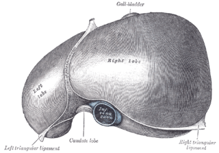
The falciform ligament is a ligament that attaches the liver to the anterior (ventral) body wall, and separates the liver into the left medial lobe and left lateral lobe. The falciform ligament, from Latin, meaning 'sickle-shaped', is a broad and thin fold of peritoneum, its base being directed downward and backward and its apex upward and backward. The falciform ligament droops down from the hilum of the liver.

The greater omentum is a large apron-like fold of visceral peritoneum that hangs down from the stomach. It extends from the greater curvature of the stomach, passing in front of the small intestines and doubles back to ascend to the transverse colon before reaching to the posterior abdominal wall. The greater omentum is larger than the lesser omentum, which hangs down from the liver to the lesser curvature. The common anatomical term "epiploic" derives from "epiploon", from the Greek epipleein, meaning to float or sail on, since the greater omentum appears to float on the surface of the intestines. It is the first structure observed when the abdominal cavity is opened anteriorly.

The broad ligament of the uterus is the wide fold of peritoneum that connects the sides of the uterus to the walls and floor of the pelvis.
The suspensory ligament of the ovary, also infundibulopelvic ligament, is a fold of peritoneum that extends out from the ovary to the wall of the pelvis.

The coronary ligament of the liver refers to parts of the peritoneal reflections that hold the liver to the inferior surface of the diaphragm.

The peritoneum of the anterior pelvic wall covers the superior surface of the bladder, and on either side of this viscus forms a depression, termed the paravesical fossa, which is limited laterally by the fold of peritoneum covering the ductus deferens.

The recto-vesical pouch is the pocket that lies between the rectum and the urinary bladder in human males and other male mammals. It is lined by peritoneum and at its base is the rectoprostatic fascia. When a man is upright or supine, the recto-vesical pouch is the lowest part of his peritoneal cavity. Because of this, peritoneal fluid and other fluids that enter the peritoneal cavity, including ascites, blood and pus, tend to collect in this pouch.
The extraperitoneal space is the portion of the abdomen and pelvis which does not lie within peritoneum.
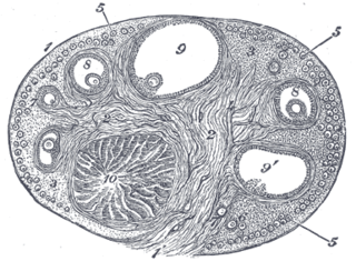
The ovarian surface epithelium, also called the germinal epithelium of Waldeyer, is a layer of simple squamous-to-cuboidal epithelial cells covering the ovary.

In human anatomy, the omental foramen, is the passage of communication, or foramen, between the greater sac, and the lesser sac.