
The endometrium is the inner epithelial layer, along with its mucous membrane, of the mammalian uterus. It has a basal layer and a functional layer: the basal layer contains stem cells which regenerate the functional layer. The functional layer thickens and then is shed during menstruation in humans and some other mammals, including apes, Old World monkeys, some species of bat, the elephant shrew and the Cairo spiny mouse. In most other mammals, the endometrium is reabsorbed in the estrous cycle. During pregnancy, the glands and blood vessels in the endometrium further increase in size and number. Vascular spaces fuse and become interconnected, forming the placenta, which supplies oxygen and nutrition to the embryo and fetus. The speculated presence of an endometrial microbiota has been argued against.

The uterus or womb is the organ in the reproductive system of most female mammals, including humans, that accommodates the embryonic and fetal development of one or more embryos until birth. The uterus is a hormone-responsive sex organ that contains glands in its lining that secrete uterine milk for embryonic nourishment.

The female reproductive system is made up of the internal and external sex organs that function in the reproduction of new offspring. In humans, the female reproductive system is immature at birth and develops to maturity at puberty to be able to produce gametes, and to carry a fetus to full term. The internal sex organs are the vagina, uterus, fallopian tubes, and ovaries. The female reproductive tract includes the vagina, uterus, and fallopian tubes and is prone to infections. The vagina allows for sexual intercourse and childbirth, and is connected to the uterus at the cervix. The uterus or womb accommodates the embryo which develops into the fetus. The uterus also produces secretions which help the transit of sperm to the fallopian tubes, where sperm fertilize ova produced by the ovaries. The external sex organs are also known as the genitals and these are the organs of the vulva including the labia, clitoris, and vaginal opening.
An abdominal pregnancy is a rare type of ectopic pregnancy where the embryo or fetus is growing and developing outside the uterus, in the abdomen, and not in a fallopian tube, an ovary, or the broad ligament.

The epoophoron or epoöphoron is a remnant of the mesonephric tubules that can be found next to the ovary and fallopian tube.
The parametrium is the fibrous and fatty connective tissue that surrounds the uterus. This tissue separates the supravaginal portion of the cervix from the bladder. The parametrium lies in front of the cervix and extends laterally between the layers of the broad ligaments. It connects the uterus to other tissues in the pelvis. It is different from the perimetrium, which is the outermost layer of the uterus.

The mesovarium is the portion of the broad ligament of the uterus that suspends the ovaries. The ovary is not covered by the mesovarium; rather, it is covered by germinal epithelium.

The uterine cavity is the inside of the uterus. It is triangular in shape, the base being formed by the internal surface of the body of the uterus between the openings of the fallopian tubes, the apex by the internal orifice of the uterus through which the cavity of the body communicates with the canal of the cervix. The uterine cavity where it enters the openings of the fallopian tubes is a mere slit, flattened antero-posteriorly.

The mesosalpinx is part of the lining of the abdominal cavity in higher vertebrates, specifically the portion of the broad ligament that stretches from the ovary to the level of the fallopian tube.
The perimetrium is the outer serosal layer of the uterus, derived from the peritoneum overlying the uterine fundus, and can be considered a visceral peritoneum. It consists of a superficial layer of mesothelium, and a thin layer of loose connective tissue beneath it.

In human female anatomy, the vesicouterine pouch, also uterovesicle pouch, is a fold of peritoneum over the uterus and the bladder. Like the rectouterine pouch, it is a female pelvic recess, but shallower and closer to the anterior fornix of the vagina.

The uterine plexuses lie along the sides and superior angles of the uterus between the two layers of the broad ligament, and communicate with the ovarian and vaginal plexuses.
Vesicular appendages of the epoöphoron are small pedunculated vesicles of the fimbriae of the uterine tube, or connected to the broad ligament. They were described by Giovanni Battista Morgagni and are remnants of the cranial part of the mesonephric duct. Typically they are asymptomatic.
The arcuate vessels of the uterus are a component of the blood supply of the uterus. They are arteries and veins that branch from the uterine arteries and veins, respectively, with additional anastomoses from the ovarian arteries and veins, and penetrate and assume a circumferential course in the myometrium.

The uterine horns(cornua of uterus) are the points in the upper uterus where the fallopian tubes or oviducts exit to meet the ovaries. They are one of the points of attachment for the round ligament of uterus (the other being the mons pubis). They also provide attachment to the ovarian ligament, which is located below the fallopian tube at the back, while the round ligament of uterus is located below the tube at the front.
Tubal reversal, also called tubal sterilization reversal, tubal ligation reversal, or microsurgical tubal reanastomosis, is a surgical procedure that can restore fertility to women after a tubal ligation. By rejoining the separated segments of the fallopian tube, tubal reversal can give women the chance to become pregnant again. In some cases, however, the separated segments cannot actually be reattached to each other. In some cases the remaining segment of tube needs to be re-implanted into the uterus. In other cases, when the end of the tube has been removed, a procedure called a neofimbrioplasty must be performed to recreate a functional end of the tube which can then act like the missing fimbria and retrieve the egg that has been released during ovulation.

Parametritis is an infection of the parametrium. It is considered a form of pelvic inflammatory disease.
The development of the reproductive system is the part of embryonic growth that results in the sex organs and contributes to sexual differentiation. Due to its large overlap with development of the urinary system, the two systems are typically described together as the urogenital or genitourinary system.
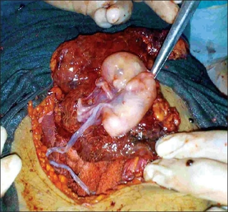
The fallopian tubes, also known as uterine tubes, oviducts or salpinges, are paired tubes in the human female body that stretch from the uterus to the ovaries. The fallopian tubes are part of the female reproductive system. In other mammals, they are only called oviducts.
An uterine orgasm, or womb orgasm, is an orgasm of the uterus, usually by stimulation of an area just outside the cervix, or the cervix itself, or the vaginal wall adjacent to the uterus. Uterine orgasms can also occur without stimulation. There is some anecdotal evidence to suggest that this is a type of sexual climax.
