Vaginal support structures

Last updated
1116 Muscle of the Female Perineum.png

The vaginal support structures are those muscles, bones, ligaments, tendons, membranes and fascia, of the pelvic floor that maintain the position of the vagina within the pelvic cavity and allow the normal functioning of the vagina and other reproductive structures in the female. Defects or injuries to these support structures in the pelvic floor leads to pelvic organ prolapse. Anatomical and congenital variations of vaginal support structures can predispose a woman to further dysfunction and prolapse later in life. [1] The urethra is part of the anterior wall of the vagina and damage to the support structures there can lead to incontinence and urinary retention. [2]

Contents

Pelvic bones

The support for the vagina is provided by muscles, membranes, tendons and ligaments. These structures are attached to the hip bones. These bones are the pubis, ilium and ischium. The interior surface of these pelvic bones and their projections and contours are used as attachment sites for the fascia, muscles, tendons and ligaments that support the vagina. These bones are then fuse and attach to the sacrum behind the vagina and anteriorly at the pubic symphysis. [3] Supporting ligaments include the sacrospinous and sacrotuberous ligaments. The sacrospinous ligament is unusual in that it is thin and triangular. [3] [4]

Pelvic diaphragm

The muscular pelvic diaphragm is composed of the bilateral levator ani and coccygeus muscles and these attach to the inner pelvic surface. The iliococcygeus and pubococcygeus make up the levator ani muscle. The muscles pass behind the rectum. The levator ani surrounds the opening which the urethra, rectum and vagina pass. The pubococcygeus muscle is subdivided into the pubourethralis, pubovaginal muscle and the puborectalis muscle. The names describe the attachments of the muscles to the urethra, vagina, anus, and rectum. The names are also called the pubourethralis, pubovaginalis, puboanalis, and puborectalis muscles and sometimes the pubovisceralis since it attaches to the viscera. [3]

Urogenital diaphragm (perineal membrane)

The urogenital diaphragm, or perineal membrane, is present over the anterior pelvic outlet below the pelvic diaphragm. [5] The exact structure description is controversial. Despite the controversy, MRI imaging studies support the existence of the structure. [3] [6]

Superficial and inferior muscles of the perineum (urogenital diaphragm):

The perineum attaches across the gap between the inferior pubic rami bilaterally and the perineal body. This grouping of muscles constricts to close the urogenital openings. The perineum supports and functions as a sphincter at the opening of the vagina. Other structures exist below the perineum that support the anus. [3] [6]

Perineal body

The bulbospongiosus muscle is highlighted in red. Bulbospongiosus-Female.png
The bulbospongiosus muscle is highlighted in red.

The perineal body is a pyramidal structure of muscle and connective tissue and part of it is located between the anus and vagina. It is a tendon that is formed at the point where the bulbospongiosus muscle, superficial transverse perineal muscle, [7] and external anal sphincter muscle converge to form this major supportive structure of the pelvis and vagina. [8] [9] [10] Below this, muscles and their fascia converge and become part of the perineal body. The lower vagina is attached to the perineal body by attachments from the pubococcygeus, perineal muscles, and the anal sphincter. The perineal body is made up of smooth muscle, elastic connective tissue fibers, and nerve endings. Above the perineal body are the vagina and the uterus. Damage and resulting weakness of the perineal body changes the length of the vagina and predisposes it to rectocele and enterocele. [3] [6]

Endopelvic fascia and connective tissue

The internal surface of the ilium showing where the muscles supporting the vagina insert and terminate Gray236.png
The internal surface of the ilium showing where the muscles supporting the vagina insert and terminate

The vagina is attached to the pelvic walls by endopelvic fascia. The peritoneum is the external layer of skin that covers the fascia. This tissue provides additional support to the pelvic floor. The endopelvic fascia is one continuous sheet of tissue and varies in thickness. It permits some shifting of the pelvic structures. The fascia contains elastic collagen fibers in a 'mesh-like' structure. The fascia also contains fibroblasts, smooth muscle, and vascular vessels. The cardinal ligament supports the apex of the vagina and derives some of its strength from vascular tissue. The endopelvic fascia attaches to the lateral pelvic wall via the arcus tendineus. [3]

Anterior vaginal support

Not all agree to the amount of supportive tissue or fascia exists in the anterior vaginal wall. The major point of contention is whether the vaginal fascial layer exists. Some texts do not describe a fascial layer. Other sources state that the fascia is present under the urethra which is embedded in the anterior vaginal wall. [3] Despite disagreement, the urethra is embedded in the anterior vaginal wall. [3]

Lateral and mid support structures

The midsection of the vagina is supported by its lateral attachments to the arcus tendineus. Some describe the pubocervical fascia as extending from the pubic symphysis to the anterior vaginal wall and cervix. Anatomists do not agree on its existence. [3] [11]


Complications

Vaginal support structures can be damaged or weakened during childbirth or pelvic surgery. Other conditions that repeatedly strain or increase pressure in the pelvic area can also compromise support. Examples are: [12]

See also

Related Research Articles

<span class="mw-page-title-main">Perineum</span> Region of the body between the genitals and anus

The perineum in mammals is the space between the anus and the genitals. The human perineum is between the anus and scrotum in the male or between the anus and vulva in the female. The perineum is the region of the body between the pubic symphysis and the coccyx, including the perineal body and surrounding structures. The perineal raphe is visible and pronounced to varying degrees. The perineum is an erogenous zone. This area is also known as the taint or gooch in American slang.

<span class="mw-page-title-main">Levator ani</span> Broad, thin muscle group, situated on either side of the pelvis

The levator ani is a broad, thin muscle group, situated on either side of the pelvis. It is formed from three muscle components: the pubococcygeus, the iliococcygeus, and the puborectalis.

<span class="mw-page-title-main">Bulbospongiosus muscle</span> Superficial muscle of the perineum

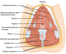
The bulbospongiosus muscles are one of the superficial muscles of the perineum. They have a slightly different origin, insertion and function in males and females. In males, these muscles cover the bulb of the penis, while in females, they cover the vestibular bulbs.

<span class="mw-page-title-main">Pelvic floor</span> Anatomical structure

The pelvic floor or pelvic diaphragm is an anatomical location in the human body, which has an important role in urinary and anal continence, sexual function and support of the pelvic organs. The pelvic floor includes muscles, both skeletal and smooth, ligaments and fascia. and separates between the pelvic cavity from above, and the perineum from below. It is formed by the levator ani muscle and coccygeus muscle, and associated connective tissue.

<span class="mw-page-title-main">Urogenital diaphragm</span> Layer of the pelvis

Older texts have asserted the existence of a urogenital diaphragm, also called the triangular ligament, which was described as a layer of the pelvis that separates the deep perineal sac from the upper pelvis, lying between the inferior fascia of the urogenital diaphragm and superior fascia of the urogenital diaphragm.

<span class="mw-page-title-main">Cystocele</span> Medical condition

The cystocele, also known as a prolapsed bladder, is a medical condition in which a woman's bladder bulges into her vagina. Some may have no symptoms. Others may have trouble starting urination, urinary incontinence, or frequent urination. Complications may include recurrent urinary tract infections and urinary retention. Cystocele and a prolapsed urethra often occur together and is called a cystourethrocele. Cystocele can negatively affect quality of life.

<span class="mw-page-title-main">External anal sphincter</span> Flat plane of skeletal muscle fibers

The external anal sphincter is an oval tube of skeletal muscle fibers. Distally, it is adherent to the skin surrounding the margin of the anus. It exhibits a resting state of tonical contraction.

<span class="mw-page-title-main">External sphincter muscle of male urethra</span>

The external sphincter muscle of male urethra, also sphincter urethrae membranaceae, sphincter urethrae externus, surrounds the whole length of the membranous urethra, and is enclosed in the fascia of the urogenital diaphragm.

<span class="mw-page-title-main">Ischium</span> Lower and back region of the hip bone

The ischium forms the lower and back region of the hip bone.

<span class="mw-page-title-main">Fascia of Colles</span>

The membranous layer of the superficial fascia of the perineum is the deeper layer of the superficial perineal fascia. It is thin, aponeurotic in structure, and of considerable strength, serving to bind down the muscles of the root of the penis. Colles' fascia emerges from the perineal membrane, which divides the base of the penis from the prostate. Colles' fascia emerges from the inferior side of the perineal membrane and continues along the ventral (inferior) penis without covering the scrotum. It separates the skin and subcutaneous fat from the superficial perineal pouch.

<span class="mw-page-title-main">Fascia of Scarpa</span> Deep membranous layer of the superficial fascia of the abdomen

The fascia of Scarpa is the deep membranous layer (stratum membranosum) of the superficial fascia of the abdomen. It is a layer of the anterior abdominal wall. It is found deep to the fascia of Camper and superficial to the external oblique muscle.

<span class="mw-page-title-main">Uterine prolapse</span> Medical condition

Uterine prolapse is a form of pelvic organ prolapse in which the uterus and a portion of the upper vagina protrude into the vaginal canal and, in severe cases, through the opening of the vagina. It is most often caused by injury or damage to structures that hold the uterus in place within the pelvic cavity. Symptoms may include vaginal fullness, pain with sexual intercourse, difficulty urinating, and urinary incontinence. Risk factors include older age, pregnancy, vaginal childbirth, obesity, chronic constipation, and chronic cough. Prevalence, based on physical exam alone, is estimated to be approximately 14%.

<span class="mw-page-title-main">Perineal membrane</span> Anatomical term

The perineal membrane is an anatomical term for a fibrous membrane in the perineum. The term "inferior fascia of urogenital diaphragm", used in older texts, is considered equivalent to the perineal membrane.

<span class="mw-page-title-main">Superficial perineal pouch</span>

The superficial perineal pouch is a compartment of the perineum.

<span class="mw-page-title-main">Deep perineal pouch</span> Anatomic space enclosed partly by the perineum

The deep perineal pouch is the anatomic space enclosed in part by the perineum and located superior to the perineal membrane.

<span class="mw-page-title-main">Urogenital triangle</span> Anterior part of the perineum

The urogenital triangle is the anterior part of the perineum. In female mammals, it contains the vagina and associated parts of the internal genitalia.

<span class="mw-page-title-main">Outline of human anatomy</span> Overview of and topical guide to human anatomy

The following outline is provided as an overview of and topical guide to human anatomy:

<span class="mw-page-title-main">Root of penis</span> Internal portion of the human penis

In human male anatomy, the radix or root of the penis is the internal and most proximal portion of the human penis that lies in the perineum. Unlike the pendulous body of the penis, which is suspended from the pubic symphysis, the root is attached to the pubic arch of the pelvis and is not visible externally. It is triradiate in form, consisting of three masses of erectile tissue; the two diverging crura, one on either side, and the median bulb of the penis or urethral bulb. Approximately one third to one half of the penis is embedded in the pelvis and can be felt through the scrotum and in the perineum.

<span class="mw-page-title-main">Pelvis</span> Lower torso of the human body

The pelvis is the lower part of the trunk, between the abdomen and the thighs, together with its embedded skeleton.

The urogenital hiatus is a gap in the anteromedial part of the pelvic floor, allowing passage of the urethra, the vagina, and deep dorsal vein of clitoris (females) or penis (males).

References

PD-icon.svgThis article incorporates text in the public domain from the 20th edition of Gray's Anatomy (1918)

  1. Craft, T. M.; Parr, M. J. A.; Nolan, Jerry P. (2004-11-10). Key Topics in Critical Care, Second Edition. CRC Press. p. 1068. ISBN   9781841843582.
  2. 1 2 "Cystocele (Prolapsed Bladder) | NIDDK". National Institute of Diabetes and Digestive and Kidney Diseases. Retrieved 2018-02-03.PD-icon.svg This article incorporates text from this source, which is in the public domain .
  3. 1 2 3 4 5 6 7 8 9 10 Herschorn, Sender (2004). "Female Pelvic Floor Anatomy: The Pelvic Floor, Supporting Structures, and Pelvic Organs". Reviews in Urology. 6 (Suppl 5): S2–S10. ISSN   1523-6161. PMC   1472875 . PMID   16985905.
  4. "Human Anatomy". ect.downstate.edu. Retrieved 2018-02-05.
  5. TA A09.5.03.002
  6. 1 2 3 Snell, Richard S. (2004). Clinical Anatomy: An Illustrated Review with Questions and Explanations. Lippincott Williams & Wilkins. p. 99. ISBN   9780781743167.
  7. Also known as transversus perinaei superficialis.
  8. Sarton, Julie (2010). "Assessment of the Pelvic Floor Muscles in Women with Sexual Pain". The Journal of Sexual Medicine. 7 (11): 3526–3529. doi:10.1111/j.1743-6109.2010.02059.x. PMID   21064249.
  9. "Superficial transverse perineal muscle". IMAIOS. Retrieved 2018-02-03.
  10. "Human Anatomy, The Female Perineum, Muscles of the Superficial Perineal Pouch". act.downstate.edu. SUNY Downstate Medical Center. Retrieved 2018-02-03.
  11. "Arcus tendineus fasciae pelvis". TheFreeDictionary.com. Retrieved 2018-02-03.
  12. "Vaginal and cervical trauma". stratog.rcog.org.uk. Retrieved 2018-02-10.