Pelvic floor

Last updated
Female pelvic muscles Pelvic Muscles (Female Side).png
Female pelvic muscles
Male pelvic muscles Pelvic Muscles (Male Side).png
Male pelvic muscles

The pelvic floor or pelvic diaphragm is an anatomical location in the human body, [1] which has an important role in urinary and anal continence, sexual function and support of the pelvic organs. [2] The pelvic floor includes muscles, both skeletal and smooth, ligaments and fascia. [3] and separates between the pelvic cavity from above, and the perineum from below.[ citation needed ] It is formed by the levator ani muscle and coccygeus muscle, and associated connective tissue. [4]

Contents

The pelvic floor has two hiatuses (gaps): (anteriorly) the urogenital hiatus through which urethra and vagina pass, and (posteriorly) the rectal hiatus through which the anal canal passes. [5]

Structure

Definition

Some sources do not consider "pelvic floor" and "pelvic diaphragm" to be identical, with the "diaphragm" consisting of only the levator ani and coccygeus, while the "floor" also includes the perineal membrane and deep perineal pouch. [6] However, other sources include the fascia as part of the diaphragm. [7] In practice, the two terms are often used interchangeably.[ citation needed ]

Relations

The pelvic cavity of the true pelvis has the pelvic floor as its inferior boundary (and the pelvic brim as its superior boundary). The perineum has the pelvic floor as its superior boundary.[ citation needed ]

Posteriorly, the pelvic floor extends into the anal triangle.[ citation needed ]

Function

It is important in providing support for pelvic viscera (organs), e.g. the bladder, intestines, the uterus (in females), and in maintenance of continence as part of the urinary and anal sphincters. It facilitates birth by resisting the descent of the presenting part, causing the fetus to rotate forwards to navigate through the pelvic girdle. It helps maintain optimal intra-abdominal pressure. [5]

Clinical significance

The female pelvic floor 1116 Muscle of the Female Perineum.png
The female pelvic floor
The male pelvic floor 1116 Muscle of the Male Perineum.png
The male pelvic floor

The pelvic floor is subject to clinically relevant changes that can result in:

Pelvic floor dysfunction can result after treatment for gynecological cancers. [9]

Damage to the pelvic floor not only contributes to urinary incontinence but can lead to pelvic organ prolapse. Pelvic organ prolapse occurs in women when pelvic organs (e.g. the vagina, bladder, rectum, or uterus) protrude into or outside of the vagina. The causes of pelvic organ prolapse are not unlike those that also contribute to urinary incontinence. These include inappropriate (asymmetrical, excessive, insufficient) muscle tone and asymmetries caused by trauma to the pelvis. Age, pregnancy, family history, and hormonal status all contribute to the development of pelvic organ prolapse. The vagina is suspended by attachments to the perineum, pelvic side wall and sacrum via attachments that include collagen, elastin, and smooth muscle. Surgery can be performed to repair pelvic floor muscles. The pelvic floor muscles can be strengthened with Kegel exercises. [10]

Disorders of the posterior pelvic floor include rectal prolapse, rectocele, perineal hernia, and a number of functional disorders including anismus. Constipation due to any of these disorders is called "functional constipation" and is identifiable by clinical diagnostic criteria. [11]

Different positions to perform pelvic floor exercises Bowel and bladder control postitions.png
Different positions to perform pelvic floor exercises

Pelvic floor exercise (PFE), also known as Kegel exercises, may improve the tone and function of the pelvic floor muscles, which is of particular benefit for women (and less commonly men) who experience stress urinary incontinence. [12] [10] However, compliance with PFE programs often is poor, [12] PFE generally is ineffective for urinary incontinence unless performed with biofeedback and trained supervision, [10] and in severe cases it may have no benefit. Pelvic floor muscle tone may be estimated using a perineometer, which measures the pressure within the vagina. [13] Medication may also be used to improve continence. [14] In severe cases, surgery may be used to repair or even to reconstruct the pelvic floor. [14] One surgery which interrupts pelvic floor musculature in males is a radical prostatectomy. With the removal of the prostate, many males experience urinary incontinence post operation; pelvic floor exercises may be used to counteract this pre and post operation. Pre-operative pelvic floor exercising significantly decreases the prevalence of urinary incontinence post radical prostatectomy. [15] Prostatitis and prostatectomies are two contributors to erectile dysfunction; following a radical prostatectomy studies show that erectile dysfunction is improved by pelvic floor muscle training under the supervision of physical therapists certified in pelvic floor rehabilitation . [16]

Perineology or pelviperineology is a specialty dealing with the functional troubles of the three axes (urological, gynecological and coloproctological) of the pelvic floor. [17]

Additional images

See also

Related Research Articles

<span class="mw-page-title-main">Perineum</span> Region of the body between the genitals and anus

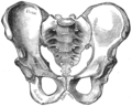
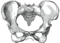
The perineum in mammals is the space between the anus and the genitals. The human perineum is between the anus and scrotum in the male or between the anus and vulva in the female. The perineum is the region of the body between the pubic symphysis and the coccyx, including the perineal body and surrounding structures. The perineal raphe is visible and pronounced to varying degrees. The perineum is an erogenous zone. This area is also known as the taint or gooch in American slang.

<span class="mw-page-title-main">Urinary incontinence</span> Uncontrolled leakage of urine

Urinary incontinence (UI), also known as involuntary urination, is any uncontrolled leakage of urine. It is a common and distressing problem, which may have a large impact on quality of life. It has been identified as an important issue in geriatric health care. The term enuresis is often used to refer to urinary incontinence primarily in children, such as nocturnal enuresis. UI is an example of a stigmatized medical condition, which creates barriers to successful management and makes the problem worse. People may be too embarrassed to seek medical help, and attempt to self-manage the symptom in secrecy from others.

<span class="mw-page-title-main">Levator ani</span> Broad, thin muscle group, situated on either side of the pelvis

The levator ani is a broad, thin muscle group, situated on either side of the pelvis. It is formed from three muscle components: the pubococcygeus, the iliococcygeus, and the puborectalis.

<span class="mw-page-title-main">Kegel exercise</span> Pelvic floor exercise

Kegel exercise, also known as pelvic floor exercise, involves repeatedly contracting and relaxing the muscles that form part of the pelvic floor, now sometimes colloquially referred to as the "Kegel muscles". The exercise can be performed many times a day, for several minutes at a time but takes one to three months to begin to have an effect.

A pessary is a prosthetic device inserted into the vagina for structural and pharmaceutical purposes. It is most commonly used to treat stress urinary incontinence to stop urinary leakage and to treat pelvic organ prolapse to maintain the location of organs in the pelvic region. It can also be used to administer medications locally in the vagina or as a method of contraception.

<span class="mw-page-title-main">Pelvic floor dysfunction</span> Medical condition

Pelvic floor dysfunction is a term used for a variety of disorders that occur when pelvic floor muscles and ligaments are impaired. The condition affects up to 50 percent of women who have given birth. Although this condition predominantly affects women, up to 16 percent of men are affected as well. Symptoms can include pelvic pain, pressure, pain during sex, urinary incontinence (UI), overactive bladder, bowel incontinence, incomplete emptying of feces, constipation, myofascial pelvic pain and pelvic organ prolapse. When pelvic organ prolapse occurs, there may be visible organ protrusion or a lump felt in the vagina or anus. Research carried out in the UK has shown that symptoms can restrict everyday life for women. However, many people found it difficult to talk about it and to seek care, as they experienced embarrassment and stigma.

<span class="mw-page-title-main">Urogenital diaphragm</span> Layer of the pelvis

Older texts have asserted the existence of a urogenital diaphragm, also called the triangular ligament, which was described as a layer of the pelvis that separates the deep perineal sac from the upper pelvis, lying between the inferior fascia of the urogenital diaphragm and superior fascia of the urogenital diaphragm.

<span class="mw-page-title-main">Cystocele</span> Medical condition

The cystocele, also known as a prolapsed bladder, is a medical condition in which a woman's bladder bulges into her vagina. Some may have no symptoms. Others may have trouble starting urination, urinary incontinence, or frequent urination. Complications may include recurrent urinary tract infections and urinary retention. Cystocele and a prolapsed urethra often occur together and is called a cystourethrocele. Cystocele can negatively affect quality of life.

<span class="mw-page-title-main">Prostatectomy</span> Surgical removal of all or part of the prostate gland

Prostatectomy is the surgical removal of all or part of the prostate gland. This operation is done for benign conditions that cause urinary retention, as well as for prostate cancer and for other cancers of the pelvis.

<span class="mw-page-title-main">Pelvic organ prolapse</span> Descent of the pelvic organs from their normal positions

Pelvic organ prolapse (POP) is characterized by descent of pelvic organs from their normal positions into the vagina. In women, the condition usually occurs when the pelvic floor collapses after gynecological cancer treatment, childbirth or heavy lifting. Injury incurred to fascia membranes and other connective structures can result in cystocele, rectocele or both. Treatment can involve dietary and lifestyle changes, physical therapy, or surgery.

<span class="mw-page-title-main">Stress incontinence</span> Form of urinary incontinence from an inadequate closure of the bladder

Stress incontinence, also known as stress urinary incontinence (SUI) or effort incontinence is a form of urinary incontinence. It is due to inadequate closure of the bladder outlet by the urethral sphincter.

<span class="mw-page-title-main">Deep perineal pouch</span> Anatomic space enclosed partly by the perineum

The deep perineal pouch is the anatomic space enclosed in part by the perineum and located superior to the perineal membrane.

<span class="mw-page-title-main">Urethral sphincters</span> Muscles keeping urine in the bladder

The urethral sphincters are two muscles used to control the exit of urine in the urinary bladder through the urethra. The two muscles are either the male or female external urethral sphincter and the internal urethral sphincter. When either of these muscles contracts, the urethra is sealed shut.

<span class="mw-page-title-main">Defecography</span> Visualisation of the mechanics of a patients defecation

Defecography is a type of medical radiological imaging in which the mechanics of a patient's defecation are visualized in real time using a fluoroscope. The anatomy and function of the anorectum and pelvic floor can be dynamically studied at various stages during defecation.

Urogynecology or urogynaecology is a surgical sub-specialty of urology and gynecology.

Descending perineum syndrome refers to a condition where the perineum "balloons" several centimeters below the bony outlet of the pelvis during strain, although this descent may happen without straining. The syndrome was first described in 1966 by Parks et al.

Urethral hypermobility is a condition of excessive movement of the female urethra due to a weakened urogenital diaphragm. It describes the instability of the urethra in relation to the pelvic floor muscles. A weakened pelvic floor muscle fails to adequately close the urethra and hence can cause stress urinary incontinence. This condition may be diagnosed by primary care providers or urologists. Treatment may include pelvic floor muscle exercises, surgery, or minimally invasive procedures.

<span class="mw-page-title-main">Vaginal support structures</span> Structures that maintain the position of the vagina within the pelvic cavity

The vaginal support structures are those muscles, bones, ligaments, tendons, membranes and fascia, of the pelvic floor that maintain the position of the vagina within the pelvic cavity and allow the normal functioning of the vagina and other reproductive structures in the female. Defects or injuries to these support structures in the pelvic floor leads to pelvic organ prolapse. Anatomical and congenital variations of vaginal support structures can predispose a woman to further dysfunction and prolapse later in life. The urethra is part of the anterior wall of the vagina and damage to the support structures there can lead to incontinence and urinary retention.

The urogenital hiatus is a gap in the anteromedial part of the pelvic floor, allowing passage of the urethra, the vagina, and deep dorsal vein of clitoris (females) or penis (males).

Transvaginal mesh, also known as vaginal mesh implant, is a net-like surgical tool that is used to treat pelvic organ prolapse (POP) and stress urinary incontinence (SUI) among female patients. The surgical mesh is placed transvaginally to reconstruct weakened pelvic muscle walls and to support the urethra or bladder.

References

PD-icon.svgThis article incorporates text in the public domain from page 420 of the 20th edition of Gray's Anatomy (1918)

  1. Bordoni B, Sugumar K, Leslie SW (2023). "Anatomy, Abdomen and Pelvis, Pelvic Floor". StatPearls. Treasure Island (FL): StatPearls Publishing. PMID   29489277 . Retrieved 2023-10-13.
  2. Fernandes AC, Palacios-Ceña D, Hay-Smith J, Pena CC, Sidou MF, de Alencar AL, et al. (July 2021). "Women report sustained benefits from attending group-based education about pelvic floor muscles: a longitudinal qualitative study". Journal of Physiotherapy. 67 (3): 210–216. doi: 10.1016/j.jphys.2021.06.010 . PMID   34147398. S2CID   235492234.
  3. Roch M, Gaudreault N, Cyr MP, Venne G, Bureau NJ, Morin M (August 2021). "The Female Pelvic Floor Fascia Anatomy: A Systematic Search and Review". Life. 11 (9): 900. Bibcode:2021Life...11..900R. doi: 10.3390/life11090900 . PMC   8467746 . PMID   34575049.
  4. "Pelvic Floor Muscles: Anatomy, Function & Conditions". Cleveland Clinic. Retrieved 2023-03-16.
  5. 1 2 Daftary S, Chakravarti S (2011). "Reproductive Anatomy". Manual of Obstetrics (3rd ed.). Elsevier. pp. 1–16. ISBN   978-81-312-2556-1.
  6. Drake RL, Vogl W, Mitchell AW (2005). Gray's Anatomy For Students. Elsevier Health Sciences TW. p. 391. ISBN   978-0-443-06612-2.
  7. Herschorn S (2004). "Female pelvic floor anatomy: the pelvic floor, supporting structures, and pelvic organs". Reviews in Urology. 6 (Suppl 5): S2–S10. PMC   1472875 . PMID   16985905.
  8. "Cystocele (Prolapsed Bladder) | NIDDK". National Institute of Diabetes and Digestive and Kidney Diseases. Retrieved 2017-12-02.
  9. Ramaseshan AS, Felton J, Roque D, Rao G, Shipper AG, Sanses TV (April 2018). "Pelvic floor disorders in women with gynecologic malignancies: a systematic review". International Urogynecology Journal. 29 (4): 459–476. doi:10.1007/s00192-017-3467-4. PMC   7329191 . PMID   28929201.
  10. 1 2 3 Harvey MA (June 2003). "Pelvic floor exercises during and after pregnancy: a systematic review of their role in preventing pelvic floor dysfunction". Journal of Obstetrics and Gynaecology Canada. 25 (6): 487–498. doi:10.1016/s1701-2163(16)30310-3. PMID   12806450.
  11. Berman L, Aversa J, Abir F, Longo WE (July 2005). "Management of disorders of the posterior pelvic floor". The Yale Journal of Biology and Medicine. 78 (4): 211–221. PMC   2259151 . PMID   16720016.
  12. 1 2 Kielb SJ (2005). "Stress incontinence: alternatives to surgery". International Journal of Fertility and Women's Medicine. 50 (1): 24–29. PMID   15971718.
  13. Barbosa PB, Franco MM, Souza Fd, Antônio FI, Montezuma T, Ferreira CH (June 2009). "Comparison between measurements obtained with three different perineometers". Clinics. 64 (6): 527–533. doi:10.1590/s1807-59322009000600007. PMC   2705146 . PMID   19578656.
  14. 1 2 "Pelvic Floor Dysfunction: Symptoms, Causes & Treatment". Cleveland Clinic. Retrieved 2023-03-16.
  15. Zhou L, Chen Y, Yuan X, Zeng L, Zhu J, Zheng J (2023). "Preoperative pelvic floor muscle exercise for continence after radical prostatectomy: a systematic review and meta-analysis". Frontiers in Public Health. 11: 1186067. doi: 10.3389/fpubh.2023.1186067 . PMC   10425962 . PMID   37588123.
  16. Wong C, Louie DR, Beach C (April 2020). "A Systematic Review of Pelvic Floor Muscle Training for Erectile Dysfunction After Prostatectomy and Recommendations to Guide Further Research". The Journal of Sexual Medicine. 17 (4): 737–748. doi:10.1016/j.jsxm.2020.01.008. PMID   32029399.
  17. Beco J, Mouchel J (2002-10-01). "Understanding the concept of perineology". International Urogynecology Journal and Pelvic Floor Dysfunction. 13 (5): 275–277. doi:10.1007/s001920200060. PMID   12355284. S2CID   12964013.