The peritoneum is the serous membrane forming the lining of the abdominal cavity or coelom in amniotes and some invertebrates, such as annelids. It covers most of the intra-abdominal organs, and is composed of a layer of mesothelium supported by a thin layer of connective tissue. This peritoneal lining of the cavity supports many of the abdominal organs and serves as a conduit for their blood vessels, lymphatic vessels, and nerves.
A body cavity is any fluid-filled space in a multicellular organism other than those of vessels. The human body cavity normally refers to the ventral body cavity, because it is by far the largest.

The thoracic diaphragm, or simply the diaphragm, is a sheet of internal skeletal muscle in humans and other mammals that extends across the bottom of the thoracic cavity. The diaphragm separates the thoracic cavity, containing the heart and lungs, from the abdominal cavity and performs an important function in respiration: as the diaphragm contracts, the volume of the thoracic cavity increases, a negative vacuum is created which draws air into the lungs.

The retroperitoneal space (retroperitoneum) is the anatomical space in the abdominal cavity behind (retro) the peritoneum. It has no specific delineating anatomical structures. Organs are retroperitoneal if they have peritoneum on their anterior side only. Structures that are not suspended by mesentery in the abdominal cavity and that lie between the parietal peritoneum and abdominal wall are classified as retroperitoneal.
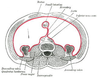
The mesentery is a contiguous set of tissues that attaches the intestines to the posterior abdominal wall in humans and is formed by the double fold of peritoneum. It helps in storing fat and allowing blood vessels, lymphatics, and nerves to supply the intestines, among other functions.

The abdomen constitutes the part of the body between the thorax (chest) and pelvis, in humans and in other vertebrates. The abdomen is the frontal part of the abdominal segment of the trunk, the dorsal part of this segment being the back of the abdomen. The region occupied by the abdomen is termed the abdominal cavity. In arthropods it is the posterior tagma of the body; it follows the thorax or cephalothorax. The abdomen stretches from the thorax at the thoracic diaphragm to the pelvis at the pelvic brim. The pelvic brim stretches from the lumbosacral joint to the pubic symphysis and is the edge of the pelvic inlet. The space above this inlet and under the thoracic diaphragm is termed the abdominal cavity. The boundary of the abdominal cavity is the abdominal wall in the front and the peritoneal surface at the rear.
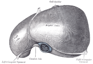
The falciform ligament is a ligament that attaches the liver to the anterior (ventral) body wall, and separates the liver into the left medial lobe and left lateral lobe. The falciform ligament, from Latin, meaning 'sickle-shaped', is a broad and thin fold of peritoneum, its base being directed downward and backward and its apex upward and backward. The falciform ligament droops down from the hilum of the liver.

The greater omentum is a large apron-like fold of visceral peritoneum that hangs down from the stomach. It extends from the greater curvature of the stomach, passing in front of the small intestines and doubles back to ascend to the transverse colon before reaching to the posterior abdominal wall. The greater omentum is larger than the lesser omentum, which hangs down from the liver to the lesser curvature. The common anatomical term "epiploic" derives from "epiploon", from the Greek epipleein, meaning to float or sail on, since the greater omentum appears to float on the surface of the intestines. It is the first structure observed when the abdominal cavity is opened anteriorly.

Lateral plate mesoderm is a type of mesoderm that is found at the periphery of the embryo.

The pelvic cavity is a body cavity that is bounded by the bones of the pelvis. Its oblique roof is the pelvic inlet. Its lower boundary is the pelvic floor.
Hemoperitoneum is the presence of blood in the peritoneal cavity. The blood accumulates in the space between the inner lining of the abdominal wall and the internal abdominal organs. Hemoperitoneum is generally classified as a surgical emergency; in most cases, urgent laparotomy is needed to identify and control the source of the bleeding. In selected cases, careful observation may be permissible. The abdominal cavity is highly distensible and may easily hold greater than five liters of blood, or more than the entire circulating blood volume for an average-sized individual. Therefore, large-scale or rapid blood loss into the abdomen will reliably induce hemorrhagic shock and, if untreated, may rapidly lead to death.

The lateral inguinal fossa is a structure described in human anatomy. It is a shallow concave stretch of peritoneum on the deep surface of the anterior abdominal wall and is best seen from the greater peritoneal cavity, looking anteriorly.
A fold of peritoneum, the phrenicocolic ligament is continued from the left colic flexure to the thoracic diaphragm opposite the tenth and eleventh ribs; it passes below and serves to support the spleen, and therefore has received the name of sustentaculum lienis.

The following outline is provided as an overview of and topical guide to human anatomy:
Peritoneal recesses are the spaces formed by peritoneum draping over viscera.
The development of the digestive system concerns the epithelium of the digestive system and the parenchyma of its derivatives, which originate from the endoderm. Connective tissue, muscular components, and peritoneal components originate in the mesoderm. Different regions of the gut tube such as the esophagus, stomach, duodenum, etc. are specified by a retinoic acid gradient that causes transcription factors unique to each region to be expressed. Differentiation of the gut and its derivatives depends upon reciprocal interactions between the gut endoderm and its surrounding mesoderm. Hox genes in the mesoderm are induced by a Hedgehog signaling pathway secreted by gut endoderm and regulate the craniocaudal organization of the gut and its derivatives. The gut system extends from the oropharyngeal membrane to the cloacal membrane and is divided into the foregut, midgut, and hindgut.

Anatomical terminology is a form of scientific terminology used by anatomists, zoologists, and health professionals such as doctors.
Peritoneal carcinomatosis (PC) is intraperitoneal dissemination (carcinosis) of any form of cancer that does not originate from the peritoneum itself. PC is most commonly seen in abdominopelvic malignancies. Computed tomography (CT) is particularly important for detailed preoperative assessment and evaluation of the radiological Peritoneal Cancer Index (PCI). The imaging findings vary from simple ascites to multifocal discrete nodules and infiltrative peritoneal masses. Various tumours and tumour like conditions can mimic PC. A systematic analysis of CT imaging features is helpful to narrow down the differential diagnosis, staging and effectively guiding the patient management.