Abdominal cavity

Last updated
Abdominal cavity
Scheme body cavities-en.svg
Gray1225.png
Front of abdomen, showing surface markings for duodenum, pancreas, and kidneys.
Details
Identifiers
Latin cavitas abdominis
MeSH D034841
TA98 A01.1.00.051
A10.1.00.001
TA2 128
FMA 12266
Anatomical terminology

The abdominal cavity is a large body cavity in humans [1] and many other animals that contain organs. It is a part of the abdominopelvic cavity. [2] It is located below the thoracic cavity, and above the pelvic cavity. Its dome-shaped roof is the thoracic diaphragm, a thin sheet of muscle under the lungs, and its floor is the pelvic inlet, opening into the pelvis.

Contents

Structure

The abdominal cavity is labeled 3 in this image, and together with the pelvic cavity (4) it makes up the abdominopelvic cavity 6. Body Cavities Frontal view labeled.jpg
The abdominal cavity is labeled 3 in this image, and together with the pelvic cavity (4) it makes up the abdominopelvic cavity 6.

Organs

Organs of the abdominal cavity include the stomach, liver, gallbladder, spleen, pancreas, small intestine, kidneys, large intestine, and adrenal glands. [1]

Peritoneum

The abdominal cavity is lined with a protective membrane termed the peritoneum. The inside wall is covered by the parietal peritoneum. The kidneys are located behind the peritoneum, in the retroperitoneum, outside the abdominal cavity. The viscera are also covered by visceral peritoneum.

Between the visceral and parietal peritoneum is the peritoneal cavity, which is a potential space. [1] It contains a serous fluid called peritoneal fluid that allows motion. This motion is apparent of the gastrointestinal tract. The peritoneum, by virtue of its connection to the two (parietal and visceral) portions, gives support to the abdominal organs.

The peritoneum divides the cavity into numerous compartments. One of these the lesser sac is located behind the stomach and joins into the greater sac via the foramen of Winslow. [1] Some of the organs are attached to the walls of the abdomen via folds of peritoneum and ligaments, such as the liver and others use broad areas of the peritoneum, such as the pancreas. The peritoneal ligaments are actually dense folds of the peritoneum that are used to connect viscera to viscera or viscera to the walls of the abdomen. [1] They are named in such a way as to show what they connect typically. For example, the gastrocolic ligament connects the stomach and colon and the splenocolic ligament connects the spleen and the colon, or sometimes by their shape as the round ligament or triangular ligament. [1]

Mesentery

Mesenteries are folds of peritoneum that are attached to the walls of the abdomen and enclose viscera completely. They are supplied with plentiful amounts of blood. The three most important mesenteries are mesentery for the small intestine, the transverse mesocolon, which attaches the back portion of the colon to the abdominal wall, and the sigmoid mesocolon which enfolds the sigmoid colon. [1]

Omenta

The omentum are specialized folds of peritoneum that enclose nerves, blood vessels, lymph channels, fatty tissue, and connective tissue. There are two omenta. First, is the greater omentum that hangs off of the transverse colon and greater curvature of the stomach. The other is the lesser omentum that extends between the stomach and the liver. [1]

Clinical significance

Ascites

When fluid collects in the abdominal cavity, this condition is called ascites. This is usually not noticeable until enough fluid has collected to distend the abdomen. The collection of fluid will cause pressure on the viscera, veins, and thoracic cavity. Treatment is directed at the cause of the fluid accumulation. One method is to decrease the portal vein pressure, especially useful in treating cirrhosis. Chylous ascites heals best if the lymphatic vessel involved is closed. Heart failure can cause recurring ascites. [1]

Inflammation

Another disorder is called peritonitis which usually accompanies inflammatory processes elsewhere. It can be caused by damage to an organ, or from a contusion to the abdominal wall from the outside or by surgery. It may be brought in by the bloodstream or the lymphatic system. The most common origin is the gastrointestinal tract. Peritonitis can be acute or chronic, generalized or localized, and may have one origin or multiple origins. The omenta can help control the spread of infection; however without treatment, the infection will spread throughout the cavity. An abscess may also form as a secondary reaction to an infection. Antibiotics have become an important tool in fighting abscesses; however, external drainage is usually required also. [1]

See also

Related Research Articles

<span class="mw-page-title-main">Peritoneum</span> Serous membrane that forms lining of abdominal cavity or coelom

The peritoneum is the serous membrane forming the lining of the abdominal cavity or coelom in amniotes and some invertebrates, such as annelids. It covers most of the intra-abdominal organs, and is composed of a layer of mesothelium supported by a thin layer of connective tissue. This peritoneal lining of the cavity supports many of the abdominal organs and serves as a conduit for their blood vessels, lymphatic vessels, and nerves.

<span class="mw-page-title-main">Body cavity</span> Internal space within a multicellular organism

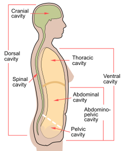
A body cavity is any space or compartment, or potential space, in an animal body. Cavities accommodate organs and other structures; cavities as potential spaces contain fluid.

<span class="mw-page-title-main">Peritonitis</span> Medical condition

Peritonitis is inflammation of the localized or generalized peritoneum, the lining of the inner wall of the abdomen and cover of the abdominal organs. Symptoms may include severe pain, swelling of the abdomen, fever, or weight loss. One part or the entire abdomen may be tender. Complications may include shock and acute respiratory distress syndrome.

<span class="mw-page-title-main">Organ (biology)</span> Collection of tissues with similar functions

In a multicellular organism, an organ is a collection of tissues joined in a structural unit to serve a common function. In the hierarchy of life, an organ lies between tissue and an organ system. Tissues are formed from same type cells to act together in a function. Tissues of different types combine to form an organ which has a specific function. The intestinal wall for example is formed by epithelial tissue and smooth muscle tissue. Two or more organs working together in the execution of a specific body function form an organ system, also called a biological system or body system.

<span class="mw-page-title-main">Mesothelium</span> Membrane lining body cavities

The mesothelium is a membrane composed of simple squamous epithelial cells of mesodermal origin, which forms the lining of several body cavities: the pleura, peritoneum and pericardium.

<span class="mw-page-title-main">Mesentery</span> Contiguous fold of tissues that supports the intestines

The mesentery is an organ that attaches the intestines to the posterior abdominal wall and is formed by the double fold of peritoneum. It helps in storing fat and allowing blood vessels, lymphatics, and nerves to supply the intestines, among other functions.

The peritoneal cavity is a potential space between the parietal peritoneum and visceral peritoneum. The parietal and visceral peritonea are layers of the peritoneum named depending on their function/location. It is one of the spaces derived from the coelomic cavity of the embryo, the others being the pleural cavities around the lungs and the pericardial cavity around the heart.

<span class="mw-page-title-main">Serous membrane</span> Smooth coating lining contents & inner walls of body cavities

The serous membrane is a smooth tissue membrane of mesothelium lining the contents and inner walls of body cavities, which secrete serous fluid to allow lubricated sliding movements between opposing surfaces. The serous membrane that covers internal organs is called a visceral membrane; while the one that covers the cavity wall is called the parietal membrane. Between the two opposing serosal surfaces is often a potential space, mostly empty except for the small amount of serous fluid.

<span class="mw-page-title-main">Lesser omentum</span>

The lesser omentum is the double layer of peritoneum that extends from the liver to the lesser curvature of the stomach, and to the first part of the duodenum. The lesser omentum is usually divided into these two connecting parts: the hepatogastric ligament, and the hepatoduodenal ligament.

<span class="mw-page-title-main">Ascending colon</span> Section of the large intestine

In the anatomy of humans and homologous primates, the ascending colon is the part of the colon located between the cecum and the transverse colon.

<span class="mw-page-title-main">Abdomen</span> Part of the body between the chest and pelvis

The abdomen is the part of the body between the thorax (chest) and pelvis, in humans and in other vertebrates. The abdomen is the front part of the abdominal segment of the torso. The area occupied by the abdomen is called the abdominal cavity. In arthropods it is the posterior tagma of the body; it follows the thorax or cephalothorax.

<span class="mw-page-title-main">Lesser sac</span> Abdominal cavity

The lesser sac, also known as the omental bursa, is a part of the peritoneal cavity that is formed by the lesser and greater omentum. Usually found in mammals, it is connected with the greater sac via the omental foramen or Foramen of Winslow. In mammals, it is common for the lesser sac to contain considerable amounts of fat.

<span class="mw-page-title-main">Transverse colon</span> Longest section of the large intestine

In human anatomy, the transverse colon is the longest and most movable part of the colon.

<span class="mw-page-title-main">Greater omentum</span> Fat sheath under abdominal wall

The greater omentum is a large apron-like fold of visceral peritoneum that hangs down from the stomach. It extends from the greater curvature of the stomach, passing in front of the small intestines and doubles back to ascend to the transverse colon before reaching to the posterior abdominal wall. The greater omentum is larger than the lesser omentum, which hangs down from the liver to the lesser curvature. The common anatomical term "epiploic" derives from "epiploon", from the Greek epipleein, meaning to float or sail on, since the greater omentum appears to float on the surface of the intestines. It is the first structure observed when the abdominal cavity is opened anteriorly.

The paracolic gutters are peritoneal recesses – spaces between the colon and the abdominal wall.

<span class="mw-page-title-main">Left triangular ligament</span>

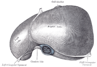
The left triangular ligament is a large peritoneal fold. It connects the posterior part of the upper surface of the left lobe of the liver to the thoracic diaphragm.

Peritoneal fluid is a serous fluid made by the peritoneum in the abdominal cavity which lubricates the surface of tissue that lines the abdominal wall and pelvic cavity. It covers most of the organs in the abdomen. An increased volume of peritoneal fluid is called ascites.

<span class="mw-page-title-main">Peritoneal recesses</span>

Peritoneal recesses are the spaces formed by peritoneum draping over viscera.

Peritoneal ligaments are folds of peritoneum that are used to connect viscera to viscera or the abdominal wall.

The development of the digestive system in the human embryo concerns the epithelium of the digestive system and the parenchyma of its derivatives, which originate from the endoderm. Connective tissue, muscular components, and peritoneal components originate in the mesoderm. Different regions of the gut tube such as the esophagus, stomach, duodenum, etc. are specified by a retinoic acid gradient that causes transcription factors unique to each region to be expressed. Differentiation of the gut and its derivatives depends upon reciprocal interactions between the gut endoderm and its surrounding mesoderm. Hox genes in the mesoderm are induced by a Hedgehog signaling pathway secreted by gut endoderm and regulate the craniocaudal organization of the gut and its derivatives. The gut system extends from the oropharyngeal membrane to the cloacal membrane and is divided into the foregut, midgut, and hindgut.

References

  1. 1 2 3 4 5 6 7 8 9 10 "Abdominal cavity" . Encyclopædia Britannica. Vol. I: A-Ak – Bayes (15th ed.). Chicago, Illinois: Encyclopædia Britannica, Inc. 2010. pp.  19–20. ISBN   978-1-59339-837-8.
  2. Wingerd, Bruce (1994). The Human Body: Concepts of Anatomy and Physiology. Fort Worth: Saunders College Publishing. pp. 11–12. ISBN   0-03-055507-8.