Peritonitis

Last updated
Peritonitis
Other namesSurgical abdomen, acute abdomen [1]
Tuberculous peritonitis (6544825621).jpg
Peritonitis from tuberculosis
Pronunciation
Specialty Emergency medicine, general surgery
Symptoms Severe pain, swelling of the abdomen, fever [2] [3]
Complications Sepsis (sepsis is likely if not quickly treated), shock, acute respiratory distress syndrome [4] [5]
Usual onsetSudden [1]
TypesPrimary, secondary, tertiary, generalized, localized [1]
Causes Perforation of the intestinal tract, pancreatitis, pelvic inflammatory disease, cirrhosis, ruptured appendix [3]
Risk factors Ascites, peritoneal dialysis [4]
Diagnostic method Examination, blood tests, medical imaging [6]
Treatment Antibiotics, intravenous fluids, pain medication, surgery [3] [4]
FrequencyRelatively common [1]

Peritonitis is inflammation of the localized or generalized peritoneum, the lining of the inner wall of the abdomen and cover of the abdominal organs. [2] Symptoms may include severe pain, swelling of the abdomen, fever, or weight loss. [2] [3] One part or the entire abdomen may be tender. [1] Complications may include shock and acute respiratory distress syndrome. [4] [5]

Contents

Causes include perforation of the intestinal tract, pancreatitis, pelvic inflammatory disease, stomach ulcer, cirrhosis, a ruptured appendix or even a perforated gallbladder. [3] Risk factors include ascites (the abnormal build-up of fluid in the abdomen) and peritoneal dialysis. [4] Diagnosis is generally based on examination, blood tests, and medical imaging. [6]

Treatment often includes antibiotics, intravenous fluids, pain medication, and surgery. [3] [4] Other measures may include a nasogastric tube or blood transfusion. [4] Without treatment death may occur within a few days. [4] About 20% of people with cirrhosis who are hospitalized have peritonitis. [1]

Signs and symptoms

Abdominal pain

The main manifestations of peritonitis are acute abdominal pain, abdominal tenderness, abdominal guarding, rigidity, which are exacerbated by moving the peritoneum, e.g., coughing (forced cough may be used as a test), flexing one's hips, or eliciting the Blumberg's sign (meaning that pressing a hand on the abdomen elicits less pain than releasing the hand abruptly, which will aggravate the pain, as the peritoneum snaps back into place). Rigidity is highly specific for diagnosing peritonitis (specificity: 76–100%). [7] The presence of these signs in a person is sometimes referred to as peritonism. [8] The localization of these manifestations depends on whether peritonitis is localized (e.g., appendicitis or diverticulitis before perforation), or generalized to the whole abdomen. In either case, pain typically starts as a generalized abdominal pain (with involvement of poorly localizing visceral innervation of the visceral peritoneal layer), and may become localized later (with involvement of the somatic innervation of the parietal peritoneal layer). Peritonitis is an example of an acute abdomen. [9]

Other symptoms

Complications

Causes

Infection

Non-infection

Risk factors

Diagnosis

A diagnosis of peritonitis is based primarily on the clinical manifestations described above. Rigidity (involuntary contraction of the abdominal muscles) is the most specific exam finding for diagnosing peritonitis. [14] If focal peritonitis is detected, further work-up should be done. If diffuse peritonitis is detected, then urgent surgical consultation should be obtained, and may warrant surgery without further investigations. Leukocytosis, hypokalemia, hypernatremia, and acidosis may be present, but they are not specific findings. Abdominal X-rays may reveal dilated, edematous intestines, although such X-rays are mainly useful to look for pneumoperitoneum, an indicator of gastrointestinal perforation. The role of whole-abdomen ultrasound examination is under study and is likely to expand in the future. Computed tomography (CT or CAT scanning) may be useful in differentiating causes of abdominal pain. If reasonable doubt still persists, an exploratory peritoneal lavage or laparoscopy may be performed. In people with ascites, a diagnosis of peritonitis is made via paracentesis (abdominal tap): More than 250 polymorphonuclear cells per μL is considered diagnostic. In addition, Gram stain is almost always negative, whereas culture of the peritoneal fluid can determine the microorganism responsible and determine their sensitivity to antimicrobial agents. [15] [16]

Pathology

In normal conditions, the peritoneum appears greyish and glistening; it becomes dull 2–4 hours after the onset of peritonitis, initially with scarce serous or slightly turbid fluid. Later on, the exudate becomes creamy and evidently suppurative; in people who are dehydrated, it also becomes very inspissated. The quantity of accumulated exudate varies widely. It may be spread to the whole peritoneum, or be walled off by the omentum and viscera. Inflammation features infiltration by neutrophils with fibrino-purulent exudation. [17]

Treatment

Depending on the severity of the person's state, the management of peritonitis may include:

Prognosis

If properly treated, typical cases of surgically correctable peritonitis (e.g., perforated peptic ulcer, appendicitis, and diverticulitis) have a mortality rate of about <10% in otherwise healthy people. The mortality rate rises to 35% in peritonitis patients who develop sepsis, and patients who have underlying renal insufficiency and complications have a higher mortality rate. [22]

Etymology

The term "peritonitis" comes from Greek περιτόναιον peritonaion "peritoneum, abdominal membrane" and -itis "inflammation". [23]

Related Research Articles

<span class="mw-page-title-main">Peritoneum</span> Serous membrane that forms lining of abdominal cavity or coelom

The peritoneum is the serous membrane forming the lining of the abdominal cavity or coelom in amniotes and some invertebrates, such as annelids. It covers most of the intra-abdominal organs, and is composed of a layer of mesothelium supported by a thin layer of connective tissue. This peritoneal lining of the cavity supports many of the abdominal organs and serves as a conduit for their blood vessels, lymphatic vessels, and nerves.

Peptic ulcer disease is a break in the inner lining of the stomach, the first part of the small intestine, or sometimes the lower esophagus. An ulcer in the stomach is called a gastric ulcer, while one in the first part of the intestines is a duodenal ulcer. The most common symptoms of a duodenal ulcer are waking at night with upper abdominal pain, and upper abdominal pain that improves with eating. With a gastric ulcer, the pain may worsen with eating. The pain is often described as a burning or dull ache. Other symptoms include belching, vomiting, weight loss, or poor appetite. About a third of older people with peptic ulcers have no symptoms. Complications may include bleeding, perforation, and blockage of the stomach. Bleeding occurs in as many as 15% of cases.

<span class="mw-page-title-main">Abdominal cavity</span> Body cavity in the abdominal area

The abdominal cavity is a large body cavity in humans and many other animals that contain organs. It is a part of the abdominopelvic cavity. It is located below the thoracic cavity, and above the pelvic cavity. Its dome-shaped roof is the thoracic diaphragm, a thin sheet of muscle under the lungs, and its floor is the pelvic inlet, opening into the pelvis.

<span class="mw-page-title-main">Appendicitis</span> Inflammation of the appendix

Appendicitis is inflammation of the appendix. Symptoms commonly include right lower abdominal pain, nausea, vomiting, and decreased appetite. However, approximately 40% of people do not have these typical symptoms. Severe complications of a ruptured appendix include widespread, painful inflammation of the inner lining of the abdominal wall and sepsis.

<span class="mw-page-title-main">Cholecystitis</span> Inflammation of the gallbladder

Cholecystitis is inflammation of the gallbladder. Symptoms include right upper abdominal pain, pain in the right shoulder, nausea, vomiting, and occasionally fever. Often gallbladder attacks precede acute cholecystitis. The pain lasts longer in cholecystitis than in a typical gallbladder attack. Without appropriate treatment, recurrent episodes of cholecystitis are common. Complications of acute cholecystitis include gallstone pancreatitis, common bile duct stones, or inflammation of the common bile duct.

<span class="mw-page-title-main">Diverticulitis</span> Digestive disease of the large intestine

Diverticulitis, also called colonic diverticulitis, is a gastrointestinal disease characterized by inflammation of abnormal pouches—diverticula—that can develop in the wall of the large intestine. Symptoms typically include lower abdominal pain of sudden onset, but the onset may also occur over a few days. There may also be nausea, diarrhea or constipation. Fever or blood in the stool suggests a complication. People may experience a single attack, repeated attacks, or ongoing "smouldering" diverticulitis.

<span class="mw-page-title-main">Peritoneal dialysis</span> Type of dialysis

Peritoneal dialysis (PD) is a type of dialysis that uses the peritoneum in a person's abdomen as the membrane through which fluid and dissolved substances are exchanged with the blood. It is used to remove excess fluid, correct electrolyte problems, and remove toxins in those with kidney failure. Peritoneal dialysis has better outcomes than hemodialysis during the first couple of years. Other benefits include greater flexibility and better tolerability in those with significant heart disease.

Spontaneous bacterial peritonitis (SBP) is the development of a bacterial infection in the peritoneum, despite the absence of an obvious source for the infection. It is specifically an infection of the ascitic fluid – an increased volume of peritoneal fluid. Ascites is most commonly a complication of cirrhosis of the liver. It can also occur in patients with nephrotic syndrome. SBP has a high mortality rate.

Colic in horses is defined as abdominal pain, but it is a clinical symptom rather than a diagnosis. The term colic can encompass all forms of gastrointestinal conditions which cause pain as well as other causes of abdominal pain not involving the gastrointestinal tract. What makes it tricky is that different causes can manifest with similar signs of distress in the animal. Recognizing and understanding these signs is pivotal, as timely action can spell the difference between a brief moment of discomfort and a life-threatening situation. The most common forms of colic are gastrointestinal in nature and are most often related to colonic disturbance. There are a variety of different causes of colic, some of which can prove fatal without surgical intervention. Colic surgery is usually an expensive procedure as it is major abdominal surgery, often with intensive aftercare. Among domesticated horses, colic is the leading cause of premature death. The incidence of colic in the general horse population has been estimated between 4 and 10 percent over the course of the average lifespan. Clinical signs of colic generally require treatment by a veterinarian. The conditions that cause colic can become life-threatening in a short period of time.

<span class="mw-page-title-main">Gastrointestinal perforation</span> Medical condition

Gastrointestinal perforation, also known as gastrointestinal rupture, is a hole in the wall of the gastrointestinal tract. The gastrointestinal tract is composed of hollow digestive organs leading from the mouth to the anus. Symptoms of gastrointestinal perforation commonly include severe abdominal pain, nausea, and vomiting. Complications include a painful inflammation of the inner lining of the abdominal wall and sepsis.

<span class="mw-page-title-main">Pneumoperitoneum</span> Medical condition

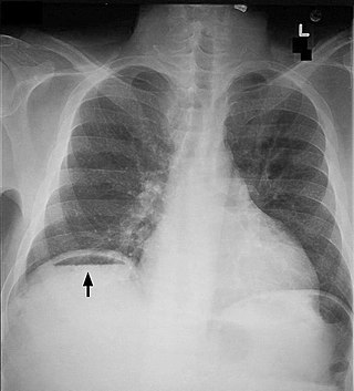
Pneumoperitoneum is pneumatosis in the peritoneal cavity, a potential space within the abdominal cavity. The most common cause is a perforated abdominal organ, generally from a perforated peptic ulcer, although any part of the bowel may perforate from a benign ulcer, tumor or abdominal trauma. A perforated appendix seldom causes a pneumoperitoneum.

<span class="mw-page-title-main">Abdominal distension</span> Physical symptom

Abdominal distension occurs when substances, such as air (gas) or fluid, accumulate in the abdomen causing its expansion. It is typically a symptom of an underlying disease or dysfunction in the body, rather than an illness in its own right. People with this condition often describe it as "feeling bloated". Affected people often experience a sensation of fullness, abdominal pressure, and sometimes nausea, pain, or cramping. In the most extreme cases, upward pressure on the diaphragm and lungs can also cause shortness of breath. Through a variety of causes, bloating is most commonly due to buildup of gas in the stomach, small intestine, or colon. The pressure sensation is often relieved, or at least lessened, by belching or flatulence. Medications that settle gas in the stomach and intestines are also commonly used to treat the discomfort and lessen the abdominal distension.

Valentino's syndrome is pain presenting in the right lower quadrant of the abdomen caused by a duodenal ulcer with perforation through the retroperitoneum.

An acute abdomen refers to a sudden, severe abdominal pain. It is in many cases a medical emergency, requiring urgent and specific diagnosis. Several causes need immediate surgical treatment.

<span class="mw-page-title-main">Bowel resection</span> Surgical procedure in which a part of an intestine is removed

A bowel resection or enterectomy is a surgical procedure in which a part of an intestine (bowel) is removed, from either the small intestine or large intestine. Often the word enterectomy is reserved for the sense of small bowel resection, in distinction from colectomy, which covers the sense of large bowel resection. Bowel resection may be performed to treat gastrointestinal cancer, bowel ischemia, necrosis, or obstruction due to scar tissue, volvulus, and hernias. Some patients require ileostomy or colostomy after this procedure as alternative means of excretion. Complications of the procedure may include anastomotic leak or dehiscence, hernias, or adhesions causing partial or complete bowel obstruction. Depending on which part and how much of the intestines are removed, there may be digestive and metabolic challenges afterward, such as short bowel syndrome.

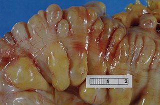
Stercoral ulcer is an ulcer of the colon due to pressure and irritation resulting from severe, prolonged constipation due to a large bowel obstruction, damage to the autonomic nervous system, or stercoral colitis. It is most commonly located in the sigmoid colon and rectum. Prolonged constipation leads to production of fecaliths, leading to possible progression into a fecaloma. These hard lumps irritate the rectum and lead to the formation of these ulcers. It results in fresh bleeding per rectum. These ulcers may be seen on imaging, such as a CT scan but are more commonly identified using endoscopy, usually a colonoscopy. Treatment modalities can include both surgical and non-surgical techniques.

Abdominal guarding is the tensing of the abdominal wall muscles to guard inflamed organs within the abdomen from the pain of pressure upon them. The tensing is detected when the abdominal wall is pressed. Abdominal guarding is also known as 'défense musculaire'.

<span class="mw-page-title-main">Abdominal trauma</span> Medical condition

Abdominal trauma is an injury to the abdomen. Signs and symptoms include abdominal pain, tenderness, rigidity, and bruising of the external abdomen. Complications may include blood loss and infection.

Tertiary peritonitis is the inflammation of the peritoneum which persists for 48 hours after a surgery that has been successfully carried out in adequate surgical conditions. Tertiary peritonitis is usually the most delayed and severe consequence of nosocomial intra-abdominal infection. Patients who acquire tertiary peritonitis are usually admitted to ICU due to the critical, life-threatening nature of the condition which can lead to multi-organ failure despite treatment and has a high mortality rate of 60%. Signs and symptoms of tertiary peritonitis include fever, hypotension and abdominal pain. Diagnosis of the condition is often difficult and treatment intervention should be as early as possible.

<span class="mw-page-title-main">Abdominal tuberculosis</span> Medical condition

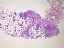
Abdominal tuberculosis is a type of extrapulmonary tuberculosis which involves the abdominal organs such as intestines, peritoneum and abdominal lymph nodes. It can either occur in isolation or along with a primary focus in patients with disseminated tuberculosis.

References

  1. 1 2 3 4 5 6 Ferri, Fred F. (2017). Ferri's Clinical Advisor 2018 E-Book: 5 Books in 1. Elsevier Health Sciences. pp. 979–980. ISBN   9780323529570. Archived from the original on 2020-10-08. Retrieved 2020-08-24.
  2. 1 2 3 "Peritonitis - National Library of Medicine". PubMed Health. Archived from the original on 2016-01-24. Retrieved 22 December 2017.
  3. 1 2 3 4 5 6 "Peritonitis". NHS. 28 September 2017. Archived from the original on 31 December 2017. Retrieved 31 December 2017.
  4. 1 2 3 4 5 6 7 8 "Acute Abdominal Pain". Merck Manuals Professional Edition. Archived from the original on 13 July 2018. Retrieved 31 December 2017.
  5. 1 2 "Acute Abdominal Pain". Merck Manuals Consumer Version. Archived from the original on 13 July 2018. Retrieved 31 December 2017.
  6. 1 2 "Encyclopaedia : Peritonitis". NHS Direct Wales. 25 April 2015. Archived from the original on 31 December 2017. Retrieved 31 December 2017.
  7. McGee, Steven R. (2018). "Abdominal Pain and Tenderness". Evidence-based physical diagnosis (4th ed.). Philadelphia, PA: Elsevier. ISBN   9780323508711. OCLC   959371826.
  8. "Biology Online's definition of peritonism". Archived from the original on 2018-06-12. Retrieved 2008-08-14.
  9. Okamoto, Koh; Hatakeyama, Shuji (2018-09-20). "Tuberculous Peritonitis". New England Journal of Medicine. 379 (12): e20. doi:10.1056/NEJMicm1713168. ISSN   0028-4793. PMID   30231225. S2CID   205088395.
  10. Ragetly, G. R.; Bennett, R. A.; Ragetly, C. A. (2012). "Therapie und Prognose der septischen Peritonitis". Tierärztliche Praxis Ausgabe K: Kleintiere / Heimtiere. 40 (5): 372–378. doi:10.1055/s-0038-1623666. ISSN   1434-1239. S2CID   73133175.
  11. "Peritonitis - Symptoms and causes". Mayo Clinic. Archived from the original on September 22, 2017. Retrieved July 2, 2016.
  12. Arfania D, Everett ED, Nolph KD, Rubin J (1981). "Uncommon causes of peritonitis in patients undergoing peritoneal dialysis". Archives of Internal Medicine. 141 (1): 61–64. doi:10.1001/archinte.141.1.61. PMID   7004371.
  13. Ljubin-Sternak, Suncanica; Mestrovic, Tomislav (2014). "Review: Chlamydia trachonmatis and Genital Mycoplasmias: Pathogens with an Impact on Human Reproductive Health". Journal of Pathogens. 2014 (183167): 183167. doi: 10.1155/2014/183167 . PMC   4295611 . PMID   25614838.
  14. Nishijima, D. K., Simel, D. L., Wisner, D. H., & Holmes, J. F. (2012). Does this adult patient have a blunt intra-abdominal injury?. JAMA, 307(14), 1517–1527. https://doi.org/10.1001/jama.2012.422
  15. Spalding, Drc; Williamson, Rcn (January 2008). "Peritonitis". British Journal of Hospital Medicine. 69 (Sup1): M12–M15. doi:10.12968/hmed.2008.69.Sup1.28050. ISSN   1750-8460. PMID   18293728.
  16. Ludlam, H A; Price, T N; Berry, A J; Phillips, I (September 1988). "Laboratory diagnosis of peritonitis in patients on continuous ambulatory peritoneal dialysis". Journal of Clinical Microbiology. 26 (9): 1757–1762. doi:10.1128/jcm.26.9.1757-1762.1988. ISSN   0095-1137. PMC   266711 . PMID   3183023.
  17. Arvind, Sharda; Raje, Shweta; Rao, Gayatri; Chawla, Latika (February 2019). "Laparoscopic Diagnosis of Peritoneal Tuberculosis". Journal of Minimally Invasive Gynecology. 26 (2): 346–347. doi: 10.1016/j.jmig.2018.04.006 . PMID   29680232. S2CID   5041460.
  18. "Peritoneal Dialysis". Brenner and Rector's The Kidney (11th ed.). Philadelphia, PA: Elsevier. 2020. pp. 2094–2118. ISBN   9780323759335.
  19. Holten, Keith B.; Onusko, Edward M. (August 1, 2000). "Appropriate Prescribing of Oral Beta-Lactam Antibiotics". American Family Physician . 62 (3): 611–620. PMID   10950216. Archived from the original on June 22, 2018. Retrieved July 22, 2019.
  20. Li, Philip Kam-Tao; Szeto, Cheuk Chun; Piraino, Beth; de Arteaga, Javier; Fan, Stanley; Figueiredo, Ana E.; Fish, Douglas N.; Goffin, Eric; Kim, Yong-Lim; Salzer, William; Struijk, Dirk G. (September 2016). "ISPD Peritonitis Recommendations: 2016 Update on Prevention and Treatment". Peritoneal Dialysis International. 36 (5): 481–508. doi:10.3747/pdi.2016.00078. ISSN   0896-8608. PMC   5033625 . PMID   27282851.
  21. "Peritonitis: Emergencies: Merck Manual Home Edition". Archived from the original on 2010-10-18. Retrieved 2007-11-25.
  22. "Peritonitis and Abdominal Sepsis: Background, Anatomy, Pathophysiology". 2021-07-14.{{cite journal}}: Cite journal requires |journal= (help)
  23. "peritonitis - Online Etymology Dictionary". Archived from the original on 2011-09-16. Retrieved 2017-05-09.