Cholecystitis

Last updated
Cholecystitis
AcuteCholMark.png
Acute cholecystitis as seen on CT. Note the fat stranding around the enlarged gallbladder.
Specialty General surgery, gastroenterology
Symptoms Intense right upper abdominal pain, nausea, vomiting, fever [1]
DurationShort term or long term [2]
CausesGallstones, severe illness [1] [3]
Risk factors Birth control pills, pregnancy, family history, obesity, diabetes, liver disease, rapid weight loss [4]
Diagnostic method Abdominal ultrasound [5]
Differential diagnosis Hepatitis, peptic ulcer disease, pancreatitis, pneumonia, angina [6]
Treatment Gallbladder removal surgery, gallbladder drainage [7] [5]
Prognosis Generally good with treatment [4]

Cholecystitis is inflammation of the gallbladder. [8] Symptoms include right upper abdominal pain, pain in the right shoulder, nausea, vomiting, and occasionally fever. [1] Often gallbladder attacks (biliary colic) precede acute cholecystitis. [1] The pain lasts longer in cholecystitis than in a typical gallbladder attack. [1] Without appropriate treatment, recurrent episodes of cholecystitis are common. [1] Complications of acute cholecystitis include gallstone pancreatitis, common bile duct stones, or inflammation of the common bile duct. [1] [8]

Contents

More than 90% of the time acute cholecystitis is caused from blockage of the cystic duct by a gallstone. [1] Risk factors for gallstones include birth control pills, pregnancy, a family history of gallstones, obesity, diabetes, liver disease, or rapid weight loss. [4] Occasionally, acute cholecystitis occurs as a result of vasculitis or chemotherapy, or during recovery from major trauma or burns. [9] Cholecystitis is suspected based on symptoms and laboratory testing. [5] Abdominal ultrasound is then typically used to confirm the diagnosis. [5]

Treatment is usually with laparoscopic gallbladder removal, within 24 hours if possible. [7] [10] Taking pictures of the bile ducts during the surgery is recommended. [7] The routine use of antibiotics is controversial. [5] [11] They are recommended if surgery cannot occur in a timely manner or if the case is complicated. [5] Stones in the common bile duct can be removed before surgery by endoscopic retrograde cholangiopancreatography (ERCP) or during surgery. [7] Complications from surgery are rare. [4] In people unable to have surgery, gallbladder drainage may be tried. [5]

About 10–15% of adults in the developed world have gallstones. [5] Women more commonly have stones than men and they occur more commonly after age 40. [4] Certain ethnic groups are more often affected; for example, 48% of American Indians have gallstones. [4] Of all people with stones, 1–4% have biliary colic each year. [5] If untreated, about 20% of people with biliary colic develop acute cholecystitis. [5] Once the gallbladder is removed outcomes are generally good. [4] Without treatment, chronic cholecystitis may occur. [2] The word is from Greek, cholecyst- meaning "gallbladder" and -itis meaning "inflammation". [12]

Signs and symptoms

Location of the gallbladder Blausen 0428 Gallbladder-Liver-Pancreas Location.png
Location of the gallbladder

Most people with gallstones do not have symptoms. [1] However, when a gallstone temporarily lodges in the cystic duct, they experience biliary colic. [1] Biliary colic is abdominal pain in the right upper quadrant or epigastric region. It is episodic, occurring after eating greasy or fatty foods, and leads to nausea and/or vomiting. [13] People with cholecystitis most commonly have symptoms of biliary colic before developing cholecystitis. The pain becomes severe and constant in cholecystitis. Nausea is common and vomiting occurs in 75% of people with cholecystitis. [14] In addition to abdominal pain, right shoulder pain can be present. [13]

On physical examination, an inflamed gallbladder is almost always tender to the touch and palpable (~25-50% of cases) in the midclavicular right lower rib margin. [13] Additionally, a fever is common. [14] A gallbladder with cholecystitis is almost always tender to touch. [13] Because of the inflammation, its size can be felt from the outside of the body in 25–50% of people with cholecystitis. [13] Pain with deep inspiration leading to termination of the breath while pressing on the right upper quadrant of the abdomen usually causes severe pain (Murphy's sign). [15] Yellowing of the skin (jaundice) may occur but is often mild. Severe jaundice suggests another cause of symptoms such as choledocholithiasis. [14] People who are old, have diabetes, chronic illness, or who are immunocompromised may have vague symptoms that may not include fever or localized tenderness. [16]

Complications

A number of complications may occur from cholecystitis if not detected early or properly treated. Signs of complications include high fever, shock and jaundice. Complications include the following: [13]

Gangrene and gallbladder rupture

Cholecystitis causes the gallbladder to become distended and firm. Distension can lead to decreased blood flow to the gallbladder, causing tissue death and eventually gangrene. [13] Once tissue has died, the gallbladder is at greatly increased risk of rupture (perforation), which can cause sharp pain. Rupture can also occur in cases of chronic cholecystitis. [13] Rupture is a rare but serious complication that leads to abscess formation or peritonitis. [14] Massive rupture of the gallbladder has a mortality rate of 30%. [13]

Empyema

Untreated cholecystitis can lead to worsened inflammation and infected bile that can lead to a collection of pus inside the gallbladder, also known as empyema. [13] The symptoms of empyema are similar to uncomplicated cholecystitis but greater severity: high fever, severe abdominal pain, more severely elevated white blood count. [13]

Fistula formation and gallstone ileus

The inflammation of cholecystitis can lead to adhesions between the gallbladder and other parts of the gastrointestinal tract, most commonly the duodenum. [13] These adhesions can lead to the formation of direct connections between the gallbladder and gastrointestinal tract, called fistulas. [13] With these direct connections, gallstones can pass from the gallbladder to the intestines. Gallstones can get trapped in the gastrointestinal tract, most commonly at the connection between the small and large intestines (ileocecal valve). When a gallstone gets trapped, it can lead to an intestinal obstruction, called gallstone ileus, leading to abdominal pain, vomiting, constipation, and abdominal distension. [13]

Causes

Cholecystitis occurs when the gallbladder becomes inflamed. [13] Gallstones are the most common cause of gallbladder inflammation but it can also occur due to blockage from a tumor or scarring of the bile duct. [13] [17] The greatest risk factor for cholecystitis is gallstones. [17] Risk factors for gallstones include female sex, increasing age, pregnancy, oral contraceptives, obesity, diabetes mellitus, ethnicity (Native North American), rapid weight loss. [13]

Acute calculous cholecystitis

Gallstones blocking the flow of bile account for 90% of cases of cholecystitis (acute calculous cholecystitis). [1] [14] Blockage of bile flow leads to thickening and buildup of bile causing an enlarged, red, and tense gallbladder. [1] The gallbladder is initially sterile but often becomes infected by bacteria, predominantly E. coli , Klebsiella , Streptococcus , and Clostridium species. [13] Inflammation can spread to the outer covering of the gallbladder and surrounding structures such as the diaphragm, causing referred right shoulder pain. [13]

Acalculous cholecystitis

In acalculous cholecystitis, no stone is in the biliary ducts. [13] It accounts for 5–10% of all cases of cholecystitis and is associated with high morbidity and mortality rates. [13] Acalculous cholecystitis is typically seen in people who are hospitalized and critically ill. [13] Males are more likely to develop acute cholecystitis following surgery in the absence of trauma. [14] [18] It is associated with many causes including vasculitis, chemotherapy, major trauma or burns. [9]

The presentation of acalculous cholecystitis is similar to calculous cholecystitis. [19] [13] Patients are more likely to have yellowing of the skin (jaundice) than in calculous cholecystitis. [20] Ultrasonography or computed tomography often shows an immobile, enlarged gallbladder. [13] Treatment involves immediate antibiotics and cholecystectomy within 24–72 hours. [20]

Chronic cholecystitis

Chronic cholecystitis occurs after repeated episodes of acute cholecystitis and is almost always due to gallstones. [13] Chronic cholecystitis may be asymptomatic, may present as a more severe case of acute cholecystitis, or may lead to a number of complications such as gangrene, perforation, or fistula formation. [13] [14]

Xanthogranulomatous cholecystitis (XGC) is a rare form of chronic cholecystitis which mimics gallbladder cancer although it is not cancerous. [21] [22] It was first reported in the medical literature in 1976 by McCoy and colleagues. [21] [23]

Mechanism

Blockage of the cystic duct by a gallstone causes a buildup of bile in the gallbladder and increased pressure within the gallbladder. Concentrated bile, pressure, and sometimes bacterial infection irritate and damage the gallbladder wall, causing inflammation and swelling of the gallbladder. [1] Inflammation and swelling of the gallbladder can reduce normal blood flow to areas of the gallbladder, which can lead to cell death due to inadequate oxygen. [13]

Diagnosis

The diagnosis of cholecystitis is suggested by the history (abdominal pain, nausea, vomiting, fever) and physical examinations in addition to laboratory and ultrasonographic testing. Boas's sign, which is pain in the area below the right scapula, can be a symptom of acute cholecystitis. [24]

Blood tests

In someone suspected of having cholecystitis, blood tests are performed for markers of inflammation (e.g. complete blood count, C-reactive protein), as well as bilirubin levels in order to assess for bile duct blockage. [14] Complete blood count typically shows an increased white blood count (12,000–15,000/mcL). [14] C-reactive protein is usually elevated although not commonly measured in the United States. [1] Bilirubin levels are often mildly elevated (1–4 mg/dL). [14] If bilirubin levels are more significantly elevated, alternate or additional diagnoses should be considered such as gallstone blocking the common bile duct (common bile duct stone). [1] Less commonly, blood aminotransferases are elevated. [13] The degree of elevation of these laboratory values may depend on the degree of inflammation of the gallbladder. [25]

Imaging

Right upper quadrant abdominal ultrasound is most commonly used to diagnose cholecystitis. [1] [26] [27] Ultrasound findings suggestive of acute cholecystitis include gallstones, pericholecystic fluid (fluid surrounding the gallbladder), gallbladder wall thickening (wall thickness over 3 mm), [28] dilation of the bile duct, and sonographic Murphy's sign. [13] Given its higher sensitivity, hepatic iminodiacetic acid (HIDA) scan can be used if ultrasound is not diagnostic. [13] [14] CT scan may also be used if complications such as perforation or gangrene are suspected. [14]

Histopathology

Histopathology is indicated if preoperative imaging and/or gross examination gives a suspicion of gallbladder cancer. [30]

Differential diagnosis

Many other diagnoses can have similar symptoms as cholecystitis. Additionally the symptoms of chronic cholecystitis are commonly vague and can be mistaken for other diseases. These alternative diagnoses include but are not limited to: [14]

Treatment

X-ray during laparoscopic cholecystectomy Laprascopy-Roentgen.jpg
X-ray during laparoscopic cholecystectomy

Surgery

For most people with acute cholecystitis, the treatment of choice is surgical removal of the gallbladder, laparoscopic cholecystectomy. [32] Laparoscopic cholecystectomy is performed using several small incisions located at various points across the abdomen. Several studies have demonstrated the superiority of laparoscopic cholecystectomy when compared to open cholecystectomy (using a large incision in the right upper abdomen under the rib cage). People undergoing laparoscopic surgery report less incisional pain postoperatively as well as having fewer long-term complications and less disability following the surgery. [33] [34] Additionally, laparoscopic surgery is associated with a lower rate of surgical site infection. [35]

During the days prior to laparoscopic surgery, studies showed that outcomes were better following early removal of the gallbladder, preferably within the first week. [36] Early laparoscopic cholecystectomy (within 7 days of visiting a doctor with symptoms) as compared to delayed treatment (more than 6 weeks) may result in shorter hospital stays and a decreased risk of requiring an emergency procedure. [37] There is no difference in terms of negative outcomes including bile duct injury or conversion to open cholecystectomy. [37] For early cholecystectomy, the most common reason for conversion to open surgery is inflammation that hides Calot's triangle. For delayed surgery, the most common reason was fibrotic adhesions. [37]

Other

Radiography of a percutaneous drainage catheter (yellow arrow). In this control, the instilled radiocontrast is filling out the gallbladder (red arrow), where the filling defects are gallstones. The cystic duct (blue arrow) is tortuous, the common bile duct (green arrow) is mildly dilated but patent, with tapering at ampulla Vateri (white arrow), but without obstruction. Contrast was seen extending into the duodenum (orange arrows), demonstrating open passage through the bile ducts. X-ray with contrast of a percutaneous gallbladder drain.jpg
Radiography of a percutaneous drainage catheter (yellow arrow). In this control, the instilled radiocontrast is filling out the gallbladder (red arrow), where the filling defects are gallstones. The cystic duct (blue arrow) is tortuous, the common bile duct (green arrow) is mildly dilated but patent, with tapering at ampulla Vateri (white arrow), but without obstruction. Contrast was seen extending into the duodenum (orange arrows), demonstrating open passage through the bile ducts.

Supportive measures may be instituted prior to surgery. These measures include fluid resuscitation. Intravenous opioids can be used for pain control. [39]

Antibiotics are often not needed. [40] If used they should target enteric organisms (e.g. Enterobacteriaceae), such as E. coli and Bacteroides . This may consist of a broad spectrum antibiotic; such as piperacillin-tazobactam, ampicillin-sulbactam, ticarcillin-clavulanate (Timentin), a third generation cephalosporin (e.g.ceftriaxone) or a quinolone antibiotic (such as ciprofloxacin) and anaerobic bacteria coverage, such as metronidazole. For penicillin allergic people, aztreonam or a quinolone with metronidazole may be used.[ citation needed ]

In cases of severe inflammation, shock, or if the person has higher risk for general anesthesia (required for cholecystectomy), an interventional radiologist may insert a percutaneous drainage catheter into the gallbladder (percutaneous cholecystostomy tube) and treat the person with antibiotics until the acute inflammation resolves. A cholecystectomy may then be warranted if the person's condition improves. [41]

Homeopathic approaches to treating cholecystitis have not been validated by evidence and should not be used in place of surgery. [42]

Epidemiology

Cholecystitis accounts for 3–10% of cases of abdominal pain worldwide. [43] Cholecystitis caused an estimated 651,829 emergency department visits and 389,180 hospital admissions in the US in 2012. [44] The 2012 US mortality rate was 0.7 per 100,000 people. [44] The frequency of cholecystitis is highest in people age 50–69 years old. [43]

Related Research Articles

<span class="mw-page-title-main">Gallbladder</span> Organ in humans and other vertebrates

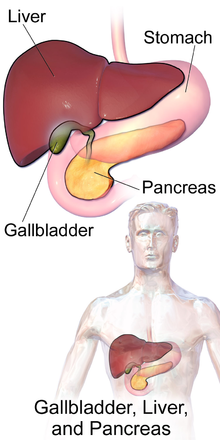
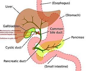
In vertebrates, the gallbladder, also known as the cholecyst, is a small hollow organ where bile is stored and concentrated before it is released into the small intestine. In humans, the pear-shaped gallbladder lies beneath the liver, although the structure and position of the gallbladder can vary significantly among animal species. It receives bile, produced by the liver, via the common hepatic duct, and stores it. The bile is then released via the common bile duct into the duodenum, where the bile helps in the digestion of fats.

<span class="mw-page-title-main">Gallstone</span> Disease where stones form in the gallbladder

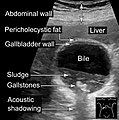
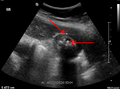
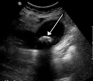
A gallstone is a stone formed within the gallbladder from precipitated bile components. The term cholelithiasis may refer to the presence of gallstones or to any disease caused by gallstones, and choledocholithiasis refers to the presence of migrated gallstones within bile ducts.

<span class="mw-page-title-main">Cholecystectomy</span> Surgical removal of the gallbladder

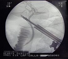
Cholecystectomy is the surgical removal of the gallbladder. Cholecystectomy is a common treatment of symptomatic gallstones and other gallbladder conditions. In 2011, cholecystectomy was the eighth most common operating room procedure performed in hospitals in the United States. Cholecystectomy can be performed either laparoscopically, or via an open surgical technique.

<span class="mw-page-title-main">Mirizzi's syndrome</span> Medical condition

Mirizzi's syndrome is a rare complication in which a gallstone becomes impacted in the cystic duct or neck of the gallbladder causing compression of the common hepatic duct, resulting in obstruction and jaundice. The obstructive jaundice can be caused by direct extrinsic compression by the stone or from fibrosis caused by chronic cholecystitis (inflammation). A cholecystocholedochal fistula can occur.

Courvoisier's principle states that a painless palpably enlarged gallbladder accompanied with mild jaundice is unlikely to be caused by gallstones. Usually, the term is used to describe the physical examination finding of the right-upper quadrant of the abdomen. This sign implicates possible malignancy of the gallbladder or pancreas and the swelling is unlikely due to gallstones.

<span class="mw-page-title-main">Gastrointestinal disease</span> Medical condition

Gastrointestinal diseases refer to diseases involving the gastrointestinal tract, namely the esophagus, stomach, small intestine, large intestine and rectum, and the accessory organs of digestion, the liver, gallbladder, and pancreas.

<span class="mw-page-title-main">Common bile duct stone</span> Medical condition

Common bile duct stone, also known as choledocholithiasis, is the presence of gallstones in the common bile duct (CBD). This condition can cause jaundice and liver cell damage. Treatments include choledocholithotomy and endoscopic retrograde cholangiopancreatography (ERCP).

<span class="mw-page-title-main">Gallbladder cancer</span> Medical condition

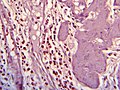
Gallbladder cancer is a relatively uncommon cancer, with an incidence of fewer than 2 cases per 100,000 people per year in the United States. It is particularly common in central and South America, central and eastern Europe, Japan and northern India; it is also common in certain ethnic groups e.g. Native American Indians and Hispanics. If it is diagnosed early enough, it can be cured by removing the gallbladder, part of the liver and associated lymph nodes. Most often it is found after symptoms such as abdominal pain, jaundice and vomiting occur, and it has spread to other organs such as the liver.

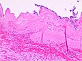
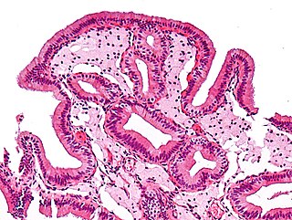
Adenomyoma is a tumor (-oma) including components derived from glands (adeno-) and muscle (-my-). It is a type of complex and mixed tumor, and several variants have been described in the medical literature. Uterine adenomyoma, the localized form of uterine adenomyosis, is a tumor composed of endometrial gland tissue and smooth muscle in the myometrium. Adenomyomas containing endometrial glands are also found outside of the uterus, most commonly on the uterine adnexa but can also develop at distant sites outside of the pelvis. Gallbladder adenomyoma, the localized form of adenomyomatosis, is a polypoid tumor in the gallbladder composed of hyperplastic mucosal epithelium and muscularis propria.

<span class="mw-page-title-main">Ascending cholangitis</span> Medical condition

Ascending cholangitis, also known as acute cholangitis or simply cholangitis, is inflammation of the bile duct, usually caused by bacteria ascending from its junction with the duodenum. It tends to occur if the bile duct is already partially obstructed by gallstones.

<span class="mw-page-title-main">Biliary colic</span> Medical condition in which gallstones cause acute pain

Biliary colic, also known as symptomatic cholelithiasis, a gallbladder attack or gallstone attack, is when a colic occurs due to a gallstone temporarily blocking the cystic duct. Typically, the pain is in the right upper part of the abdomen, and can be severe. Pain usually lasts from 15 minutes to a few hours. Often, it occurs after eating a heavy meal, or during the night. Repeated attacks are common. Cholecystokinin - a gastrointestinal hormone - plays a role in the colic, as following the consumption of fatty meals, the hormone triggers the gallbladder to contract, which may expel stones into the duct and temporarily block it until being successfully passed.

<span class="mw-page-title-main">Biliary tract</span> Organ system

The biliary tract refers to the liver, gallbladder and bile ducts, and how they work together to make, store and secrete bile. Bile consists of water, electrolytes, bile acids, cholesterol, phospholipids and conjugated bilirubin. Some components are synthesized by hepatocytes ; the rest are extracted from the blood by the liver.

<span class="mw-page-title-main">Gallbladder disease</span> Medical condition

Gallbladder diseases are diseases involving the gallbladder and is closely linked to biliary disease, with the most common cause being gallstones (cholelithiasis).

<span class="mw-page-title-main">Gallbladder polyp</span> Medical condition

Gallbladder polyps are growths or lesions resembling growths in the wall of the gallbladder. True polyps are abnormal accumulations of mucous membrane tissue that would normally be shed by the body.

Biliary dyskinesia is a disorder of some component of biliary part of the digestive system in which bile cannot physically move in the proper direction through the tubular biliary tract. It most commonly involves abnormal biliary tract peristalsis muscular coordination within the gallbladder in response to dietary stimulation of that organ to squirt the liquid bile through the common bile duct into the duodenum. Ineffective peristaltic contraction of that structure produces postprandial right upper abdominal pain (cholecystodynia) and almost no other problem. When the dyskinesia is localized at the biliary outlet into the duodenum just as increased tonus of that outlet sphincter of Oddi, the backed-up bile can cause pancreatic injury with abdominal pain more toward the upper left side. In general, biliary dyskinesia is the disturbance in the coordination of peristaltic contraction of the biliary ducts, and/or reduction in the speed of emptying of the biliary tree into the duodenum.

Biliary injury is the traumatic damage of the bile ducts. It is most commonly an iatrogenic complication of cholecystectomy, but can also be caused by other operations or by major trauma. The risk of biliary injury is higher during laparoscopic cholecystectomy than during open cholecystectomy. Biliary injury may lead to several complications and may even cause death if not diagnosed in time and managed properly. Ideally biliary injury should be managed at a center with facilities and expertise in endoscopy, radiology and surgery.

<span class="mw-page-title-main">Biliary sludge</span> Medical condition

Biliary sludge refers to a viscous mixture of small particles derived from bile. These sediments consist of cholesterol crystals, calcium salts, calcium bilirubinate, mucin, and other materials.

<span class="mw-page-title-main">Biloma</span> Circumscribed abdominal collection of bile outside the biliary tree

A biloma is a circumscribed abdominal collection of bile outside the biliary tree. It occurs when there is excess bile in the abdominal cavity. It can occur during or after a bile leak. There is an increased chance of a person developing biloma after having a gallbladder removal surgery, known as laparoscopic cholecystectomy. This procedure can be complicated by biloma with incidence of 0.3–2%. Other causes are liver biopsy, abdominal trauma, and, rarely, spontaneous perforation. The formation of biloma does not occur frequently. Biliary fistulas are also caused by injury to the bile duct and can result in the formation of bile leaks. Biliary fistulas are abnormal communications between organs and the biliary tract. Once diagnosed, they usually require drainage. The term "biloma" was first coined in 1979 by Gould and Patel. They discovered it in a case with extrahepatic bile leakage. The cause of this was trauma to the upper right quadrant of the abdomen. Originally, biloma was described as an "encapsulated collection" of extrahepatic bile. Biloma is now described as extrabiliary collections of bile that can be either intrahepatic or extrahepatic. The most common cause of biloma is trauma to the liver. There are other causes such as abdominal surgery, endoscopic surgery and percutaneous catheter drainage. Injury and abdominal trauma can cause damage to the biliary tree. The biliary tree is a system of vessels that direct secreations from the liver, gallbladder, and pancreas through a series of ducts into the duodenum. This can result in a bile leak which is a common cause of the formation of biloma. It is possible for biloma to be associated with mortality, though it is not common. Bile leaks occur in about one percent of causes.

Canine gallbladder mucocele (GBM) is an emerging biliary disease in dogs described as the excessive and abnormal accumulation of thick, gelatinous mucus in the lumen, which results in an enlarged gallbladder. GBMs have been diagnosed more frequently in comparison to prior to the 2000s when it was considered rare. The mucus is usually pale yellow to dark green in appearance.

<span class="mw-page-title-main">Choledochoduodenostomy</span>

Choledochoduodenostomy (CDD) is a surgical procedure to create an anastomosis, a surgical connection, between the common bile duct (CBD) and an alternative portion of the duodenum. In healthy individuals, the CBD meets the pancreatic duct at the ampulla of Vater, which drains via the major duodenal papilla to the second part of duodenum. In cases of benign conditions such as narrowing of the distal CBD or recurrent CBD stones, performing a CDD provides the diseased patient with CBD drainage and decompression. A side-to-side anastomosis is usually performed.

References

  1. 1 2 3 4 5 6 7 8 9 10 11 12 13 14 15 16 Strasberg, SM (26 June 2008). "Clinical practice. Acute calculous cholecystitis". The New England Journal of Medicine. 358 (26): 2804–11. doi:10.1056/nejmcp0800929. PMID   18579815.
  2. 1 2 Feldman, Mark (2010). Sleisenger & Fordtran's Gastrointestinal and liver disease pathophysiology, diagnosis, management (9 ed.). [S.l.]: MD Consult. p. 1065. ISBN   9781437727678. Archived from the original on 2017-09-08.
  3. Levy, Angela D.; Mortele, Koenraad J.; Yeh, Benjamin M. (2015). Gastrointestinal Imaging. Oxford University Press. p. 456. ISBN   9780199392148. Archived from the original on 2017-09-08.
  4. 1 2 3 4 5 6 7 "Gallstones". NIDDK. November 2013. Archived from the original on 28 July 2016. Retrieved 27 July 2016.
  5. 1 2 3 4 5 6 7 8 9 10 Ansaloni, L (2016). "2016 WSES guidelines on acute calculous cholecystitis". World Journal of Emergency Surgery. 11: 25. doi: 10.1186/s13017-016-0082-5 . PMC   4908702 . PMID   27307785.
  6. Ferri, Fred F. (2010). Ferri's differential diagnosis : a practical guide to the differential diagnosis of symptoms, signs, and clinical disorders (2nd ed.). Philadelphia, PA: Elsevier/Mosby. p. Chapter C. ISBN   978-0323076999.
  7. 1 2 3 4 Patel, PP; Daly, SC; Velasco, JM (18 October 2015). "Training vs practice: A tale of opposition in acute cholecystitis". World Journal of Hepatology. 7 (23): 2470–3. doi: 10.4254/wjh.v7.i23.2470 . PMC   4606202 . PMID   26483868.
  8. 1 2 Internal Clinical Guidelines Team (October 2014). "Gallstone Disease: Diagnosis and Management of Cholelithiasis, Cholecystitis and Choledocholithiasis. Clinical Guideline 188": 101. PMID   25473723.{{cite journal}}: Cite journal requires |journal= (help)
  9. 1 2 "Section VIII". Sleisenger and Fordtran's Gastrointestinal and Liver Disease: Pathophysiology, Diagnosis, Management (10 ed.). Elsevier Health Sciences. 2015. p. 1154. ISBN   9781455749898. Archived from the original on 2017-09-08.
  10. Schuld, J; Glanemann, M (June 2015). "Acute Cholecystitis". Viszeralmedizin. 31 (3): 163–5. doi:10.1159/000431275. PMC   4569253 . PMID   26468309.
  11. van Dijk, AH; de Reuver, PR; Tasma, TN; van Dieren, S; Hugh, TJ; Boermeester, MA (June 2016). "Systematic review of antibiotic treatment for acute calculous cholecystitis". The British Journal of Surgery. 103 (7): 797–811. doi:10.1002/bjs.10146. PMID   27027851. S2CID   205507793.
  12. Collins, Edwards (2013). A Short Course in Medical Terminology. Lippincott Williams & Wilkins. p. 246. ISBN   9781469835785. Archived from the original on 2017-09-08.
  13. 1 2 3 4 5 6 7 8 9 10 11 12 13 14 15 16 17 18 19 20 21 22 23 24 25 26 27 28 29 30 Greenberger N.J., Paumgartner G (2012). Chapter 311. Diseases of the Gallbladder and Bile Ducts. In Longo D.L., Fauci A.S., Kasper D.L., Hauser S.L., Jameson J, Loscalzo J (Eds), Harrison's Principles of Internal Medicine, 18e
  14. 1 2 3 4 5 6 7 8 9 10 11 12 13 Friedman L.S. (2015). Liver, Biliary Tract, & Pancreas Disorders. In Papadakis M.A., McPhee S.J., Rabow M.W. (Eds), Current Medical Diagnosis & Treatment 2015
  15. Singer, AJ; McCracken, G; Henry, MC; Thode HC, Jr; Cabahug, CJ (September 1996). "Correlation among clinical, laboratory, and hepatobiliary scanning findings in patients with suspected acute cholecystitis". Annals of Emergency Medicine. 28 (3): 267–72. doi:10.1016/s0196-0644(96)70024-0. PMID   8780468.
  16. Demehri, FR; Alam, HB (15 October 2014). "Evidence-Based Management of Common Gallstone-Related Emergencies". Journal of Intensive Care Medicine. 31 (1): 3–13. doi:10.1177/0885066614554192. PMID   25320159. S2CID   8792764.
  17. 1 2 "Cholecystitis". Mayo Clinic. 28 August 2014. Archived from the original on 25 November 2014. Retrieved 13 November 2014.
  18. Barie PS. Acalculous and postoperative cholecystitis. In: Surgical intensive care, Barie PS, Shires GT. (Eds), Little Brown & Co, Boston 1993. p.837.
  19. Savoca PE; Longo WE; Zucker KA; et al. (1990). "The increasing prevalence of acalculous cholecystitis in outpatients. Results of a 7-year study". Ann Surg. 211 (4): 433–7. doi:10.1097/00000658-199004000-00009. PMC   1358029 . PMID   2322038.
  20. 1 2 Butler, Kathryn; Harisinghani, Mukesh (2015). "Chapter 15: Acute Cholecystitis". Acute Care Surgery: Imaging Essentials for Rapid Diagnosis. McGraw-Hill Education. ISBN   978-0-07-183120-8.
  21. 1 2 Makino I, Yamaguchi T, Sato N, Yasui T, Kita I (August 2009). "Xanthogranulomatous cholecystitis mimicking gallbladder carcinoma with a false-positive result on fluorodeoxyglucose PET". World J. Gastroenterol. 15 (29): 3691–3. doi: 10.3748/wjg.15.3691 . PMC   2721248 . PMID   19653352.
  22. Rao RV, Kumar A, Sikora SS, Saxena R, Kapoor VK (2005). "Xanthogranulomatous cholecystitis: differentiation from associated gall bladder carcinoma". Trop Gastroenterol. 26 (1): 31–3. PMID   15974235.
  23. McCoy JJ, Vila R, Petrossian G, McCall RA, Reddy KS (March 1976). "Xanthogranulomatous cholecystitis. Report of two cases". J S C Med Assoc. 72 (3): 78–9. PMID   1063276.
  24. "Boas' sign" (web). GPnotebook. Archived from the original on 2007-02-07. Retrieved 2007-01-21.
  25. Vroon, D. H.; Israili, Z.; Walker, H. K.; Hall, W. D.; Hurst, J. W. (1990). Aminotransferases. Butterworths. ISBN   9780409900774. PMID   21250265 . Retrieved 27 June 2021 via National Center for Biotechnology Information, U.S. National Library of Medicine.
  26. Shea JA, Berlin JA, Escarce JJ, Clarke JR, Kinosian BP, Cabana MD, Tsai WW, Horangic N, Malet PF, Schwartz JS (November 1994). "Revised estimates of diagnostic test sensitivity and specificity in suspected biliary tract disease". Arch. Intern. Med. 154 (22): 2573–81. doi:10.1001/archinte.154.22.2573. PMID   7979854.
  27. Fink-Bennett D, Freitas JE, Ripley SD, Bree RL (August 1985). "The sensitivity of hepatobiliary imaging and real-time ultrasonography in the detection of acute cholecystitis". Arch Surg. 120 (8): 904–6. doi:10.1001/archsurg.1985.01390320028004. PMID   3893388.
  28. van Breda Vriesman, Adriaan C.; Engelbrecht, Marc R.; Smithuis, Robin H. M.; Puylaert, Julien B. C. M. (2007). "Diffuse Gallbladder Wall Thickening: Differential Diagnosis". American Journal of Roentgenology. 188 (2): 495–501. doi:10.2214/AJR.05.1712. ISSN   0361-803X. PMID   17242260.
  29. 1 2 "UOTW #30 - Ultrasound of the Week". Ultrasound of the Week. 23 December 2014. Archived from the original on 9 May 2017. Retrieved 27 May 2017.
  30. Talreja, Vikash; Ali, Aun; Khawaja, Rabel; Rani, Kiran; Samnani, Sunil Sadruddin; Farid, Farah Naz (2016). "Surgically Resected Gall Bladder: Is Histopathology Needed for All?". Surgery Research and Practice. 2016: 1–4. doi: 10.1155/2016/9319147 . ISSN   2356-7759. PMC   4829732 . PMID   27123469.
  31. Kuzu, MA; Oztürk, Y; Ozbek, H; Soran, A (October 1996). "Acalculous cholecystitis: ascariasis as an unusual cause". Journal of Gastroenterology. 31 (5): 747–9. doi:10.1007/bf02347629. PMID   8887047. S2CID   19385620.
  32. Strasberg SM (2008). "Acute Calculous Cholecystitis". New England Journal of Medicine. 358 (26): 2804–2811. doi:10.1056/NEJMcp0800929. PMID   18579815.
  33. Velanovich V (2000). "Laparoscopic vs open surgery: A preliminary comparison of quality-of-life outcomes". Surgical Endoscopy. 14 (1): 16–21. doi:10.1007/s004649900003. PMID   10653229. S2CID   26868091.
  34. Chen L, Tao SF, Xu Y, Fang F, Peng SY (2005). "Patients' quality of life after laparoscopic or open cholecystectomy". Journal of Zhejiang University Science. 6B (7): 678–681. doi:10.1631/jzus.2005.B0678. PMC   1389804 . PMID   15973772.
  35. Richards C, Edwards J, Culver D, Emori TG, Tolson J, Gaynes R (2003). "Does Using a Laparoscopic Approach to Cholecystectomy Decrease the Risk of Surgical Site Infection?". Annals of Surgery. 237 (3): 358–362. doi:10.1097/01.SLA.0000055221.50062.7A. PMC   1514308 . PMID   12616119.
  36. Banz V, Gsponer T, Candinas D, Güller U (2011). "Population-Based Analysis of 4113 Patients with Acute Cholecystitis". Annals of Surgery. 254 (6): 964–970. doi:10.1097/SLA.0b013e318228d31c. PMID   21817893. S2CID   875668.
  37. 1 2 3 Gurusamy, Kurinchi Selvan; Davidson, Christopher; Gluud, Christian; Davidson, Brian R. (2013-06-30). "Early versus delayed laparoscopic cholecystectomy for people with acute cholecystitis". The Cochrane Database of Systematic Reviews (6): CD005440. doi:10.1002/14651858.CD005440.pub3. ISSN   1469-493X. PMID   23813477.
  38. Padilla, Rebekah M; Hulsberg, Paul C; Soule, Erik; Harmon, Taylor S; Eadie, Erik; Hood, Preston; Shabandi, Michael; Matteo, Jerry (2018). "Against the Odds: A Novel Technique to Perform Cholangiography from a Percutaneous Approach through the Cystic Duct". Cureus. 10 (11): e3577. doi: 10.7759/cureus.3577 . ISSN   2168-8184. PMC   6333255 . PMID   30656081.
  39. "Cholecystitis". The Lecturio Medical Concept Library. Retrieved 8 July 2021.
  40. van Dijk, AH; de Reuver, PR; Tasma, TN; van Dieren, S; Hugh, TJ; Boermeester, MA (30 March 2016). "Systematic review of antibiotic treatment for acute calculous cholecystitis". The British Journal of Surgery. 103 (7): 797–811. doi:10.1002/bjs.10146. PMID   27027851. S2CID   205507793.
  41. "Acute Cholangitis". The Lecturio Medical Concept Library. Retrieved 27 June 2021.
  42. Tuomela, R (1987). "Science, Protoscience, and Pseudoscience". In Pitt JC, Marcello P (eds.). Rational Changes in Science. Boston Studies in the Philosophy of Science. Vol. 98. Springer. pp. 83–101. doi:10.1007/978-94-009-3779-6_4. ISBN   978-94-010-8181-8.
  43. 1 2 Kimura, Yasutoshi; Takada, Tadahiro; Kawarada, Yoshifumi; Nimura, Yuji; Hirata, Koichi; Sekimoto, Miho; Yoshida, Masahiro; Mayumi, Toshihiko; Wada, Keita (2016-12-12). "Definitions, pathophysiology, and epidemiology of acute cholangitis and cholecystitis: Tokyo Guidelines". Journal of Hepato-Biliary-Pancreatic Surgery. 14 (1): 15–26. doi:10.1007/s00534-006-1152-y. ISSN   0944-1166. PMC   2784509 . PMID   17252293.
  44. 1 2 Peery, Anne F.; Crockett, Seth D.; Barritt, Alfred S.; Dellon, Evan S.; Eluri, Swathi; Gangarosa, Lisa M.; Jensen, Elizabeth T.; Lund, Jennifer L.; Pasricha, Sarina (2015). "Burden of Gastrointestinal, Liver, and Pancreatic Diseases in the United States". Gastroenterology. 149 (7): 1731–1741.e3. doi:10.1053/j.gastro.2015.08.045. PMC   4663148 . PMID   26327134.