Biliary colic

Last updated
Biliary colic
Other namesGallstone attack, gallbladder attack
Gallstones.PNG
Biliary colic is often related to a stone in the gallbladder
Specialty Gastroenterology   OOjs UI icon edit-ltr-progressive.svg

Biliary colic, also known as symptomatic cholelithiasis, a gallbladder attack or gallstone attack, is when a colic (sudden pain) occurs due to a gallstone temporarily blocking the cystic duct. [1] Typically, the pain is in the right upper part of the abdomen, and can be severe. [2] Pain usually lasts from 15 minutes to a few hours. [1] Often, it occurs after eating a heavy meal, or during the night. [1] Repeated attacks are common. [3] Cholecystokinin - a gastrointestinal hormone - plays a role in the colic, as following the consumption of fatty meals, the hormone triggers the gallbladder to contract, which may expel stones into the duct and temporarily block it until being successfully passed. [4]

Contents

Gallstone formation occurs from the precipitation of crystals that aggregate to form stones. The most common form is cholesterol gallstones. [5] Other forms include calcium, bilirubin, pigment, and mixed gallstones. [5] Other conditions that produce similar symptoms include appendicitis, stomach ulcers, pancreatitis, and gastroesophageal reflux disease. [1]

Treatment for gallbladder attacks is typically surgery to remove the gallbladder. [1] This can be either done through small incisions or through a single larger incision. [1] Open surgery through a larger incision is associated with more complications than surgery through small incisions. [6] Surgery is typically done under general anesthesia. [1] In those who are unable to have surgery, medication to try to dissolve the stones or shock wave lithotripsy may be tried. [1] As of 2017, it is not clear whether surgery is indicated for everyone with biliary colic. [6]

In the developed world, 10 to 15% of adults have gallstones. [3] Of those with gallstones, biliary colic occurs in 1 to 4% each year. [3] Nearly 30% of people have further problems related to gallstones in the year following an attack. [3] About 15% of people with biliary colic eventually develop inflammation of the gallbladder if not treated. [3] Other complications include inflammation of the pancreas. [3]

Signs and symptoms

Pain is the most common presenting symptom. It is usually described as sharp, crampy, dull or severe right upper quadrant pain, which may radiate to the right shoulder, or less commonly, behind the breastbone. [7] Nausea and vomiting can be associated with biliary colic. Individuals may also present with pain that is induced following a fatty meal and the symptom of indigestion. The pain often lasts longer than 30 minutes, up to a few hours. [7] The pain caused by biliary colic can become so extreme that sufferers may admit themselves to emergency rooms and hospitals to seek treatment. In general, the pain subsides once the gallstone is successfully passed, [8] but soreness may persist for around 24 hours after the worst of the pain passes. [9]

Biliary colic can be distinguished from other digestive conditions with similar symptoms, such as indigestion, gastric reflux or heartburn, in that the pain caused by biliary colic is not relieved by vomiting, bowel movements or flatulence. [10] The pain is also not affected by changes in posture or antacid medicine. [11] Episodes of biliary colic are generally intermittent, and sufferers may experience several weeks or months without an attack before experiencing it again. [10]

Patients usually have normal vital signs with biliary colic, whereas patients with cholecystitis are usually febrile and more ill appearing. Lab studies that should be ordered include a complete blood count, liver function tests and lipase. In biliary colic, lab findings are usually within normal limits. Alanine aminotransferase and aspartate transaminase are usually suggestive of liver disease whereas elevation of bilirubin and alkaline phosphatase suggests common bile duct obstruction. [12] Pancreatitis should be considered if the lipase value is elevated; gallstone disease is the major cause of pancreatitis.[ citation needed ]

Complications

The presence of gallstones can lead to inflammation of the gallbladder (cholecystitis) or the biliary tree (cholangitis) or acute inflammation of the pancreas (pancreatitis). Rarely, a gallstone can become impacted in the ileocecal valve that joins the caecum and the ileum, causing gallstone ileus (mechanical ileus). [7]

Complications from delayed surgery include pancreatitis, empyema, and perforation of the gallbladder, cholecystitis, cholangitis, and obstructive jaundice. [13]

Biliary pain in the absence of gallstones, known as postcholecystectomy syndrome, may severely affect the patient's quality of life, even in the absence of disease progression. [14]

Causes

Biliary pain is most frequently caused by obstruction of the common bile duct or the cystic duct by a gallstone. However, the presence of gallstones is a frequent incidental finding and does not always necessitate treatment, in the absence of identifiable disease. Furthermore, biliary pain may be associated with functional disorders of the biliary tract, so-called acalculous biliary pain (pain without stones), and can even be found in patients post-cholecystectomy (removal of the gallbladder), possibly as a consequence of dysfunction of the biliary tree and the sphincter of Oddi. Acute episodes of biliary pain may be induced or exacerbated by certain foods, most commonly those high in fat. [15]

Risk factors

Cholesterol gallstone formation risk factors include age, female sex, family history, race, [7] [16] pregnancy, parity, obesity, hormonal birth control, diabetes mellitus, cirrhosis, prolonged fasting, rapid weight loss, total parenteral nutrition, ileal disease and impaired gallbladder emptying. [17]

Patients that have gallstones and biliary colic are at increased risk for complications, including cholecystitis. [18] Complications from gallstone disease is 0.3% per year and therefore prophylactic cholecystectomy are rarely indicated unless part of a special population that includes porcelain gallbladder, individuals eligible for organ transplant, diabetics and those with sickle cell anemia. [7]

Diagnosis

Diagnosis is guided by the person's presenting symptoms and laboratory findings. The gold standard imaging modality for the presence of gallstones is ultrasound of the right upper quadrant. There are many reasons for this choice, including no exposure to radiation, low cost, and availability in city, urban, and rural hospitals. Gallstones are detected with a specificity and sensitivity of greater than 95% with ultrasound. [19] [ page needed ] Further signs on ultrasound may suggest cholecystitis or choledocholithiasis. [13] Computed Tomography (CT) is not indicated when investigating for gallbladder disease as 60% of stones are not radiopaque. [13] CT should only be utilized if other intra-abdominal pathology exists or the diagnosis is uncertain. [20] Endoscopic retrograde cholangiopancreatography (ERCP) should be used only if lab tests suggest the existence of a gallstone in the bile duct. [13] ERCP is then both diagnostic and therapeutic.[ citation needed ]

Management

Medications

Initial management includes the relief of symptoms and correcting electrolyte and fluid imbalance that may occur with vomiting. [12] Antiemetics, such as dimenhydrinate, are used to treat the nausea. [12] Pain may be treated with anti-inflammatories, NSAIDs such as ketorolac or diclofenac. [21] Opioids, such as morphine, less commonly may be used. [22] NSAIDs are more or less equivalent to opioids. [23] Hyoscine butylbromide, an antispasmodic, is also indicated in biliary colic. [24]

In biliary colic, the risk of infection is minimal and therefore antibiotics are not required. [25] Presence of infection indicates cholecystitis. [25]

Surgery

It is unclear whether those experiencing a gallstone attack should receive surgical treatment or not. [6] The scientific basis to assess whether surgery outperformed other treatment was insufficient and better studies were needed as of a SBU report in 2017. [6] Treatment of biliary colic is dictated by the underlying cause. [26] The presence of gallstones, usually visualized by ultrasound, generally necessitates a surgical treatment (removal of the gall bladder, typically via laparoscopy). [26] Removal of the gallbladder with surgery, known as a cholecystectomy, is the definitive surgical treatment for biliary colic. [27] A 2013 Cochrane review found tentative evidence to suggest that early gallbladder removal may be better than delayed removal. [28] Early laparoscopic cholecystectomy happens within 72 hours of diagnosis. [13] In a Cochrane review that evaluated receiving early versus delayed surgery, they found that 23% of those who waited on average 4 months ended up in hospital for complications, compared to none with early intervention with surgery. [13] [28] Early intervention has other advantages including a reduced number of visits to the emergency department, fewer conversions to open surgery, less operating time required, and reduced time in hospital postoperatively. [13] The Swedish agency SBU estimated in 2017 that increasing acute phase surgeries could free multiple in-hospital days per patient and would additionally spare pain and suffering in wait of receiving an operation. [6] The report found that those with acute inflammation of the gallbladder can be surgically treated in the acute phase, within a few days of symptom debut, without increasing the risk for complications (compared to when the surgery is done later in an asymptomatic stage). [6]

Epidemiology

The annual risk of developing biliary colic is 2 to 3%. [7] [18]

Related Research Articles

<span class="mw-page-title-main">Pancreatitis</span> Inflammation of the pancreas

Pancreatitis is a condition characterized by inflammation of the pancreas. The pancreas is a large organ behind the stomach that produces digestive enzymes and a number of hormones. There are two main types: acute pancreatitis, and chronic pancreatitis.

<span class="mw-page-title-main">Gallbladder</span> Organ in humans and other vertebrates

In vertebrates, the gallbladder, also known as the cholecyst, is a small hollow organ where bile is stored and concentrated before it is released into the small intestine. In humans, the pear-shaped gallbladder lies beneath the liver, although the structure and position of the gallbladder can vary significantly among animal species. It receives bile, produced by the liver, via the common hepatic duct, and stores it. The bile is then released via the common bile duct into the duodenum, where the bile helps in the digestion of fats.

<span class="mw-page-title-main">Gallstone</span> Disease where stones form in the gallbladder

A gallstone is a stone formed within the gallbladder from precipitated bile components. The term cholelithiasis may refer to the presence of gallstones or to any disease caused by gallstones, and choledocholithiasis refers to the presence of migrated gallstones within bile ducts.

<span class="mw-page-title-main">Cholecystitis</span> Inflammation of the gallbladder

Cholecystitis is inflammation of the gallbladder. Symptoms include right upper abdominal pain, pain in the right shoulder, nausea, vomiting, and occasionally fever. Often gallbladder attacks precede acute cholecystitis. The pain lasts longer in cholecystitis than in a typical gallbladder attack. Without appropriate treatment, recurrent episodes of cholecystitis are common. Complications of acute cholecystitis include gallstone pancreatitis, common bile duct stones, or inflammation of the common bile duct.

<span class="mw-page-title-main">Cholecystectomy</span> Surgical removal of the gallbladder

Cholecystectomy is the surgical removal of the gallbladder. Cholecystectomy is a common treatment of symptomatic gallstones and other gallbladder conditions. In 2011, cholecystectomy was the eighth most common operating room procedure performed in hospitals in the United States. Cholecystectomy can be performed either laparoscopically, or via an open surgical technique.

<span class="mw-page-title-main">Mirizzi's syndrome</span> Medical condition

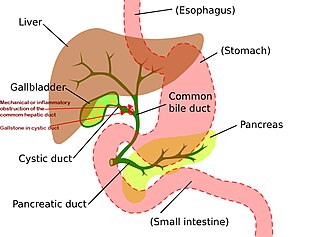
Mirizzi's syndrome is a rare complication in which a gallstone becomes impacted in the cystic duct or neck of the gallbladder causing compression of the common hepatic duct, resulting in obstruction and jaundice. The obstructive jaundice can be caused by direct extrinsic compression by the stone or from fibrosis caused by chronic cholecystitis (inflammation). A cholecystocholedochal fistula can occur.

Courvoisier's principle states that a painless palpably enlarged gallbladder accompanied with mild jaundice is unlikely to be caused by gallstones. Usually, the term is used to describe the physical examination finding of the right-upper quadrant of the abdomen. This sign implicates possible malignancy of the gallbladder or pancreas and the swelling is unlikely due to gallstones.

<span class="mw-page-title-main">Common bile duct stone</span> Medical condition

Common bile duct stone, also known as choledocholithiasis, is the presence of gallstones in the common bile duct (CBD). This condition can cause jaundice and liver cell damage. Treatments include choledocholithotomy and endoscopic retrograde cholangiopancreatography (ERCP).

<span class="mw-page-title-main">Gallbladder cancer</span> Medical condition

Gallbladder cancer is a relatively uncommon cancer, with an incidence of fewer than 2 cases per 100,000 people per year in the United States. It is particularly common in central and South America, central and eastern Europe, Japan and northern India; it is also common in certain ethnic groups e.g. Native American Indians and Hispanics. If it is diagnosed early enough, it can be cured by removing the gallbladder, part of the liver and associated lymph nodes. Most often it is found after symptoms such as abdominal pain, jaundice and vomiting occur, and it has spread to other organs such as the liver.

<span class="mw-page-title-main">Porcelain gallbladder</span> Medical condition

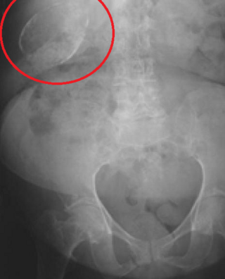
Porcelain gallbladder is a calcification of the gallbladder believed to be brought on by excessive gallstones, although the exact cause is not clear. As with gallstone disease in general, this condition occurs mostly in overweight female patients of middle age. It is a morphological variant of chronic cholecystitis. Inflammatory scarring of the wall, combined with dystrophic calcification within the wall transforms the gallbladder into a porcelain-like vessel. Removal of the gallbladder (cholecystectomy) is the recommended treatment.

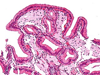
Adenomyoma is a tumor (-oma) including components derived from glands (adeno-) and muscle (-my-). It is a type of complex and mixed tumor, and several variants have been described in the medical literature. Uterine adenomyoma, the localized form of uterine adenomyosis, is a tumor composed of endometrial gland tissue and smooth muscle in the myometrium. Adenomyomas containing endometrial glands are also found outside of the uterus, most commonly on the uterine adnexa but can also develop at distant sites outside of the pelvis. Gallbladder adenomyoma, the localized form of adenomyomatosis, is a polypoid tumor in the gallbladder composed of hyperplastic mucosal epithelium and muscularis propria.

<span class="mw-page-title-main">Ascending cholangitis</span> Medical condition

Ascending cholangitis, also known as acute cholangitis or simply cholangitis, is inflammation of the bile duct, usually caused by bacteria ascending from its junction with the duodenum. It tends to occur if the bile duct is already partially obstructed by gallstones.

<span class="mw-page-title-main">Biliary tract</span> Organ system

The biliary tract refers to the liver, gallbladder and bile ducts, and how they work together to make, store and secrete bile. Bile consists of water, electrolytes, bile acids, cholesterol, phospholipids and conjugated bilirubin. Some components are synthesized by hepatocytes ; the rest are extracted from the blood by the liver.

<span class="mw-page-title-main">Gallbladder disease</span> Medical condition

Gallbladder diseases are diseases involving the gallbladder and is closely linked to biliary disease, with the most common cause being gallstones (cholelithiasis).

<span class="mw-page-title-main">Cholescintigraphy</span> Medical imaging of hepatobiliary tract using radiotracers

Cholescintigraphy or hepatobiliary scintigraphy is scintigraphy of the hepatobiliary tract, including the gallbladder and bile ducts. The image produced by this type of medical imaging, called a cholescintigram, is also known by other names depending on which radiotracer is used, such as HIDA scan, PIPIDA scan, DISIDA scan, or BrIDA scan. Cholescintigraphic scanning is a nuclear medicine procedure to evaluate the health and function of the gallbladder and biliary system. A radioactive tracer is injected through any accessible vein and then allowed to circulate to the liver, where it is excreted into the bile ducts and stored by the gallbladder until released into the duodenum.

Biliary injury is the traumatic damage of the bile ducts. It is most commonly an iatrogenic complication of cholecystectomy, but can also be caused by other operations or by major trauma. The risk of biliary injury is higher during laparoscopic cholecystectomy than during open cholecystectomy. Biliary injury may lead to several complications and may even cause death if not diagnosed in time and managed properly. Ideally biliary injury should be managed at a center with facilities and expertise in endoscopy, radiology and surgery.

<span class="mw-page-title-main">Sphincter of Oddi dysfunction</span> Medical condition

Sphincter of Oddi dysfunction refers to a group of functional disorders leading to abdominal pain due to dysfunction of the Sphincter of Oddi: functional biliary sphincter of Oddi and functional pancreatic sphincter of Oddi disorder. The sphincter of Oddi is a sphincter muscle, a circular band of muscle at the bottom of the biliary tree which controls the flow of pancreatic juices and bile into the second part of the duodenum. The pathogenesis of this condition is recognized to encompass stenosis or dyskinesia of the sphincter of Oddi ; consequently the terms biliary dyskinesia, papillary stenosis, and postcholecystectomy syndrome have all been used to describe this condition. Both stenosis and dyskinesia can obstruct flow through the sphincter of Oddi and can therefore cause retention of bile in the biliary tree and pancreatic juice in the pancreatic duct.

Cholecystostomy or (cholecystotomy) is a medical procedure used to drain the gallbladder through either a percutaneous or endoscopic approach. The procedure involves creating a stoma in the gallbladder, which can facilitate placement of a tube or stent for drainage, first performed by American surgeon, Dr. John Stough Bobbs, in 1867. It is sometimes used in cases of cholecystitis or other gallbladder disease where the person is ill, and there is a need to delay or defer cholecystectomy. The first endoscopic cholecystostomy was performed by Drs. Todd Baron and Mark Topazian in 2007 using ultrasound guidance to puncture the stomach wall and place a plastic biliary catheter for gallbladder drainage.

<span class="mw-page-title-main">Biliary sludge</span> Medical condition

Biliary sludge refers to a viscous mixture of small particles derived from bile. These sediments consist of cholesterol crystals, calcium salts, calcium bilirubinate, mucin, and other materials.

<span class="mw-page-title-main">Choledochoduodenostomy</span>

Choledochoduodenostomy (CDD) is a surgical procedure to create an anastomosis, a surgical connection, between the common bile duct (CBD) and an alternative portion of the duodenum. In healthy individuals, the CBD meets the pancreatic duct at the ampulla of Vater, which drains via the major duodenal papilla to the second part of duodenum. In cases of benign conditions such as narrowing of the distal CBD or recurrent CBD stones, performing a CDD provides the diseased patient with CBD drainage and decompression. A side-to-side anastomosis is usually performed.

References

  1. 1 2 3 4 5 6 7 8 "Gallstones". NIDDK.NIH.gov. Washington DC: National Institute of Diabetes and Digestive and Kidney Diseases. November 2013. Archived from the original on 16 August 2016. Retrieved 27 July 2016.
  2. Internal Clinical Guidelines Team (October 2014). "Gallstone Disease: Diagnosis and Management of Cholelithiasis, Cholecystitis and Choledocholithiasis". NICE.org: 21. PMID   25473723. Clinical Guideline 188. Retrieved 24 June 2018.
  3. 1 2 3 4 5 6 Ansaloni, L. (2016). "2016 WSES guidelines on acute calculous cholecystitis". World Journal of Emergency Surgery. 11: 25. doi: 10.1186/s13017-016-0082-5 . PMC   4908702 . PMID   27307785.
  4. Sigmon, David F.; Dayal, Nalin; Meseeha, Marcelle (2023), "Biliary Colic", StatPearls, Treasure Island (FL): StatPearls Publishing, PMID   28613523 , retrieved 2023-11-18
  5. 1 2 Sabiston, David C.; Townsend, Courtney M. (2012). Sabiston Textbook of Surgery: The Biological Basis of Modern Surgical Practice. Philadelphia: Elsevier/Saunders. pp. 328–358. ISBN   978-1-4377-1560-6.
  6. 1 2 3 4 5 6 "Surgery to treat gallstones and acute inflammation of the gallbladder". SBU.se. Swedish Agency for Health Technology Assessment and Assessment of Social Services (SBU). 2016-12-16. Retrieved 2017-06-01.
  7. 1 2 3 4 5 6 Portincasa, P.; Moschetta, A.; Petruzzelli, M.; Palasciano, G.; Di Ciaula, A.; Pezzolla, A. (2006). "Gallstone disease: Symptoms and diagnosis of gallbladder stones". Best Practice & Research: Clinical Gastroenterology. 20 (6): 1017–1029. doi:10.1016/j.bpg.2006.05.005. PMID   17127185.
  8. "What Causes a Gallbladder Attack?". www.hopkinsmedicine.org. 2023-11-30. Retrieved 2024-01-08.
  9. "Biliary Colic". Harvard Health. 2019-04-22. Retrieved 2024-03-09.
  10. 1 2 "Gallstones". National Health Service . 2017-10-20. Retrieved 2023-12-11.
  11. "Biliary Dyskinesia: Symptoms, Causes & Treatment". Cleveland Clinic . Retrieved 2023-12-12.
  12. 1 2 3 Rosen, Peter; Marx, John A. (2013). Rosen's Emergency Medicine: Concepts and Clinical Practice. Philadelphia: Elsevier/Saunders. pp. 1186–1206. ISBN   978-1-4557-0605-1.
  13. 1 2 3 4 5 6 7 Duncan, C. B.; Riall, T. S. (Nov 2012). "Evidence-based current surgical practice: calculous gallbladder disease". Journal of Gastrointestinal Surgery. 16 (11): 2011–2025. doi:10.1007/s11605-012-2024-1. PMC   3496004 . PMID   22986769.
  14. Postcholecystectomy Syndrome at eMedicine
  15. Rodriguez, Diana (4 January 2023). "When Gallbladder Problems Lead to Biliary Colic". Everyday Health.
  16. Stinton, Laura M.; Shaffer, Eldon A. (15 April 2012). "Epidemiology of Gallbladder Disease: Cholelithiasis and Cancer". Gut and Liver. 6 (2): 172–187. doi:10.5009/gnl.2012.6.2.172. PMC   3343155 . PMID   22570746.
  17. Walton, Thomas J.; Lobo, Dileep N. (2009). "Gallstones". Surgery. 27 (1): 19–24. doi:10.1016/j.mpsur.2008.12.001.
  18. 1 2 Afdhal, Nezam H. (2011). Goldman's Cecil Medicine (24th ed.). Philadelphia: Elsevier/Saunders. pp. 1011–1020. ISBN   978-1-4377-1604-7.
  19. Fischer, J. E., ed. (2007). Master of Surgery (5th ed.). Philadelphia: Lippincott Williams & Wilkins.
  20. Shakespear, J. S.; Shaaban, A. M.; Rezvani, M. (2010). "CT findings of acute cholecystitis and its complications". American Journal of Roentgenology. 194 (6): 1523–1529. doi:10.2214/ajr.09.3640. PMID   20489092.
  21. Fraquelli, M.; Casazza, G.; Conte, D.; Colli, A. (9 September 2016). "Non-steroid anti-inflammatory drugs for biliary colic". The Cochrane Database of Systematic Reviews. 2016 (9): CD006390. doi:10.1002/14651858.CD006390.pub2. PMC   6457716 . PMID   27610712.
  22. Rosen, Peter; Marx, John A. (2013). Rosen's Emergency Medicine: Concepts and Clinical Practice. Philadelphia: Elsevier/Saunders. pp. 223–233. ISBN   978-1-4557-0605-1.
  23. Colli, A.; Conte, D.; Valle, S. D.; Sciola, V.; Fraquelli, M. (June 2012). "Meta-analysis: nonsteroidal anti-inflammatory drugs in biliary colic". Alimentary Pharmacology & Therapeutics. 35 (12): 1370–1378. doi: 10.1111/j.1365-2036.2012.05115.x . PMID   22540869.
  24. "Hyoscine butylbromide (Buscopan) injection: Risk of serious adverse effects in patients with underlying cardiac disease". Gov.uk. Retrieved 23 September 2017.
  25. 1 2 Cecil, Russell L. (Russell La Fayette); Goldman, Lee; Schafer, Andrew I. (2012). Goldman's Cecil Medicine. Philadelphia: Elsevier/Saunders. pp. 1011–1021. ISBN   978-1-4377-1604-7.
  26. 1 2 Sigmon S, Dayal N, Meseeha M (2021). "Biliary Colic". National Center for Biotechnology Information, U.S. National Library of Medicine. PMID   28613523 . Retrieved 8 July 2021.
  27. "Cholecystectomy: Approaches and Technique". The Lecturio Medical Concept Library. Retrieved 8 July 2021.
  28. 1 2 Gurusamy, K. S.; Koti, R.; Fusai, G.; Davidson, B. R. (2013). "Early versus delayed laparoscopic cholecystectomy for uncomplicated biliary colic". Cochrane Database Syst Rev. 6 (6): CD007196. doi:10.1002/14651858.CD007196.pub3. PMID   23813478.