Esophagitis

Last updated
Esophagitis
Other namesOesophagitis
Esophageal ulcer.jpg
An esophageal ulcer visualized by esophagoscopy: the reddened area at 10 o'clock on the surface of the mucosa.
Specialty Gastroenterology   OOjs UI icon edit-ltr-progressive.svg
Complications Cancer

Esophagitis, also spelled oesophagitis, is a disease characterized by inflammation of the esophagus. The esophagus is a tube composed of a mucosal lining, and longitudinal and circular smooth muscle fibers. It connects the pharynx to the stomach; swallowed food and liquids normally pass through it. [1]

Contents

Esophagitis can be asymptomatic; or can cause epigastric and/or substernal burning pain, especially when lying down or straining; and can make swallowing difficult (dysphagia). The most common cause of esophagitis is the reverse flow of acid from the stomach into the lower esophagus: gastroesophageal reflux disease (GERD). [2]

Signs and symptoms

The symptoms of esophagitis include: [2]

Complications

If the disease remains untreated, it can cause scarring and discomfort in the esophagus. If the irritation is not allowed to heal, esophagitis can result in esophageal ulcers. Esophagitis can develop into Barrett's esophagus and can increase the risk of esophageal cancer. [3]

Causes

Infectious esophagitis cannot be spread. However, infections can be spread by those who have infectious esophagitis. Esophagitis can develop due to many causes. GERD is the most common cause of esophagitis because of the backflow of acid from the stomach, which can irritate the lining of the esophagus.

Other causes include:

Mechanism

The esophagus is a muscular tube made of both voluntary and involuntary muscles. It is responsible for peristalsis of food. It is about 8 inches long and passes through the diaphragm before entering the stomach. The esophagus is made up of three layers: from the inside out, they are the mucosa, submucosa, muscularis externa. The mucosa, the inner most layer and lining of the esophagus, is composed of stratified squamous epithelium, lamina propria, and muscularis mucosae. At the end of the esophagus is the lower esophageal sphincter, which normally prevents stomach acid from entering the esophagus.

If the sphincter is not sufficiently tight, it may allow acid to enter the esophagus, causing inflammation of one or more layers. Esophagitis may also occur if an infection is present, which may be due to bacteria, viruses, or fungi; or by diseases that affect the immune system. [4]

Irritation can be caused by GERD, vomiting, surgery, medications, hernias, and radiation injury. [4] Inflammation can cause the esophagus to narrow, which makes swallowing food difficult and may result in food bolus impaction.

Diagnosis

Esophagitis can be diagnosed by upper endoscopy, biopsy, upper GI series (or barium swallow), and laboratory tests. [4]

An upper endoscopy is a procedure to look at the esophagus by using an endoscope. While looking at the esophagus, the doctor is able to take a small biopsy. The biopsy can be used to confirm inflammation of the esophagus.

An upper GI series uses a barium contrast, fluoroscopy, and an X-ray. During a barium X-ray, a solution with barium or pill is taken before getting an X-ray. The barium makes the organs more visible and can detect if there is any narrowing, inflammation, or other abnormalities that can be causing the disease. The upper GI series can be used to find the cause of GI symptoms. An esophagram is if only the throat and esophagus are looked at. [5]

Laboratory tests can be done on biopsies removed from the esophagus and can help determine the cause of the esophagitis. Laboratory tests can help diagnose a fungal, viral, or bacterial infection. Scanning for white blood cells can help diagnose eosinophil esophagitis.

Some lifestyle indicators for this disease include stress, unhealthy eating, smoking, drinking, family history, allergies, and immunodeficiency.

Types

Reflux esophagitis

Although it usually assumed that inflammation from acid reflux is caused by the irritant action on the mucosa by hydrochloric acid, one study suggests that the pathogenesis of reflux esophagitis may be cytokine-mediated. [6]

Microscopic Slide of Infectious Esophagitis Herpes esophagitis - high mag.jpg
Microscopic Slide of Infectious Esophagitis

Infectious esophagitis

Esophagitis happens due to a viral, fungal, parasitic or bacterial infection. More likely to happen to people who have an immunodeficiency. Types include:

Fungal

Viral

Drug-induced esophagitis

Damage to the esophagus due to medications. If the esophagus is not coated or if the medicine is not taken with enough liquid, it can damage the tissues.

Microscopic Slide of Eosinophilic Esophagitis Eosinophilic esophagitis - very high mag.jpg
Microscopic Slide of Eosinophilic Esophagitis

Eosinophilic esophagitis

Eosinophilic esophagitis is caused by a high concentration of eosinophils in the esophagus. The presence of eosinophils in the esophagus may be due to an allergen and is often correlated with GERD. The direction of cause and effect between inflammation and acid reflux is poorly established, with recent studies (in 2016) hinting that reflux does not cause inflammation. [6] This esophagitis can be triggered by allergies to food or to inhaled allergens. This type is still poorly understood.

Lymphocytic esophagitis

Lymphocytic esophagitis is a rare and poorly understood entity associated with an increased amount of lymphocytes in the lining of the esophagus. [1] It was first described in 2006. Disease associations may include Crohn's disease, gastroesophageal reflux disease and coeliac disease. It causes similar changes on endoscopy as eosinophilic esophagitis including esophageal rings, narrow-lumen esophagus, and linear furrows.

Caustic esophagitis

Caustic esophagitis is the damage of tissue via chemical origin. This occasionally occurs through occupational exposure (via breathing of fumes that mix into the saliva which is then swallowed) or through pica. It occurred in some teenagers during the fad of intentionally eating Tide pods.

By severity

The severity of reflux esophagitis is commonly classified into four grades according to the Los Angeles Classification: [7] [8]

Grade AOne or more mucosal breaks < 5 mm in maximal length
Grade BOne or more mucosal breaks > 5mm, but without continuity across mucosal folds
Grade CMucosal breaks continuous between ≥ 2 mucosal folds but involving less than 75% of the esophageal circumference
Grade DMucosal breaks involving more than 75% of esophageal circumference

Prevention

Since there can be many causes underlying esophagitis, it is important to try to find the cause to help to prevent esophagitis. To prevent reflux esophagitis, avoid acidic foods, caffeine, eating before going to bed, alcohol, fatty meals, and smoking. To prevent drug-induced esophagitis, drink plenty of liquids when taking medicines, take an alternative drug, and do not take medicines while lying down, before sleeping, or too many at one time. Esophagitis is more prevalent in adults and does not discriminate.

Treatment

Lifestyle changes

Losing weight, stop smoking and alcohol, lowering stress, avoid sleeping/lying down after eating, raising the head of the bed, taking medicines correctly, avoiding certain medications, and avoiding foods that cause the reflux that might be causing the esophagitis. [9]

Medications

Antacids

To treat reflux esophagitis, over the counter antacids, medications that reduce acid production (H-2 receptor blockers), and proton pump inhibitors are recommended to help block acid production and to let the esophagus heal. Some prescription medications to treat reflux esophagitis include higher dose H-2 receptor blockers, proton pump inhibitors, and prokinetics, which help with the emptying of the stomach. However prokinetics are no longer licensed for GERD because their evidence of efficacy is poor, and following a safety review, licensed use of domperidone and metoclopramide is now restricted to short-term use in nausea and vomiting only. [10]

For subtypes

To treat eosinophilic esophagitis, avoiding any allergens that may be stimulating the eosinophils is recommended. As for medications, proton pump inhibitors and steroids can be prescribed. Steroids that are used to treat asthma can be swallowed to treat eosinophil esophagitis due to nonfood allergens. The removal of food allergens from the diet is included to help treat eosinophilic esophagitis.

For infectious esophagitis, medicine is prescribed based on what type of infection is causing the esophagitis. These medicines are prescribed to treat bacterial, fungal, viral, and/or parasitic infections.

Procedures

As of 2020 evidence for magnetic sphincter augmentation is poor. [11]

Prognosis

The prognosis for a person with esophagitis depends on the underlying causes and conditions. If a patient has a more serious underlying cause such as a digestive system or immune system issue, it may be more difficult to treat. Normally, the prognosis would be good with no serious illnesses. If there are more causes than one, the prognosis could move to fair.

Terminology

The term is from Greek οἰσοφάγος "gullet" and -itis "inflammation".

Related Research Articles

<span class="mw-page-title-main">Gastroenterology</span> Branch of medicine focused on the digestive system and its disorders

Gastroenterology is the branch of medicine focused on the digestive system and its disorders. The digestive system consists of the gastrointestinal tract, sometimes referred to as the GI tract, which includes the esophagus, stomach, small intestine and large intestine as well as the accessory organs of digestion which include the pancreas, gallbladder, and liver. The digestive system functions to move material through the GI tract via peristalsis, break down that material via digestion, absorb nutrients for use throughout the body, and remove waste from the body via defecation. Physicians who specialize in the medical specialty of gastroenterology are called gastroenterologists or sometimes GI doctors. Some of the most common conditions managed by gastroenterologists include gastroesophageal reflux disease, gastrointestinal bleeding, irritable bowel syndrome, inflammatory bowel disease (IBD) which includes Crohn's disease and ulcerative colitis, peptic ulcer disease, gallbladder and biliary tract disease, hepatitis, pancreatitis, colitis, colon polyps and cancer, nutritional problems, and many more.

<span class="mw-page-title-main">Esophagus</span> Vertebrate organ through which food passes to the stomach

The esophagus or oesophagus, colloquially known also as the food pipe, food tube, or gullet, is an organ in vertebrates through which food passes, aided by peristaltic contractions, from the pharynx to the stomach. The esophagus is a fibromuscular tube, about 25 cm (10 in) long in adults, that travels behind the trachea and heart, passes through the diaphragm, and empties into the uppermost region of the stomach. During swallowing, the epiglottis tilts backwards to prevent food from going down the larynx and lungs. The word oesophagus is from Ancient Greek οἰσοφάγος (oisophágos), from οἴσω (oísō), future form of φέρω + ἔφαγον.

<span class="mw-page-title-main">Esophageal achalasia</span> Rare, incurable, progressive motility disorder due to failure of esophogeal motor neurons

Esophageal achalasia, often referred to simply as achalasia, is a failure of smooth muscle fibers to relax, which can cause the lower esophageal sphincter to remain closed. Without a modifier, "achalasia" usually refers to achalasia of the esophagus. Achalasia can happen at various points along the gastrointestinal tract; achalasia of the rectum, for instance, may occur in Hirschsprung's disease. The lower esophageal sphincter is a muscle between the esophagus and stomach that opens when food comes in. It closes to avoid stomach acids from coming back up. A fully understood cause to the disease is unknown, as are factors that increase the risk of its appearance. Suggestions of a genetically transmittable form of achalasia exist, but this is neither fully understood, nor agreed upon.

Heartburn, also known as pyrosis, cardialgia or acid indigestion, is a burning sensation in the central chest or upper central abdomen. Heartburn is usually due to regurgitation of gastric acid into the esophagus. It is the major symptom of gastroesophageal reflux disease (GERD).

An esophageal motility disorder (EMD) is any medical disorder resulting from dysfunction of the coordinated movement of esophagus, which causes dysphagia.

<span class="mw-page-title-main">Gastroesophageal reflux disease</span> Medical condition

Gastroesophageal reflux disease (GERD) or gastro-oesophageal reflux disease (GORD) is a chronic upper gastrointestinal disease in which stomach content persistently and regularly flows up into the esophagus, resulting in symptoms and/or complications. Symptoms include dental corrosion, dysphagia, heartburn, odynophagia, regurgitation, non-cardiac chest pain, extraesophageal symptoms such as chronic cough, hoarseness, reflux-induced laryngitis, or asthma. In the long term, and when not treated, complications such as esophagitis, esophageal stricture, and Barrett's esophagus may arise.

<span class="mw-page-title-main">Hiatal hernia</span> Type of hernia

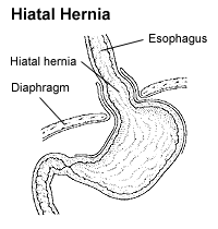
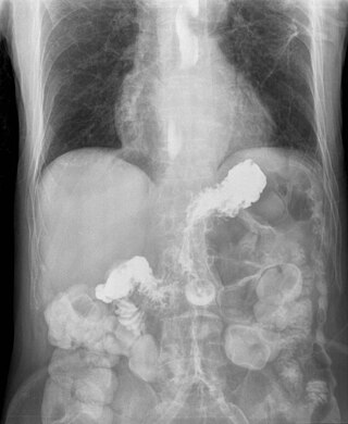
A hiatal hernia or hiatus hernia is a type of hernia in which abdominal organs slip through the diaphragm into the middle compartment of the chest. This may result in gastroesophageal reflux disease (GERD) or laryngopharyngeal reflux (LPR) with symptoms such as a taste of acid in the back of the mouth or heartburn. Other symptoms may include trouble swallowing and chest pains. Complications may include iron deficiency anemia, volvulus, or bowel obstruction.

Coffee ground vomitus refers to a particular appearance of vomit. Within organic heme molecules of red blood cells is the element iron, which oxidizes following exposure to gastric acid. This reaction causes the vomitus to look like ground coffee.

<span class="mw-page-title-main">Nissen fundoplication</span> Surgical procedure to treat gastric reflux and hiatal hernia

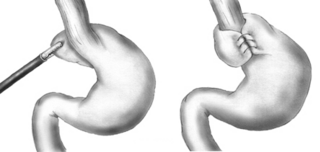
A Nissen fundoplication, or laparoscopic Nissen fundoplication when performed via laparoscopic surgery, is a surgical procedure to treat gastroesophageal reflux disease (GERD) and hiatal hernia. In GERD, it is usually performed when medical therapy has failed; but, with a Type II (paraesophageal) hiatus hernia, it is the first-line procedure. The Nissen fundoplication is total (360°), but partial fundoplications known as Thal, Belsey, Dor, Lind, and Toupet fundoplications are alternative procedures with somewhat different indications and outcomes.

<span class="mw-page-title-main">Gastrointestinal disease</span> Medical condition

Gastrointestinal diseases refer to diseases involving the gastrointestinal tract, namely the esophagus, stomach, small intestine, large intestine and rectum, and the accessory organs of digestion, the liver, gallbladder, and pancreas.

<span class="mw-page-title-main">Eosinophilic esophagitis</span> Allergic inflammatory condition of the esophagus

Eosinophilic esophagitis (EoE) is an allergic inflammatory condition of the esophagus that involves eosinophils, a type of white blood cell. In healthy individuals, the esophagus is typically devoid of eosinophils. In EoE, eosinophils migrate to the esophagus in large numbers. When a trigger food is eaten, the eosinophils contribute to tissue damage and inflammation. Symptoms include swallowing difficulty, food impaction, vomiting, and heartburn.

Esophageal dysphagia is a form of dysphagia where the underlying cause arises from the body of the esophagus, lower esophageal sphincter, or cardia of the stomach, usually due to mechanical causes or motility problems.

<span class="mw-page-title-main">Nutcracker esophagus</span> Medical condition

Nutcracker esophagus, jackhammer esophagus, or hypercontractile peristalsis, is a disorder of the movement of the esophagus characterized by contractions in the smooth muscle of the esophagus in a normal sequence but at an excessive amplitude or duration. Nutcracker esophagus is one of several motility disorders of the esophagus, including achalasia and diffuse esophageal spasm. It causes difficulty swallowing, or dysphagia, with both solid and liquid foods, and can cause significant chest pain; it may also be asymptomatic. Nutcracker esophagus can affect people of any age but is more common in the sixth and seventh decades of life.

<span class="mw-page-title-main">Diffuse esophageal spasm</span> Medical condition

Diffuse esophageal spasm (DES), also known as distal esophageal spasm, is a condition characterized by uncoordinated contractions of the esophagus, which may cause difficulty swallowing (dysphagia) or regurgitation. In some cases, it may cause symptoms such as chest pain, similar to heart disease. In many cases, the cause of DES remains unknown.

<span class="mw-page-title-main">Esophageal stricture</span> Medical condition

A benign esophageal stricture, or peptic stricture, is a narrowing or tightening of the esophagus that causes swallowing difficulties.

<span class="mw-page-title-main">Esophageal food bolus obstruction</span> Medical condition

An esophageal food bolus obstruction is a medical emergency caused by the obstruction of the esophagus by an ingested foreign body.

<span class="mw-page-title-main">Esophageal pH monitoring</span>

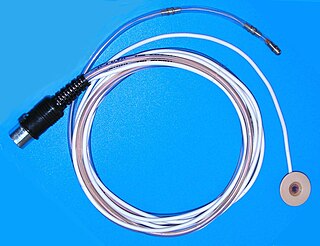
Esophageal pH monitoring is the current gold standard for diagnosis of gastroesophageal reflux disease (GERD). It provides direct physiologic measurement of acid in the esophagus and is the most objective method to document reflux disease, assess the severity of the disease and monitor the response of the disease to medical or surgical treatment. It can also be used in diagnosing laryngopharyngeal reflux.

<span class="mw-page-title-main">Esophageal intramural pseudodiverticulosis</span> Medical condition

Esophageal intramucosal pseudodiverticulosis (EIPD) is a rare condition wherein the wall of the esophagus develops numerous small outpouchings (pseudodiverticulae). Individuals with the condition typically develop difficulty swallowing. The outpouchings represent the ducts of submucosal glands of the esophagus. It typically affects individuals in their sixth and seventh decades of life. While it is associated with certain chronic conditions, particularly alcoholism, diabetes and gastroesophageal reflux disease, the cause of the condition is unknown. Treatment involves medications to treat concomitant conditions such as reflux and esophageal spasm, and dilation of strictures in the esophagus.

<span class="mw-page-title-main">Lymphocytic esophagitis</span> Medical condition

Lymphocytic esophagitis is a rare and poorly understood medical disorder involving inflammation in the esophagus. The disease is named from the primary inflammatory process, wherein lymphocytes are seen within the esophageal mucosa. Symptoms of the condition include difficulty swallowing, heartburn and food bolus obstruction. The condition was first described in 2006 by Rubio and colleagues. Initial reports questioned whether this was a true medical disorder, or whether the inflammation was secondary to another condition, such as gastroesophageal reflux disease.

Esophagogastric junction outflow obstruction (EGJOO) is an esophageal motility disorder characterized by increased pressure where the esophagus connects to the stomach at the lower esophageal sphincter. EGJOO is diagnosed by esophageal manometry. However, EGJOO has a variety of etiologies; evaluating the cause of obstruction with additional testing, such as upper endoscopy, computed tomography, or endoscopic ultrasound may be necessary. When possible, treatment of EGJOO should be directed at the cause of obstruction. When no cause for obstruction is found, observation alone may be considered if symptoms are minimal. Functional EGJOO with significant or refractory symptoms may be treated with pneumatic dilation, per-oral endoscopic myotomy (POEM), or botulinum toxin injection.

References

  1. 1 2 "Esophagitis – Symptoms and causes – Mayo Clinic". www.mayoclinic.org. Retrieved 2017-12-13.
  2. 1 2 "Esophagitis-Topic Overview". WebMD. Retrieved 2017-12-13.
  3. "Esophagitis". Johns Hopkins Medicine. 19 November 2019. Retrieved 22 July 2021.
  4. 1 2 3 4 "Understanding Esophagitis". WebMD. Retrieved 2017-11-07.
  5. "Upper Gastrointestinal (UGI) Series". WebMD. Retrieved 2017-12-13.
  6. 1 2 "Inflammation, Not Acid, Cause of GERD, Study Suggests".
  7. Farivar M. "Los Angeles Classification of Esophagitis". webgerd.com. Archived from the original on 2015-01-30. Retrieved 2010-10-27. In turn citing: Lundell LR, Dent J, Bennett JR, et al. (August 1999). "Endoscopic assessment of oesophagitis: clinical and functional correlates and further validation of the Los Angeles classification". Gut. 45 (2): 172–80. doi:10.1136/gut.45.2.172. PMC   1727604 . PMID   10403727.
  8. Laparoscopic bariatric surgery, Volume 1. William B. Inabnet, Eric J. DeMaria, Sayeed Ikramuddin. ISBN   0-7817-4874-7.[ page needed ]
  9. "Esophagitis". The Lecturio Medical Concept Library. Retrieved 22 July 2021.
  10. Medicines and Healthcare Products Regulatory  Agency (MHRA), 2013. Metoclopramide: risk of neurological adverse effects. Drug Safety Update 7 (1), S2. Medicines and Healthcare Products Regulatory  Agency, 2014. Domperidone: risks of cardiac side effects. Drug Safety Update 7 (10),  A1. Moayyedi, P., Santana, J., Khan, M., et al., 2011. Medical treatments  in the short-term management of relux oesophagitis. Cochrane DB Syst. Rev. 2011 (2),  Art. No. CD003244.
  11. Kirkham, EN; Main, BG; Jones, KJB; Blazeby, JM; Blencowe, NS (January 2020). "Systematic review of the introduction and evaluation of magnetic augmentation of the lower oesophageal sphincter for gastro-oesophageal reflux disease". The British Journal of Surgery. 107 (1): 44–55. doi:10.1002/bjs.11391. PMC   6972716 . PMID   31800095.