Viral transformation

Last updated

Viral transformation is the change in growth, phenotype, or indefinite reproduction of cells caused by the introduction of inheritable material. Through this process, a virus causes harmful transformations of an in vivo cell or cell culture. The term can also be understood as DNA transfection using a viral vector.

Contents

Figure 1: Hepatitis-B virions Hepatitis-B virions.jpg
Figure 1: Hepatitis-B virions

Viral transformation can occur both naturally and medically. Natural transformations can include viral cancers, such as human papillomavirus (HPV) and T-cell Leukemia virus type I. Hepatitis B and C are also the result of natural viral transformation of the host cells. Viral transformation can also be induced for use in medical treatments.

Cells that have been virally transformed can be differentiated from untransformed cells through a variety of growth, surface, and intracellular observations. The growth of transformed cells can be impacted by a loss of growth limitation caused by cell contact, less oriented growth, and high saturation density. Transformed cells can lose their tight junctions, increase their rate of nutrient transfer, and increase their protease secretion. Transformation can also affect the cytoskeleton and change in the quantity of signal molecules.

Type

There are three types of viral infections that can be considered under the topic of viral transformation. These are cytocidal, persistent, and transforming infections. Cytocidal infections can cause fusion of adjacent cells, disruption of transport pathways including ions and other cell signals, disruption of DNA, RNA and protein synthesis, and nearly always leads to cell death. Persistent infections involve viral material that lays dormant within a cell until activated by some stimulus. This type of infection usually causes few obvious changes within the cell but can lead to long chronic diseases. Transforming infections are also referred to as malignant transformation. This infection causes a host cell to become malignant and can be either cytocidal (usually in the case of RNA viruses) or persistent (usually in the case of DNA viruses). Cells with transforming infections undergo immortalization and inherit the genetic material to produce tumors. Since the term cytocidal, or cytolytic, refers to cell death, these three infections are not mutually exclusive. Many transforming infections by DNA tumor viruses are also cytocidal. [1]

Table 1: Cellular effects of viral infections [1]

GeneticCell FateMorphologicalBiochemicalPhysiological
Cytocidal
Productive
-----------------
Abortive
DNA degradation
-----------------
Possible mutation
Death
-----------------
Usually death

Rounding of the cell
Fusion with adjacent cells
Appearance of inclusion bodies

Inhibit DNA, RNA, and protein synthesis
Interfere with sub-cellular interactions

Insufficient movement of ions
Formation of secondary messengers
Activation of cellular cascades

Persistent
Latent
-----------------
Chronic
-----------------
Slow
Possible Mutation
-----------------
Possible Mutation
-----------------
Possible Mutation
Survival
-----------------
Variable
-----------------
Variable

Fusion with adjacent cells
Appearance of inclusion bodies
Budding

Immune responses limit viral spread
Antigen-antibody complexes can incorporate
viral antigens causing inflammation

Rare until stimulated

Transforming
DNA viruses
-----------------
RNA viruses
Mutation
-----------------
Mutation
Survival
-----------------
Survival

Unlimited cell replication

Inactivates tumor suppressor proteins
Impairs cell cycle regulation

Unlimited cell replication

Cytocidal infections

Cytocidal infections are often associated with changes in cell morphology, physiology and are thus important for the complete viral replication and transformation. Cytopathic Effects, often include a change in cell's morphology such as fusion with adjacent cells to form polykaryocytes as well as the synthesis of nuclear and cytoplasmic inclusion bodies. Physiological changes include the insufficient movement of ions, formation of secondary messengers, and activation of cellular cascades to continue cellular activity. Biochemically, many viruses inhibit the synthesis of host DNA, RNA, proteins directly or even interfere with protein-protein, DNA-protein, RNA-protein interactions at the subcellular level. Genotoxicity involves breaking, fragmenting, or rearranging chromosomes of the host. Lastly, biologic effects include the viruses' ability to affect the activity of antigens and immunologlobulins in the host cell. [1]

There are two types of cytocidal infections, productive and abortive. In productive infections, additional infectious viruses are produced. Abortive infections do not produce infectious viruses. One example of a productive cytocidal infection is the herpes virus. [2]

Persistent infections

There are three types of persistent infections, latent, chronic and slow, in which the virus stays inside the host cell for prolonged periods of time. During latent infections there is minimal to no expression of infected viral genome. The genome remains within the host cell until the virus is ready for replication. Chronic infections have similar cellular effects as acute cytocidal infections but there is a limited number of progeny and viruses involved in transformation. Lastly, slow infections have a longer incubation period in which no physiological, morphological or subcellular changes may be involved. [1]

Transforming infections

Transformation infections is limited to abortive or restrictive infections. [1] This constitutes the broadest category of infections as it can include both cytocidal and persistent infection. Viral transformation is most commonly understood as transforming infections, so the remainder of the article focuses on detailing transforming infections.

Process

Figure 2: Phage injecting its genome into bacterial cell Phage injecting its genome into bacterial cell.png
Figure 2: Phage injecting its genome into bacterial cell

In order for a cell to be transformed by a virus, the viral DNA must be entered into the host cell. The simplest consideration is viral transformation of a bacterial cell. This process is called lysogeny. As shown in Figure 2, a bacteriophage lands on a cell and pins itself to the cell. The phage can then penetrate the cell membrane and inject the viral DNA into the host cell. The viral DNA can then either lay dormant until stimulated by a source such as UV light or it can be immediately taken up by the host's genome. In either case the viral DNA will replicate along with the original host DNA during cell replication causing two cells to now be infected with the virus. The process will continue to propagate more and more infected cells. [3] This process is in contrast to the lytic cycle where a virus only uses the host cell's replication machinery to replicate itself before destroying the host cell. [4]

Figure 3: Examples of endocytosis Endocytosis types.svg
Figure 3: Examples of endocytosis

The process is similar in animal cells. In most cases, rather than viral DNA being injected into an animal cell, a section of the membrane encases the virus and the cell then absorbs both the virus and the encasing section of the membrane into the cell. This process, called endocytosis, is shown in Figure 3. [5]

Transformation of the host cell

Viral transformation disrupts the normal expression of the host cell's genes in favor of expressing a limited number of viral genes. The virus also can disrupt communication between cells and cause cells to divide at an increased rate. [6]

Physiological

Viral transformation can impose characteristically determinable features upon a cell. Typical phenotypic changes include high saturation density, anchorage-independent growth, loss of contact inhibition, loss of orientated growth, immortalization, disruption of the cell's cytoskeleton.

Biochemical

Viral genes are expressed through the use of the host cell's replication machinery; therefore, many viral genes have promoters that support binding of many transcription factors found naturally in the host cells. These transcription factors along with the virus' own proteins can repress or activate genes from both the virus and the host cell's genome. Many viruses can also increase the production of the cell's regulatory proteins. [1]

Genetic

Depending on the virus, a variety of genetic changes can occur in the host cell. In the case of a lytic cycle virus, the cell will only survive long enough to the replication machinery to be used to create additional viral units. In other cases, the viral DNA will persist within the host cell and replicate as the cell replicates. This viral DNA can either be incorporated into the host cell's genetic material or persist as a separate genetic vector. Either case can lead to damage of the host cell's chromosomes. It is possible that the damage can be repaired; however, the most common result is an instability in the original genetic material or suppression or alteration of the gene expression. [1]

Assays

An assay is an analytic tool often used in a laboratory setting in order to assess or measure some quality of a target entity. [7] In virology, assays can be used to differentiate between transformed and non-transformed cells. Varying the assay used, changes the selective pressure on the cells and therefore can change what properties are selected in the transformed cells. [6]

Three common assays used are the focus forming assay, the Anchorage independent growth assay, and the reduced serum assay.

The focus forming assay (FFA) is used to grow cells containing a transforming oncogene on a monolayer of non-transformed cells. The transformed cells will form raised, dense spots on the sample as they grow without contact inhibition. [8] This assay is highly sensitive compared to other assays used for viral analysis, such as the yield reduction assay. [9]

An example of the Anchorage independent growth assay is the soft agar assay. The assay is assessing the cells' ability to grow in a gel or viscous fluid. Transformed cells can grow in this environment and are considered anchorage independent. Cells that can only grow when attached to a solid surface are anchorage dependent untransformed cells. This assay is considered one of the most stringent for detection of malignant transformation [10]

In a reduced serum assay, cells are assayed by exploiting the changes in cell serum requirements. Non-transformed cells require at least a 5% serum medium in order to grow; however, transformed cells can grow in an environment with significantly less serum. [6]

Examples of Natural Transformation

Natural transformation is the viral transformation of cells without the interference of medical science. This is the most commonly considered form of viral transformation and includes many cancers and diseases, such as HIV, Hepatitis B, and T-cell Leukemia virus type I.

Viral oncogenesis

As many as 20% of human tumors are caused by viruses. [11] Some such viruses that are commonly recognized include HPV, T-cell Leukemia virus type I, and hepatitis B.

Viral oncogenesis are most common with DNA and RNA tumor viruses, most frequently the retroviruses. [12] There are two types of oncogenic retroviruses: acute transforming viruses and non-acute transforming viruses. Acute transforming viruses induce a rapid tumor growth since they carry viral oncogenes in their DNA/RNA to induce such growth. An example of an acute transforming virus is the Rous Sarcoma Virus (RSV) that carry the v-src oncogene. v-Src is part of the c-src, which is a cellular proto-oncogene that stimulates rapid cell growth and expansion. A non-acute transforming virus on the other hand induces a slow tumor growth, since it does not carry any viral oncogenes. It induces tumor growth by transcriptionally activating the proto-oncogenes particularly the long terminal repeat (LTR) in the proto-oncogenes. [12]

Viral Oncogenesis through transformation can occur via 2 mechanisms: [1]

  1. The tumor virus can introduce and express a "transforming" gene either through the integration of DNA or RNA into the host genome.
  2. The tumor virus can alter expression on preexisting genes of the host.

One or both of these mechanisms can occur in the same host cell.

Hepatitis B

The Hepatitis B viral protein X is believed to cause hepatocellular carcinoma through transformation, typically of liver cells. The viral DNA is incorporated into the host cell's genome causing rapid cell replication and tumor growth. [13]

Papillomaviruses

Papillomaviruses typically target epithelial cells and cause everything from warts to cervical cancer. When human papillomavirus (HPV) transforms a cell, it interferes with the function of cellular proteins while degrading other cellular proteins. [14]

Herpesviruses

The herpesviruses, Kaposi's sarcoma-associated herpesvirus and Epstein-Barr virus, are believed to cause cancer in humans, such as Kaposi's sarcoma, Burkitt's lymphoma, and nasopharyngeal carcinoma. Although genes have been identified in these viruses that cause transformation, the manner in which the virus transforms and replicates the host cell is not understood. [14]

Retroviruses

The retroviruses include T-cell Leukemia virus type I, HIV, and Rous Sarcoma Virus (RSV). The viral gene tax is expressed when the T-cell Leukemia virus transforms a cell altering the expression of cellular growth control genes and causing the transformed cells to become cancerous. HIV works differently by not directly causing cells to become cancerous but by instead making those infected more susceptible to lymphoma and Kaposi's sarcoma. Many other retroviruses contain the three genes, gag, pol, and env, which do not directly cause transformation or tumor formation. [14]

HIV

Human immunodeficiency virus is a viral infection that targets the lymph nodes. HIV binds to the immune CD4 cell and reverse transcriptase alters the host cell genome to allow integration of the viral DNA via integrase. The virus replicates using the host cell's machinery and then leaves the cell to infect additional cells via budding. [15]

Medical Applications

There are many applications in which viral transformation can be artificially induced in a cell culture in order to treat an illness or other condition. A cell culture is infected with a virus causing the transformation; transformed cells can then be used to either produce treatments or be directly introduced into the body.

Personalized type I interferons

Figure 4: Type I Human Interferon 1AU1 Human Interferon-Beta01.png
Figure 4: Type I Human Interferon

Type I interferons (IFNs) are used to treat a wide variety of medical conditions including hepatitis C, cancers, viral and inflammatory diseases. IFNs can either be extracted from a natural source, such as cultured human cells or blood leukocytes, or they can be manufactured with recombinant DNA technologies. Most of these IFN treatments have a low response rate. [16]

The use of viral transformation of the Epstein-Barr virus (EBV) has been recommended to create personalized IFNs. In this process, primary B lymphocytes are transformed with EBV. These cells can then be used to produce IFNs specific for the patient from which the B lymphocytes were extracted. This personalization decreases the likelihood of an antibody response and therefore increases the effectiveness of the treatment. [16]

Cancer treatments

When a virus transforms a cell it often causes cancer by either altering the cells' existing genome or introducing additional genetic material which causes cells to uncontrollably replicate. [11] It is rarely considered that what causes so much harm also has the capability of reversing the process and slowing the cancer growth or even leading to remission. Viruses transform host cells in order to survive and replicate; however, the immune responses of the host cell are typically compromised during transformation making transformed cells more susceptible to other viruses. [17]

The idea of using viruses to treat cancers was first introduced in 1951 when a 4-year-old boy suddenly went into a temporary remission from leukemia while he had chickenpox. This led to research in the 1990s where scientists worked to create a strain of the herpes simplex virus strong enough to infect and transform tumor cells but weak enough to leave healthy cells unharmed. Treating patients with viral transformation has the possibility of treating patients more safely and more effectively than using traditional methods, such as chemotherapy. Viruses used in the treatment of cancer gain strength and increase their effectiveness as the multiply in the body while causing only minor side effects, such as nausea, fatigue, and aches. [17]

See also

Related Research Articles

<span class="mw-page-title-main">Interferon</span> Signaling proteins released by host cells in response to the presence of pathogens

Interferons are a group of signaling proteins made and released by host cells in response to the presence of several viruses. In a typical scenario, a virus-infected cell will release interferons causing nearby cells to heighten their anti-viral defenses.

<span class="mw-page-title-main">Retrovirus</span> Family of viruses

A retrovirus is a type of virus that inserts a DNA copy of its RNA genome into the DNA of a host cell that it invades, thus changing the genome of that cell. After invading a host cell's cytoplasm, the virus uses its own reverse transcriptase enzyme to produce DNA from its RNA genome, the reverse of the usual pattern, thus retro (backwards). The new DNA is then incorporated into the host cell genome by an integrase enzyme, at which point the retroviral DNA is referred to as a provirus. The host cell then treats the viral DNA as part of its own genome, transcribing and translating the viral genes along with the cell's own genes, producing the proteins required to assemble new copies of the virus. Many retroviruses cause serious diseases in humans, other mammals, and birds.

Gene silencing is the regulation of gene expression in a cell to prevent the expression of a certain gene. Gene silencing can occur during either transcription or translation and is often used in research. In particular, methods used to silence genes are being increasingly used to produce therapeutics to combat cancer and other diseases, such as infectious diseases and neurodegenerative disorders.

<i>Hepadnaviridae</i> Family of viruses

Hepadnaviridae is a family of viruses. Humans, apes, and birds serve as natural hosts. There are currently 18 species in this family, divided among 5 genera. Its best-known member is hepatitis B virus. Diseases associated with this family include: liver infections, such as hepatitis, hepatocellular carcinomas, and cirrhosis. It is the sole accepted family in the order Blubervirales.

<i>Papillomaviridae</i> Family of viruses

Papillomaviridae is a family of non-enveloped DNA viruses whose members are known as papillomaviruses. Several hundred species of papillomaviruses, traditionally referred to as "types", have been identified infecting all carefully inspected mammals, but also other vertebrates such as birds, snakes, turtles and fish. Infection by most papillomavirus types, depending on the type, is either asymptomatic or causes small benign tumors, known as papillomas or warts. Papillomas caused by some types, however, such as human papillomaviruses 16 and 18, carry a risk of becoming cancerous.

<i>Polyomaviridae</i> Family of viruses

Polyomaviridae is a family of viruses whose natural hosts are primarily mammals and birds. As of 2024, there are eight recognized genera. 14 species are known to infect humans, while others, such as Simian Virus 40, have been identified in humans to a lesser extent. Most of these viruses are very common and typically asymptomatic in most human populations studied. BK virus is associated with nephropathy in renal transplant and non-renal solid organ transplant patients, JC virus with progressive multifocal leukoencephalopathy, and Merkel cell virus with Merkel cell cancer.

<span class="mw-page-title-main">SV40</span> Species of virus

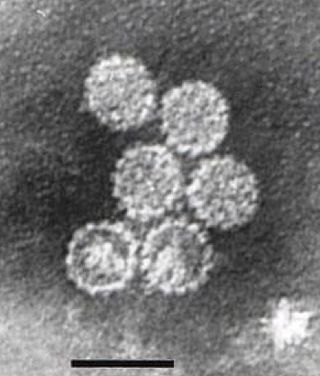
SV40 is an abbreviation for simian vacuolating virus 40 or simian virus 40, a polyomavirus that is found in both monkeys and humans. Like other polyomaviruses, SV40 is a DNA virus that sometimes causes tumors in animals, but most often persists as a latent infection. SV40 has been widely studied as a model eukaryotic virus, leading to many early discoveries in eukaryotic DNA replication and transcription.

<span class="mw-page-title-main">Oncovirus</span> Viruses that can cause cancer

An oncovirus or oncogenic virus is a virus that can cause cancer. This term originated from studies of acutely transforming retroviruses in the 1950–60s, when the term "oncornaviruses" was used to denote their RNA virus origin. With the letters "RNA" removed, it now refers to any virus with a DNA or RNA genome causing cancer and is synonymous with "tumor virus" or "cancer virus". The vast majority of human and animal viruses do not cause cancer, probably because of longstanding co-evolution between the virus and its host. Oncoviruses have been important not only in epidemiology, but also in investigations of cell cycle control mechanisms such as the retinoblastoma protein.

<span class="mw-page-title-main">Viral replication</span> Formation of biological viruses during the infection process

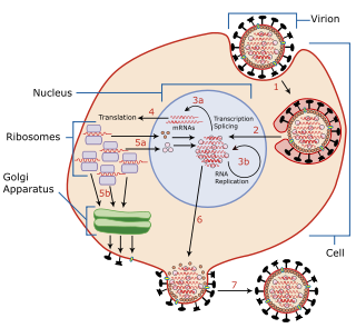
Viral replication is the formation of biological viruses during the infection process in the target host cells. Viruses must first get into the cell before viral replication can occur. Through the generation of abundant copies of its genome and packaging these copies, the virus continues infecting new hosts. Replication between viruses is greatly varied and depends on the type of genes involved in them. Most DNA viruses assemble in the nucleus while most RNA viruses develop solely in cytoplasm.

<span class="mw-page-title-main">SV40 large T antigen</span> Proto-oncogene derived from polyomavirus SV40

SV40 large T antigen is a hexamer protein that is a dominant-acting oncoprotein derived from the polyomavirus SV40. TAg is capable of inducing malignant transformation of a variety of cell types. The transforming activity of TAg is due in large part to its perturbation of the retinoblastoma (pRb) and p53 tumor suppressor proteins. In addition, TAg binds to several other cellular factors, including the transcriptional co-activators p300 and CBP, which may contribute to its transformation function. Similar proteins from related viruses are known as large tumor antigen in general.

Rous sarcoma virus (RSV) is a retrovirus and is the first oncovirus to have been described. It causes sarcoma in chickens.

The murine leukemia viruses are retroviruses named for their ability to cause cancer in murine (mouse) hosts. Some MLVs may infect other vertebrates. MLVs include both exogenous and endogenous viruses. Replicating MLVs have a positive sense, single-stranded RNA (ssRNA) genome that replicates through a DNA intermediate via the process of reverse transcription.

<span class="mw-page-title-main">Viral vector</span> Biotechnology to deliver genetic material into a cell

Viral vectors are tools commonly used by molecular biologists to deliver genetic material into cells. This process can be performed inside a living organism or in cell culture. Viruses have evolved specialized molecular mechanisms to efficiently transport their genomes inside the cells they infect. Delivery of genes or other genetic material by a vector is termed transduction and the infected cells are described as transduced. Molecular biologists first harnessed this machinery in the 1970s. Paul Berg used a modified SV40 virus containing DNA from the bacteriophage λ to infect monkey kidney cell maintained in culture.

<i>Murine respirovirus</i> Sendai virus, virus of rodents

Murine respirovirus, formerly Sendai virus (SeV) and previously also known as murine parainfluenza virus type 1 or hemagglutinating virus of Japan (HVJ), is an enveloped, 150-200 nm–diameter, negative sense, single-stranded RNA virus of the family Paramyxoviridae. It typically infects rodents and it is not pathogenic for humans or domestic animals.

The gag-onc fusion protein is a general term for a fusion protein formed from a group-specific antigen ('gag') gene and that of an oncogene ('onc'), a gene that plays a role in the development of a cancer. The name is also written as Gag-v-Onc, with "v" indicating that the Onc sequence resides in a viral genome. Onc is a generic placeholder for a given specific oncogene, such as C-jun..

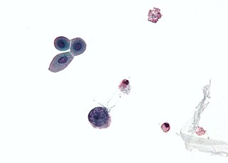
Merkel cell polyomavirus was first described in January 2008 in Pittsburgh, Pennsylvania. It was the first example of a human viral pathogen discovered using unbiased metagenomic next-generation sequencing with a technique called digital transcriptome subtraction. MCV is one of seven currently known human oncoviruses. It is suspected to cause the majority of cases of Merkel cell carcinoma, a rare but aggressive form of skin cancer. Approximately 80% of Merkel cell carcinoma (MCC) tumors have been found to be infected with MCV. MCV appears to be a common—if not universal—infection of older children and adults. It is found in respiratory secretions, suggesting that it might be transmitted via a respiratory route. However, it has also been found elsewhere, such as in shedded healthy skin and gastrointestinal tract tissues, thus its precise mode of transmission remains unknown. In addition, recent studies suggest that this virus may latently infect the human sera and peripheral blood mononuclear cells.

<span class="mw-page-title-main">Virus</span> Infectious agent that replicates in cells

A virus is a submicroscopic infectious agent that replicates only inside the living cells of an organism. Viruses infect all life forms, from animals and plants to microorganisms, including bacteria and archaea. Viruses are found in almost every ecosystem on Earth and are the most numerous type of biological entity. Since Dmitri Ivanovsky's 1892 article describing a non-bacterial pathogen infecting tobacco plants and the discovery of the tobacco mosaic virus by Martinus Beijerinck in 1898, more than 11,000 of the millions of virus species have been described in detail. The study of viruses is known as virology, a subspeciality of microbiology.

<span class="mw-page-title-main">HBx</span>

HBx is a hepatitis B viral protein. It is 154 amino acids long and interferes with transcription, signal transduction, cell cycle progress, protein degradation, apoptosis and chromosomal stability in the host. It forms a heterodimeric complex with its cellular target protein, and this interaction dysregulates centrosome dynamics and mitotic spindle formation. It interacts with DDB1 redirecting the ubiquitin ligase activity of the CUL4-DDB1 E3 complexes, which are intimately involved in the intracellular regulation of DNA replication and repair, transcription and signal transduction.

<span class="mw-page-title-main">Large tumor antigen</span>

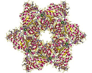
The large tumor antigen is a protein encoded in the genomes of polyomaviruses, which are small double-stranded DNA viruses. LTag is expressed early in the infectious cycle and is essential for viral proliferation. Containing four well-conserved protein domains as well as several intrinsically disordered regions, LTag is a fairly large multifunctional protein; in most polyomaviruses, it ranges from around 600-800 amino acids in length. LTag has two primary functions, both related to replication of the viral genome: it unwinds the virus's DNA to prepare it for replication, and it interacts with proteins in the host cell to dysregulate the cell cycle so that the host's DNA replication machinery can be used to replicate the virus's genome. Some polyomavirus LTag proteins - most notably the well-studied SV40 large tumor antigen from the SV40 virus - are oncoproteins that can induce neoplastic transformation in the host cell.

HSV epigenetics is the epigenetic modification of herpes simplex virus (HSV) genetic code.

References

  1. 1 2 3 4 5 6 7 8 Baron, S.; Albrecht, T.; Fons, M.; Boldogh, I.; Rabson, A. S. (1996). "Effects on Cells". In Baron, Samuel (ed.). Medical microbiology (4. ed.). Galveston, Texas: Univ. of Texas Medical Branch. ISBN   0963117211. PMID   21413282.
  2. Huang, CR; Lin, SS; Chou, MY; Ho, CC; Wang, L; Lee, YL; Chen, CS; Yang, CC (2005). "Demonstration of different modes of cell death upon herpes simplex virus 1 infection in different types of oral cells". Acta Virologica. 49 (1): 7–15. PMID   15929393.
  3. "Lysogeny". Encyclopædia Britannica. Retrieved 8 April 2014.
  4. "Two Life Cycles of a Virus" . Retrieved 8 April 2014.
  5. "The cycle of infection". Encyclopædia Britannica. Retrieved 8 April 2014.
  6. 1 2 3 Heaphy, Shaun. "Viral Transformation of Cells". University of Cape Town. Archived from the original on 17 February 2014. Retrieved 25 March 2014.
  7. Assay. Wikipedia, The Free Encyclopedia. Accessed 2014-03-25.
  8. Andrews, David. "Transformation assays: focus forming assay" (PDF). Andrews Lab. Archived from the original (PDF) on 25 March 2014. Retrieved 25 March 2014.
  9. Winship, Timothy R (11 Dec 1979). "A Sensitive Method for Quantification of Vesicular Stomatitis Virus Defective Interfering Particles: Focus Forming Assay" (PDF). Journal of General Virology. 48 (1): 237–240. doi: 10.1099/0022-1317-48-1-237 . PMID   6247438 . Retrieved 25 March 2014.[ permanent dead link ]
  10. Provost, Joseph. "Soft Agar Assay for Cology Formation" (PDF). Wallert and Provost Lab. Archived from the original (PDF) on 10 August 2013. Retrieved 25 March 2014.
  11. 1 2 Dayaram, T; Marriott, SJ (August 2008). "Effect of transforming viruses on molecular mechanisms associated with cancer". Journal of Cellular Physiology. 216 (2): 309–14. doi:10.1002/jcp.21439. PMC   4160108 . PMID   18366075.
  12. 1 2 Fan, Hung (June 15, 2011). "Cell Transformation by RNA Viruses: An Overview". Viruses. 3 (12): 858–860. doi: 10.3390/v3060858 . PMC   3185770 . PMID   21994757.
  13. Schaefer, S; Gerlich, WH (1995). "In vitro transformation by hepatitis B virus DNA". Intervirology. 38 (3–4): 143–54. doi:10.1159/000150425. PMID   8682609.
  14. 1 2 3 Cooper, Geoffrey M. (2000). The cell : a molecular approach (2nd ed.). Washington: ASM Press. ISBN   0878931023.
  15. "HIV Life Cycle". U.S. Department of Health and Human Services. Retrieved 7 May 2014.
  16. 1 2 Xu, Dongsheng; Zhang, Luwen (June 2010). "Viral transformation for production of personalized type I interferons". Biotechnology Journal. 5 (6): 578–581. doi:10.1002/biot.201000038. PMC   3023146 . PMID   20518060.
  17. 1 2 Nuwer, Rachel (19 Mar 2012). "Viruses Recruited as Killers of Tumors". New York Times. Retrieved 6 May 2014.