Von Hippel–Lindau disease (VHL), also known as VonHippel–Lindau syndrome, is a rare genetic disorder with multisystem involvement. It is characterized by visceral cysts and benign tumors with potential for subsequent malignant transformation. It is a type of phakomatosis that results from a mutation in the Von Hippel–Lindau tumor suppressor gene on chromosome 3p25.3.
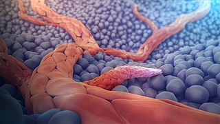
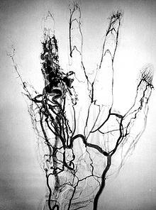
Angiogenesis is the physiological process through which new blood vessels form from pre-existing vessels, formed in the earlier stage of vasculogenesis. Angiogenesis continues the growth of the vasculature mainly by processes of sprouting and splitting, but processes such as coalescent angiogenesis, vessel elongation and vessel cooption also play a role. Vasculogenesis is the embryonic formation of endothelial cells from mesoderm cell precursors, and from neovascularization, although discussions are not always precise. The first vessels in the developing embryo form through vasculogenesis, after which angiogenesis is responsible for most, if not all, blood vessel growth during development and in disease.
Cerebral cavernous malformation (CCM) is a cavernous hemangioma that arises in the central nervous system. It can be considered to be a variant of hemangioma, and is characterized by grossly large dilated blood vessels and large vascular channels, less well circumscribed, and more involved with deep structures, with a single layer of endothelium and an absence of neuronal tissue within the lesions. These thinly walled vessels resemble sinusoidal cavities filled with stagnant blood. Blood vessels in patients with cerebral cavernous malformations (CCM) can range from a few millimeters to several centimeters in diameter. Most lesions occur in the brain, but any organ may be involved.

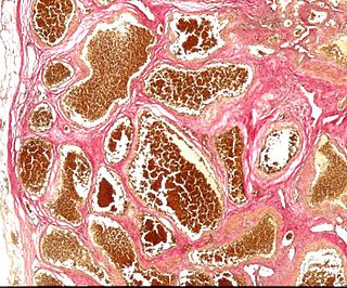
An infantile hemangioma (IH), sometimes called a strawberry mark due to appearance, is a type of benign vascular tumor or anomaly that affects babies. Other names include capillary hemangioma, "strawberry hemangioma", strawberry birthmark and strawberry nevus. and formerly known as a cavernous hemangioma. They appear as a red or blue raised lesion on the skin. Typically, they begin during the first four weeks of life, growing until about five months of life, and then shrinking in size and disappearing over the next few years. Often skin changes remain after they shrink. Complications may include pain, bleeding, ulcer formation, disfigurement, or heart failure. It is the most common tumor of orbit and periorbital areas in childhood. It may occur in the skin, subcutaneous tissues and mucous membranes of oral cavities and lips as well as in extracutaneous locations including the liver and gastrointestinal tract.
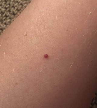
Cherry angioma, also called cherry hemangioma or Campbell de Morgan Spot, is a small bright red dome-shaped bump on the skin. It ranges between 0.5 – 6 mm in diameter and usually several are present, typically on the chest and arms, and increasing in number with age. If scratched, they may bleed.

Sturge–Weber syndrome, sometimes referred to as encephalotrigeminal angiomatosis, is a rare congenital neurological and skin disorder. It is one of the phakomatoses and is often associated with port-wine stains of the face, glaucoma, seizures, intellectual disability, and ipsilateral leptomeningeal angioma. Sturge–Weber syndrome can be classified into three different types. Type 1 includes facial and leptomeningeal angiomas as well as the possibility of glaucoma or choroidal lesions. Normally, only one side of the brain is affected. This type is the most common. Type 2 involvement includes a facial angioma with a possibility of glaucoma developing. There is no evidence of brain involvement. Symptoms can show at any time beyond the initial diagnosis of the facial angioma. The symptoms can include glaucoma, cerebral blood flow abnormalities and headaches. More research is needed on this type of Sturge–Weber syndrome. Type 3 has leptomeningeal angioma involvement exclusively. The facial angioma is absent and glaucoma rarely occurs. This type is only diagnosed via brain scan.

Hemangioendotheliomas are a family of vascular neoplasms of intermediate malignancy.

Lymphangiomas are malformations of the lymphatic system characterized by lesions that are thin-walled cysts; these cysts can be macroscopic, as in a cystic hygroma, or microscopic. The lymphatic system is the network of vessels responsible for returning to the venous system excess fluid from tissues as well as the lymph nodes that filter this fluid for signs of pathogens. These malformations can occur at any age and may involve any part of the body, but 90% occur in children less than 2 years of age and involve the head and neck. These malformations are either congenital or acquired. Congenital lymphangiomas are often associated with chromosomal abnormalities such as Turner syndrome, although they can also exist in isolation. Lymphangiomas are commonly diagnosed before birth using fetal ultrasonography. Acquired lymphangiomas may result from trauma, inflammation, or lymphatic obstruction.
Angiomatosis is a non-neoplastic condition characterised by nests of proliferating capillaries arranged in a lobular pattern, displacing adjacent muscle and fat. It consists of many angiomas.
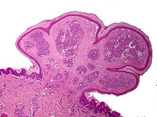
A pyogenic granuloma or lobular capillary hemangioma is a vascular tumor that occurs on both mucosa and skin, and appears as an overgrowth of tissue due to irritation, physical trauma, or hormonal factors. It is often found to involve the gums, skin, or nasal septum, and has also been found far from the head, such as in the thigh.
A vascular tumor is a tumor of vascular origin; a soft tissue growth that can be either benign or malignant, formed from blood vessels or lymph vessels. Examples of vascular tumors include hemangiomas, lymphangiomas, hemangioendotheliomas, Kaposi's sarcomas, angiosarcomas, and hemangioblastomas. An angioma refers to any type of benign vascular tumor.
A vascular malformation is a blood vessel or lymph vessel abnormality. Vascular malformations are one of the classifications of vascular anomalies, the other grouping is vascular tumors. They may cause aesthetic problems as they have a growth cycle, and can continue to grow throughout life.

A tufted angioma, also known as an acquired tufted angioma, angioblastoma, angioblastoma of Nakagawa, hypertrophic hemangioma, progressive capillary hemangioma, and tufted hemangioma usually develops in infancy or early childhood on the neck and upper trunk, and is an ill-defined, dull red macule with a mottled appearance, varying from 2 to 5 cm in diameter.
Microvenular hemangioma is an acquired benign vascular tumor that presents as an asymptomatic, slowly growing, 0.5- to 2.0 cm reddish lesion on the forearms or other sites of young to middle-aged adults. The cause of microvenular hemangioma is unknown, however it has been associated with immunosuppression.
Targetoid hemosiderotic hemangioma, also known as a hobnail hemangioma is a skin condition characterized by a central brown or purplish papule that is surrounded by an ecchymotic halo. It may appear similar to melanoma. It was first described by Santa Cruz and Aronberg in 1988.
Glomeruloid hemangioma is a distinctive vascular tumor first described in 1990 when found to be associated with POEMS syndrome and Castleman disease. Glomeruloid hemangiomas can manifest as wine-red sessile or pedunculated papules, papulonodules, subcutaneous bluish compressible tumors, or small, firm, reddish-violaceous, dome-shaped papules.
A vascular anomaly is any of a range of lesions from a simple birthmark to a large tumor that may be disfiguring. They are caused by a disorder of the vascular system. A vascular anomaly is a localized defect in blood or lymph vessels. These defects are characterized by an increased number of vessels, and vessels that are both enlarged and sinuous. Some vascular anomalies are congenital, others appear within weeks to years after birth, and others are acquired by trauma or during pregnancy. Inherited vascular anomalies are also described and often present with a number of lesions that increase with age. Vascular anomalies can also be a part of a syndrome.

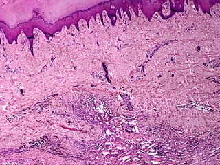
Cavernous hemangioma, also called cavernous angioma, venous malformation, or cavernoma, is a type of venous malformation due to endothelial dysmorphogenesis from a lesion which is present at birth. A cavernoma in the brain is called a cerebral cavernous malformation or CCM. Despite its designation as a hemangioma, a cavernous hemangioma is not a tumor as it does not display endothelial hyperplasia. The abnormal tissue causes a slowing of blood flow through the cavities, or "caverns". The blood vessels do not form the necessary junctions with surrounding cells, and the structural support from the smooth muscle is hindered, causing leakage into the surrounding tissue. It is the leakage of blood, referred to as hemorrhage, that causes a variety of symptoms known to be associated with the condition.
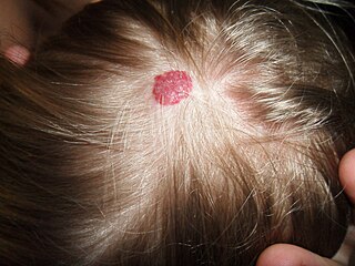
A hemangioma or haemangioma is a usually benign vascular tumor derived from blood vessel cell types. The most common form, seen in infants, is an infantile hemangioma, known colloquially as a "strawberry mark", most commonly presenting on the skin at birth or in the first weeks of life. A hemangioma can occur anywhere on the body, but most commonly appears on the face, scalp, chest or back. They tend to grow for up to a year before gradually shrinking as the child gets older. A hemangioma may need to be treated if it interferes with vision or breathing or is likely to cause long-term disfigurement. In rare cases internal hemangiomas can cause or contribute to other medical problems. They usually disappear in 10 years. The first line treatment option is beta blockers, which are highly effective in the majority of cases. Hemangiomas that form at birth are called congenital hemangiomas, while those that form later in life are called infantile hemangiomas.

Hereditary neurocutaneous angioma is a rare genetic disorder characterized by the appearance of angiomas on cutaneous and neurological areas of the body in multiple members of a single family.