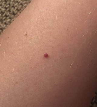
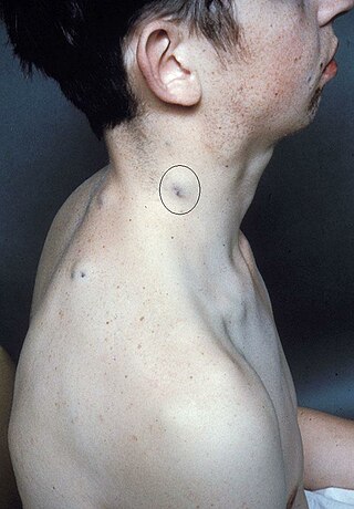
A venous lake (also known as phlebectasis[1]) is a generally solitary, soft, compressible, dark blue to violaceous, 0.2- to 1-cm papule commonly found on sun-exposed surfaces of the vermilion border of the lip, face and ears.[2][3][4] Lesions generally occur among the elderly.[5][6]
Though these lesions may resemble nodular melanoma, the lack of induration, slow growth, and lightening appearance upon diascopy suggest against it, and indicate a vascular lesion.[7] Additionally, lack of pulsation distinguishes this lesion of the lower lip from a tortuous segment of the inferior labial artery.[4]
Cause
The cause is unknown; however it is thought to be associated with sun exposure, leading to a dilated blood-filled vascular channel[2] "...lined with a singled layer of flattened endothelial cells and a thin wall of fibrous tissue filled with red blood cells."[7]
Treatment
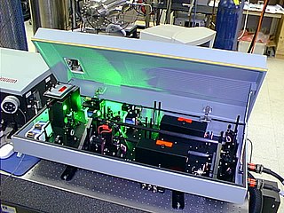
Treatment may be requested for cosmetic reasons. Traditional techniques such as surgical excision are effective but will leave a scar. Laser therapy has become the mainstay of therapy. Published research suggests that the Long Pulsed Nd:YAG laser is a very effective, with a clearance rate of 94% following a single treatment. In this study no scarring or other complications were reported. [8]
Intense pulsed light (IPL) is a technology used by cosmetic and medical practitioners to perform various skin treatments for aesthetic and therapeutic purposes, including hair removal, photorejuvenation as well as to alleviate dermatologic diseases such as acne. IPL is increasingly used in optometry and ophthalmology as well, to treat evaporative dry eye disease due to meibomian gland dysfunction.
Nd:YAG (neodymium-doped yttrium aluminum garnet; Nd:Y3Al5O12) is a crystal that is used as a lasing medium for solid-state lasers. The dopant, triply ionized neodymium, Nd(III), typically replaces a small fraction (1%) of the yttrium ions in the host crystal structure of the yttrium aluminum garnet (YAG), since the two ions are of similar size. It is the neodymium ion which provides the lasing activity in the crystal, in the same fashion as red chromium ion in ruby lasers.
Laser hair removal is the process of hair removal by means of exposure to pulses of laser light that destroy the hair follicle. It had been performed experimentally for about twenty years before becoming commercially available in 1995 and 1996. One of the first published articles describing laser hair removal was authored by the group at Massachusetts General Hospital in 1998. Laser hair removal is widely practiced in clinics, and even in homes using devices designed and priced for consumer self-treatment. Many reviews of laser hair removal methods, safety, and efficacy have been published in the dermatology literature.
An infantile hemangioma (IH), sometimes called a strawberry mark due to appearance, is a type of benign vascular tumor or anomaly that affects babies. Other names include capillary hemangioma, strawberry hemangioma, strawberry birthmark and strawberry nevus. and formerly known as a cavernous hemangioma. They appear as a red or blue raised lesion on the skin. Typically, they begin during the first four weeks of life, growing until about five months of life, and then shrinking in size and disappearing over the next few years. Often skin changes remain after they shrink. Complications may include pain, bleeding, ulcer formation, disfigurement, or heart failure. It is the most common tumor of orbit and periorbital areas in childhood. It may occur in the skin, subcutaneous tissues and mucous membranes of oral cavities and lips as well as in extracutaneous locations including the liver and gastrointestinal tract.
Hyperpigmentation is the darkening of an area of skin or nails caused by increased melanin.
A port-wine stain is a discoloration of the human skin caused by a vascular anomaly. They are so named for their coloration, which is similar in color to port wine, a fortified red wine from Portugal.
Cherry angioma, also called cherry hemangioma, is a small bright red dome-shaped bump on the skin. It ranges between 0.5 – 6 mm in diameter and usually several are present, typically on the chest and arms, and increasing in number with age. If scratched, they may bleed.
Telangiectasias, also known as spider veins, are small dilated blood vessels that can occur near the surface of the skin or mucous membranes, measuring between 0.5 and 1 millimeter in diameter. These dilated blood vessels can develop anywhere on the body, but are commonly seen on the face around the nose, cheeks and chin. Dilated blood vessels can also develop on the legs, although when they occur on the legs, they often have underlying venous reflux or "hidden varicose veins". When found on the legs, they are found specifically on the upper thigh, below the knee joint and around the ankles.
Pseudofolliculitis barbae (PFB) is a persistent irritation caused by shaving. It was first described in 1956.
Sclerotherapy is a procedure used to treat blood vessel malformations and also malformations of the lymphatic system. A medicine is injected into the vessels, which makes them shrink. It is used for children and young adults with vascular or lymphatic malformations. In adults, sclerotherapy is often used to treat spider veins, smaller varicose veins, hemorrhoids, and hydroceles.
Laser surgery is a type of surgery that uses a laser to cut tissue.
Angiokeratoma is a benign cutaneous lesion of capillaries, resulting in small marks of red to blue color and characterized by hyperkeratosis. Angiokeratoma corporis diffusum refers to Fabry's disease, but this is usually considered a distinct condition.
Tattoo removal is the process of removing an unwanted tattoo. The process of tattooing generally creates permanent markings in the skin, but people have attempted many methods to try to hide or destroy tattoos.
Nevus of Ota is a hyperpigmentation that occurs on the face, most often appearing on the white of the eye. It also occurs on the forehead, nose, cheek, periorbital region, and temple.
Actinic cheilitis is cheilitis caused by long term sunlight exposure. Essentially it is a burn, and a variant of actinic keratosis which occurs on the lip. It is a premalignant condition, as it can develop into squamous cell carcinoma.
Photorejuvenation is a skin treatment that uses lasers, intense pulsed light, or photodynamic therapy to treat skin conditions and remove effects of photoaging such as wrinkles, spots, and textures. The process induces controlled wounds to the skin. This prompts the skin to heal itself, by creating new cells. This process—to a certain extent—removes the signs of photoaging. The technique was invented by Thomas L Roberts, III using CO2 lasers in the 1990s. Observed complications have included scarring, hyperpigmentation, acne, and herpes.
Caviar tongue is a condition characterized by the purplish nodular swelling of veins found on the undersurface of the tongue.
Blue rubber bleb nevus syndrome is a rare disorder that consists mainly of abnormal blood vessels affecting the skin or internal organs – usually the gastrointestinal tract. The disease is characterized by the presence of fluid-filled blisters (blebs) as visible, circumscribed, chronic lesions (nevi).
A vascular anomaly is any of a range of lesions from a simple birthmark to a large tumor that may be disfiguring. They are caused by a disorder of the vascular system. A vascular anomaly is a localized defect in blood or lymph vessels. These defects are characterized by an increased number of vessels, and vessels that are both enlarged and sinuous. Some vascular anomalies are congenital, others appear within weeks to years after birth, and others are acquired by trauma or during pregnancy. Inherited vascular anomalies are also described and often present with a number of lesions that increase with age. Vascular anomalies can also be a part of a syndrome.
Diffuse capillary malformation with overgrowth (DCMO) is a subset of capillary malformations (CM) associated with hypertrophy, i.e. increased size of body structures. CM can be considered an umbrella term for various vascular anomalies caused by increased diameter or number of capillary blood vessels. It is commonly referred to as "port-wine stain", and is thought to affect approximately 0.5% of the population. Typically capillaries in the papillary dermis are involved, and this gives rise to pink or violaceous colored lesions. The majority of DCMO lesions are diffuse, reticulated pale-colored stains.
This page is based on this Wikipedia article Text is available under the CC BY-SA 4.0 license; additional terms may apply. Images, videos and audio are available under their respective licenses.