Veins are blood vessels in the circulatory system of humans and most other animals that carry blood towards the heart. Most veins carry deoxygenated blood from the tissues back to the heart; exceptions are those of the pulmonary and fetal circulations which carry oxygenated blood to the heart. In the systemic circulation, arteries carry oxygenated blood away from the heart, and veins return deoxygenated blood to the heart, in the deep veins.

The left and right brachiocephalic veins are major veins in the upper chest, formed by the union of the ipsilateral internal jugular vein and subclavian vein behind the sternoclavicular joint. The left brachiocephalic vein is more than twice the length of the right brachiocephalic vein.

In human anatomy, the subclavian arteries are paired major arteries of the upper thorax, below the clavicle. They receive blood from the aortic arch. The left subclavian artery supplies blood to the left arm and the right subclavian artery supplies blood to the right arm, with some branches supplying the head and thorax. On the left side of the body, the subclavian comes directly off the aortic arch, while on the right side it arises from the relatively short brachiocephalic artery when it bifurcates into the subclavian and the right common carotid artery.

The foramen spinosum is a small open hole in the greater wing of the sphenoid bone that gives passage to the middle meningeal artery and vein, and the meningeal branch of the mandibular nerve.

In human anatomy, the internal thoracic vein is the vein that drains the chest wall and breasts.

The perforating cutaneous nerve is a cutaneous nerve of the sacral plexus that provides sensory innervation to the skin of the buttocks.

A jugular foramen is one of the two large foramina (openings) in the base of the skull, located behind the carotid canal. It is formed by the temporal bone and the occipital bone. It allows many structures to pass, including the inferior petrosal sinus, three cranial nerves, the sigmoid sinus, and meningeal arteries.

The round ligament of the uterus is a ligament that connects the uterus to the labia majora. It originates at the junction of the uterus and uterine tube. It passes through the inguinal canal to insert at the labium majus.
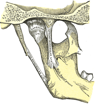
The sphenomandibular ligament is one of the three ligaments of the temporomandibular joint. It is situated medially to - and generally separate from - the articular capsule of the joint. Superiorly, it is attached to the spine of the sphenoid bone; inferiorly, it is attached to the lingula of mandible. The SML acts to limit inferior-ward movement of the mandible.

In human anatomy, the falciform ligament is a ligament that attaches the liver to the front body wall and divides the liver into the left lobe and right lobe. The falciform ligament is a broad and thin fold of peritoneum, its base being directed downward and backward and its apex upward and forward. It droops down from the hilum of the liver.

The maxillary vein or internal maxillary vein is a vein of the head. It is a short trunk which accompanies the maxillary artery. It is formed by a confluence of the veins of the pterygoid plexus. It and passes posterior-ward between the sphenomandibular ligament and the neck of the mandible to enter the parotid gland where unites with the superficial temporal vein to form the retromandibular vein.

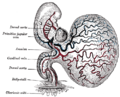
The sinus venosus is a large quadrangular cavity which precedes the atrium on the venous side of the chordate heart.
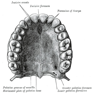
In the human mouth, the incisive foramen is the opening of the incisive canals on the hard palate immediately behind the incisor teeth. It gives passage to blood vessels and nerves. The incisive foramen is situated within the incisive fossa of the maxilla.

The broad ligament of the uterus is the wide fold of peritoneum that connects the sides of the uterus to the walls and floor of the pelvis.
The suspensory ligament of the ovary, also infundibulopelvic ligament, is a fold of peritoneum that extends out from the ovary to the wall of the pelvis.

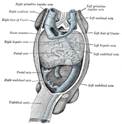
The common cardinal veins, also known as the ducts of Cuvier, are veins that drain into the sinus venosus during embryonic development. These drain an anterior cardinal vein and a posterior cardinal vein on each side. Each of the ducts of Cuvier receives an ascending vein. The ascending veins return the blood from the parietes of the trunk and from the Wolffian bodies, and are called cardinal veins. Part of the left common cardinal vein persists after birth to form the coronary sinus.

The tela choroidea is a region of meningeal pia mater that adheres to the underlying ependyma, and gives rise to the choroid plexus in each of the brain’s four ventricles. Tela is Latin for woven and is used to describe a web-like membrane or layer. The tela choroidea is a very thin part of the loose connective tissue of pia mater overlying and closely adhering to the ependyma. It has a rich blood supply. The ependyma and vascular pia mater – the tela choroidea, form regions of minute projections known as a choroid plexus that projects into each ventricle. The choroid plexus produces most of the cerebrospinal fluid of the central nervous system that circulates through the ventricles of the brain, the central canal of the spinal cord, and the subarachnoid space. The tela choroidea in the ventricles forms from different parts of the roof plate in the development of the embryo.

The following outline is provided as an overview of and topical guide to human anatomy:
The posterior cardinal veins or postcardinal veins join with the corresponding right and left cardinal veins to form the left common cardinal veins, which empty in the sinus venosus. In the development of a human embryo, most of the posterior cardinal veins regress, and what remains of them forms the renal segment of the inferior vena cava and the common iliac veins. Later in the development stages, the posterior cardinal veins are replaced by the subcardinal and supracardinal veins. The subcardinal veins form part of the inferior vena cava, the renal veins and the gonadal veins. The supracardinal veins form part of the inferior vena cava, the intercostal veins, the hemiazygos vein and the azygos vein.

Franklin Paine Mall was an American anatomist and pathologist known for his research and literature in the fields of anatomy and embryology. Mall was granted a fellowship for the Department of Pathology at the Johns Hopkins University and after positions at other universities, later returned to be the head of the first Anatomy Department at the Johns Hopkins School of Medicine. There, he reformed the field of anatomy and its educational curriculum. Mall was the founder and the first chief of the Department of Embryology at the Carnegie Institution for Science. He later donated his collection of human embryos that he started as a postgraduate student to the Carnegie Institution for Science. He was an elected member of the American Academy of Arts and Sciences, the American Philosophical Society, and the United States National Academy of Sciences.