Heart development

Last updated
Heart development
2037 Embryonic Development of Heart.jpg
Development of the human heart during the first five weeks (top), and the formation of the heart chambers (bottom) at four and eight weeks. In this figure, the blue and red colors represent blood inflow and outflow (not venous and arterial blood). Initially, all venous blood flows from the tail/atria to the ventricles/head, a very different pattern from that of an adult. [1] [2]
Details
Gives rise to Heart
System Fetal circulation, circulatory system
Anatomical terminology

Heart development, also known as cardiogenesis, refers to the prenatal development of the heart. This begins with the formation of two endocardial tubes which merge to form the tubular heart, also called the primitive heart tube. The heart is the first functional organ in vertebrate embryos.

Contents

The tubular heart quickly differentiates into the truncus arteriosus, bulbus cordis, primitive ventricle, primitive atrium, and the sinus venosus. The truncus arteriosus splits into the ascending aorta and the pulmonary trunk. The bulbus cordis forms part of the ventricles. The sinus venosus connects to the fetal circulation.

The heart tube elongates on the right side, looping and becoming the first visual sign of left-right asymmetry of the body. Septa form within the atria and ventricles to separate the left and right sides of the heart. [3]

Early development

The heart derives from embryonic mesodermal germ layer cells that differentiate after gastrulation into mesothelium, endothelium, and myocardium. Heart induction occurs in the anterior mesoderm during gastrulation through interactions with adjacent endoderm (both extra-embryonic and definitive) mediated primarily by endogenous inhibitors of WNT signaling such as DKK1. [4] [5] Mesothelial pericardium forms the outer lining of the heart. The inner lining of the heart – the endocardium, lymphatic and blood vessels, develop from endothelium. [6] [2]

Endocardial tubes

In the splanchnopleuric mesenchyme on either side of the neural plate, a horseshoe-shaped area develops as the cardiogenic region. This has formed from cardiac myoblasts and blood islands as forerunners of blood cells and vessels. [7] By day 19, an endocardial tube begins to develop in each side of this region. These two tubes grow and by the third week have converged towards each other to merge, using programmed cell death to form a single tube, the tubular heart. [8]

From splanchnopleuric mesenchyme, the cardiogenic region develops cranially and laterally to the neural plate. In this area, two separate angiogenic cell clusters form on either side and coalesce to form the endocardial tubes. As embryonic folding starts, the two endocardial tubes are pushed into the thoracic cavity, where they begin to fuse together, and this is completed at about 22 days. [9] [2]

At around 18 to 19 days after fertilisation, the heart begins to form. The heart begins to develop near the head of the embryo in the cardiogenic area. [1] Following cell signalling, two strands or cords begin to form in the cardiogenic region [1] As these form, a lumen develops within them, at which point, they are referred to as endocardial tubes. [1] At the same time that the tubes are forming other major heart components are also being formed. [8] The two tubes migrate together and fuse to form a single primitive heart tube which quickly forms five distinct regions. [1] From head to tail, these are the truncus arteriosus, bulbus cordis, primitive ventricle, primitive atrium, and the sinus venosus. [1] Initially, all venous blood flows into the sinus venosus, and contractions propel the blood from tail to head, or from the sinus venosus to the truncus arteriosus. [1] The truncus arteriosus will divide to form the aorta and pulmonary artery; the bulbus cordis will develop into the right ventricle; the primitive ventricle will form the left ventricle; the primitive atrium will become the front parts of the left and right atria and their appendages, and the sinus venosus will develop into the posterior part of the right atrium, the sinoatrial node and the coronary sinus. [1]

Heart tube position

The central part of cardiogenic area is in front of the oropharyngeal membrane and the neural plate. The growth of the brain and the cephalic folds push the oropharyngeal membrane forward, while the heart and the pericardial cavity move first to the cervical region and then into the chest. The curved portion of the horseshoe-shaped area expands to form the future ventricular infundibulum and the ventricular regions, as the heart tube continues to expand. The tube starts receiving venous drainage in its caudal pole and will pump blood out of the first aortic arch and into the dorsal aorta through its polar head. Initially the tube remains attached to the dorsal part of the pericardial cavity by a mesodermal tissue fold called the dorsal mesoderm. This mesoderm disappears to form the two pericardial sinuses the transverse and the oblique pericardial sinuses, which connect both sides of the pericardial cavity. [7]

The myocardium thickens and secretes a thick layer of rich extracellular matrix containing hyaluronic acid which separates the endothelium. Then mesothelial cells form the pericardium and migrate to form most of the epicardium. Then the heart tube is formed by the endocardium, which is the inner endothelial lining of the heart, and the myocardial muscle wall which is the epicardium that covers the outside of the tube. [7]

Heart folding and turning

The heart tube continues stretching and by day 23, in a process called morphogenesis, cardiac looping begins. The cephalic portion curves in a frontal clockwise direction. The atrial portion starts moving in a cephalically and then moves to the left from its original position. This curved shape approaches the heart and finishes its growth on day 28. The conduit forms the atrial and ventricular junctions which connect the common atrium and the common ventricle in the early embryo. The arterial bulb forms the trabecular portion of the right ventricle. A cone will form the infundibula blood of both ventricles. The arterial trunk and the roots will form the proximal portion of the aorta and the pulmonary artery. The junction between the ventricle and the arterial bulb will be called the primary intra-ventricular hole. The tube is divided into cardiac regions along its craniocaudal axis: the primitive ventricle, called primitive left ventricle, and the trabecular proximal arterial bulb, called the primitive right ventricle. [10] This time no septum is present in heart.

A functional explanation for the enigmatic turning of the heart and bowels is lacking, but one theory does give an explanation for the evolution and development of this phenomenon. According to this axial twist theory, this is due to a twist in the body of all vertebrates that occurs in the early embryo. The twist turns the anterior head (with the face and cerebrum) clockwise and the rest of the exterior body anticlockwise, such that the vertebrate body is symmetric on the outside. Since there is no evolutionary pressure on the heart and inner organs for bilateral symmetry, these body parts are excluded from the twisting and remain asymmetric. [11]

Heart chambers

Sinus venosus

In the middle of the fourth week, the sinus venosus receives venous blood from the poles of right and left sinus. Each pole receives blood from three major veins: the vitelline vein, the umbilical vein and the common cardinal vein. The sinus opening moves clockwise. This movement is caused mainly by the left to right shunt of blood, which occurs in the venous system during the fourth and fifth week of development. [12]

When the left common cardinal vein disappears in the tenth week only the oblique vein of the left atrium and the coronary sinus remain. The right pole joins the right atrium to form the wall portion of the right atrium. The right and left venous valves fuse and form a peak known as the septum spurium. At the beginning, these valves are large, but over time the left venous valve and the septum spurium fuse with the developing atrial septum. The upper right venous valve disappears, while the bottom venous valve evolves into the inferior valve of the vena cava and the coronary sinus valve. [12]

Heart wall

The main walls of the heart are formed between day 27 and 37 of the development of the early embryo. The growth consists of two tissue masses actively growing that approach one another until they merge and split light into two separate conduits. Tissue masses called endocardial cushions develop into atrioventricular and conotruncal regions. In these places, the cushions will help in the formation of auricular septum, ventricular conduits, atrio-ventricular valves and aortic and pulmonary channels. [13]

Atria

The developing heart at day 30. The septum primum (top, middle) develops downwards to separate the initially joined primitive atrium into left and right atria. Gray465.png
The developing heart at day 30. The septum primum (top, middle) develops downwards to separate the initially joined primitive atrium into left and right atria.

At the end of the fourth week, a crest grows that leaves the cephalic part. This crest is the first part of the septum primum. The two ends of the septum extend into the interior of the endocardial cushions in the atrioventricular canal. The opening between the bottom edge of the septum primum and endocardial cushions is the ostium primum (first opening). The extensions of the upper and lower endocardial pads grow along the margin of the septum primum and close the ostium primum. Coalescence of these perforations will form the ostium secundum (second opening), which allows blood to flow freely from the right atrium to the left.

When the right of the atrium expands due to the incorporation of the pole of the sinus, a new fold appears, called the septum secundum. At its right side it is fused with the left venous valve and the septum spurium. A free opening will then appear, called the foramen ovale. The remains of the upper septum primum, will become the valves of the foramen ovale. The passage between the two atrial chambers consists of a long oblique slit through which blood flows from the right atrium to the left. [13]

Ventricles

Initially, a single pulmonary vein develops in the form of a bulge in the back wall of the left atrium. This vein will connect with the veins of the developing lung buds. As development proceeds, the pulmonary vein and its branches are incorporated into the left atrium and they both form the smooth wall of the atrium. The embryonic left atrium remains as the trabecular left atrial appendage, and the embryonic right atrium remains as the right atrial appendage. [14]

Septum formation of the atrioventricular canal

At the end of the fourth week, two atrioventricular endocardial cushions appear. Initially the atrioventricular canal gives access to the primitive left ventricle, and is separated from arterial bulb by the edge of the ventricular bulb. In the fifth week, the posterior end terminates in the center part of the upper endocardial cushion. Because of this, blood can access both the left primitive ventricle and the right primitive ventricle. As the anterior and posterior pads project inwardly, they merge to form a right and left atrioventricular orifice. [15]

Atrioventricular valves

When forming intra-atrial septa, atrio-ventricular valves will begin to grow. A muscular interventricular septum begins to grow from the common ventricle to the atrio-ventricular endocardial cushions. The division begins in the common ventricle where a furrow in the outer surface of the heart will appear the interventricular foramen eventually disappears. This closure is achieved by further growth of the muscular interventricular septum, a contribution of trunk crest-conal tissue and a membranous component. [16]

Valves and outflow tracts

Truncus septum formation and arterial cone

The arterial cone is closed by the infundibular cushions. The trunk cones are closed by the forming of an infundibulotroncal septum, which is made from a straight proximal portion and distal spiral portion. Then, the narrowest portion of the aorta is in the left and dorsal portion. The distal portion of the aorta is pushed forward to the right. The proximal pulmonary artery is right and ventral, and the distal portion of the pulmonary artery is in the left dorsal portion. [13]

Pacemaker and conduction system

The rhythmic electrical depolarization waves that trigger myocardial contraction is myogenic, which means that they begin in the heart muscle spontaneously and are then responsible for transmitting signals from cell to cell. Myocytes that were obtained in the primitive heart tube, start beating as they connect together by their walls in a syncytium. Myocytes initiate rhythmic electrical activity, before the fusion of the endocardial tubes. The heartbeat begins in the region of the pacemaker which has a spontaneous depolarization time faster than the rest of myocardium. [17]

The primitive ventricle acts as initial pacemaker. But this pacemaker activity is actually made by a group of cells that derive from the sinoatrial right venous sinus. These cells form an ovoid sinoatrial node (SAN), on the left venous valve. After the development of the SAN, the superior endocardial cushions begin to form a pacemaker also known as the atrioventricular node. With the development of the SAN, a band of specialized conducting cells start to form creating the bundle of His that sends a branch to the right ventricle and one to the left ventricle. Most conduction pathways originate from the cardiogenic mesoderm but the sinus node may be derived from the neural crest. [17]

The human embryonic heart displays cardiac activity approximately 21 days after fertilization, or five weeks after the last normal menstrual period (LMP), which is the date normally used to date pregnancy in the medical community. The electrical depolarizations that trigger cardiac myocytes to contract arise spontaneously within the myocyte itself. The heartbeat is initiated in the pacemaker regions and spreads to the rest of the heart through a conduction pathway. Pacemaker cells develop in the primitive atrium and the sinus venosus to form the sinoatrial node and the atrioventricular node respectively. Conductive cells develop the bundle of His and carry the depolarization into the lower heart. Cardiac activity is visible beginning at approximately 5 weeks of pregnancy.

The human heart begins beating at a rate near the mother's, about 75-80 beats per minute (BPM). The embryonic heart rate (EHR) then accelerates linearly for the first month of beating, peaking at 165-185 BPM during the early 7th week, (early 9th week after the LMP). This acceleration is approximately 3.3 BPM per day, or about 10 BPM every three days, an increase of 100 BPM in the first month. [18]

After peaking at about 9.2 weeks after the LMP, it decelerates to about 150 BPM (+/-25 BPM) during the 15th week after the LMP. After the 15th week the deceleration slows reaching an average rate of about 145 (+/-25 BPM) BPM at term.

Imaging

Device for obstetric ultrasonography including usage in 1st trimester. Transvaginal ultrasonography device.png
Device for obstetric ultrasonography including usage in 1st trimester.
Transvaginal ultrasonography of an embryo at 5 weeks and 5 days of gestational age, with discernible cardiac activity (arrow). Embryo at 5 weeks 5 days with heartbeat - annotated.gif
Transvaginal ultrasonography of an embryo at 5 weeks and 5 days of gestational age, with discernible cardiac activity (arrow).

In the first trimester, cardiac activity can be visualized and the fetal heart motion quantified by obstetric ultrasonography. A study of 32 normal pregnancies showed that fetal heart motion was visible at a mean human chorionic gonadotropin (hCG) level of 10,000 UI/L (range 8650–12,200). [19] Obstetric ultrasonography can also use Doppler technique on key vessels such as the umbilical artery to detect abnormal flow.

Doppler fetal monitor Doptone.jpg
Doppler fetal monitor

In later stages of pregnancy, a simple Doppler fetal monitor can be used to quantify the fetal heart rate.

A fetal heartbeat can be detected at around 17 to 20 weeks of gestation when the chambers of the heart have become sufficiently developed. [20]

During childbirth, the parameter is part of cardiotocography, which is where the fetal heartbeat and uterine contractions are continuously recorded.

Additional images

Related Research Articles

<span class="mw-page-title-main">Heart</span> Organ found inside most animals

The heart is a muscular organ in most animals. This organ pumps blood through the blood vessels of the circulatory system. The pumped blood carries oxygen and nutrients to the body, while carrying metabolic waste such as carbon dioxide to the lungs. In humans, the heart is approximately the size of a closed fist and is located between the lungs, in the middle compartment of the chest, called the mediastinum.

<span class="mw-page-title-main">Heart valve</span> A flap of tissue that prevent backflow of blood around the heart

A heart valve is a biological one-way valve that allows blood to flow in one direction through the chambers of the heart. Four valves are usually present in a mammalian heart and together they determine the pathway of blood flow through the heart. A heart valve opens or closes according to differential blood pressure on each side.

<span class="mw-page-title-main">Systole</span> Part of the cardiac cycle when a heart chamber contracts

Systole is the part of the cardiac cycle during which some chambers of the heart contract after refilling with blood.

<span class="mw-page-title-main">Ventricle (heart)</span> Chamber of the heart

A ventricle is one of two large chambers located toward the bottom of the heart that collect and expel blood towards the peripheral beds within the body and lungs. The blood pumped by a ventricle is supplied by an atrium, an adjacent chamber in the upper heart that is smaller than a ventricle. Interventricular means between the ventricles, while intraventricular means within one ventricle.

<span class="mw-page-title-main">Cardiac conduction system</span> Aspect of heart function

The cardiac conduction system transmits the signals generated by the sinoatrial node – the heart's pacemaker, to cause the heart muscle to contract, and pump blood through the body's circulatory system. The pacemaking signal travels through the right atrium to the atrioventricular node, along the bundle of His, and through the bundle branches to Purkinje fibers in the walls of the ventricles. The Purkinje fibers transmit the signals more rapidly to stimulate contraction of the ventricles.

<span class="mw-page-title-main">Atrial septal defect</span> Human heart defect present at birth

Atrial septal defect (ASD) is a congenital heart defect in which blood flows between the atria of the heart. Some flow is a normal condition both pre-birth and immediately post-birth via the foramen ovale; however, when this does not naturally close after birth it is referred to as a patent (open) foramen ovale (PFO). It is common in patients with a congenital atrial septal aneurysm (ASA).

<span class="mw-page-title-main">Ostium primum atrial septal defect</span> Medical condition

The ostium primum atrial septal defect is a defect in the atrial septum at the level of the tricuspid and mitral valves. This is sometimes known as an endocardial cushion defect because it often involves the endocardial cushion, which is the portion of the heart where the atrial septum meets the ventricular septum and the mitral valve meets the tricuspid valve.

<span class="mw-page-title-main">Congenital heart defect</span> Defect in the structure of the heart that is present at birth

A congenital heart defect (CHD), also known as a congenital heart anomaly, congenital cardiovascular malformation, and congenital heart disease, is a defect in the structure of the heart or great vessels that is present at birth. A congenital heart defect is classed as a cardiovascular disease. Signs and symptoms depend on the specific type of defect. Symptoms can vary from none to life-threatening. When present, symptoms are variable and may include rapid breathing, bluish skin (cyanosis), poor weight gain, and feeling tired. CHD does not cause chest pain. Most congenital heart defects are not associated with other diseases. A complication of CHD is heart failure.

<span class="mw-page-title-main">Atrium (heart)</span> Part of the human heart

The atrium is one of the two upper chambers in the heart that receives blood from the circulatory system. The blood in the atria is pumped into the heart ventricles through the atrioventricular mitral and tricuspid heart valves.

<span class="mw-page-title-main">Cardiac cycle</span> Performance of the human heart

The cardiac cycle is the performance of the human heart from the beginning of one heartbeat to the beginning of the next. It consists of two periods: one during which the heart muscle relaxes and refills with blood, called diastole, following a period of robust contraction and pumping of blood, called systole. After emptying, the heart relaxes and expands to receive another influx of blood returning from the lungs and other systems of the body, before again contracting to pump blood to the lungs and those systems.

<span class="mw-page-title-main">Atrioventricular septal defect</span> Medical condition

Atrioventricular septal defect (AVSD) or atrioventricular canal defect (AVCD), also known as "common atrioventricular canal" or "endocardial cushion defect" (ECD), is characterized by a deficiency of the atrioventricular septum of the heart that creates connections between all four of its chambers. It is a very specific combination of 3 defects:

<span class="mw-page-title-main">Coronary sinus</span> Set of veins which drain blood from the myocardium (heart muscle)

The coronary sinus is the largest vein of the heart. It drains over half of the deoxygenated blood from the heart muscle into the right atrium. It begins on the backside of the heart, in between the left atrium, and left ventricle; it begins at the junction of the great cardiac vein, and oblique vein of the left atrium. It receives multiple tributaries. It passes across the backside of the heart along a groove between left atrium and left ventricle, then drains into the right atrium at the orifice of the coronary sinus.

Cor triatriatum is a congenital heart defect where the left atrium or right atrium is subdivided by a thin membrane, resulting in three atrial chambers.

<span class="mw-page-title-main">Sinus venosus</span>

The sinus venosus is a large quadrangular cavity which precedes the atrium on the venous side of the chordate heart.

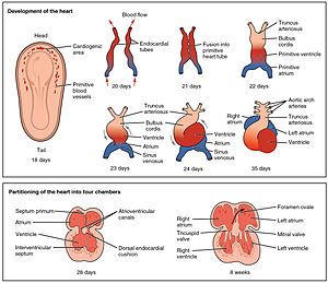
The proper development of the atrioventricular canal into its prospective components to create a clear division between the four compartments of the heart and ensure proper blood movement through the heart, are essential for proper heart function. When this process does not happen correctly, a child will develop atrioventricular canal defect which occurs in 2 out of every 10,000 births. It also has a correlation with Down syndrome because 20% of children with Down syndrome have atrioventricular canal disease as well. This is a very serious condition and surgery is necessary within the first six months of life for a child. Half of the children who are untreated with this condition die during their first year due to heart failure or pneumonia.

<span class="mw-page-title-main">Valve of inferior vena cava</span>

The valve of the inferior vena cava is a venous valve that lies at the junction of the inferior vena cava and right atrium.

<span class="mw-page-title-main">Truncus arteriosus</span> Structure during embryonic development

The truncus arteriosus is a structure that is present during embryonic development. It is an arterial trunk that originates from both ventricles of the heart that later divides into the aorta and the pulmonary trunk.

The heart is the first functional organ in a vertebrate embryo. There are 5 stages to heart development.

Cardiac physiology or heart function is the study of healthy, unimpaired function of the heart: involving blood flow; myocardium structure; the electrical conduction system of the heart; the cardiac cycle and cardiac output and how these interact and depend on one another.

The heart is a muscular organ situated in the mediastinum. It consists of four chambers, four valves, two main arteries, and the conduction system. The left and right sides of the heart have different functions: the right side receives de-oxygenated blood through the superior and inferior venae cavae and pumps blood to the lungs through the pulmonary artery, and the left side receives saturated blood from the lungs.

References

  1. 1 2 3 4 5 6 7 8 Betts JG (2013). Anatomy & physiology. pp. 787–846. ISBN   978-1938168130 . Retrieved 11 August 2014.
  2. 1 2 3 Hosseini HS, Garcia KE, Taber LA (July 2017). "A new hypothesis for foregut and heart tube formation based on differential growth and actomyosin contraction". Development. 144 (13): 2381–2391. doi:10.1242/dev.145193. PMC   5536863 . PMID   28526751.
  3. Anderson RH, Webb S, Brown NA, Lamers W, Moorman A (August 2003). "Development of the heart: (2) Septation of the atriums and ventricles". Heart. 89 (8): 949–958. doi:10.1136/heart.89.8.949. PMC   1767797 . PMID   12860885.
  4. Schneider VA, Mercola M (February 2001). "Wnt antagonism initiates cardiogenesis in Xenopus laevis". Genes & Development. 15 (3): 304–315. doi:10.1101/gad.855601. PMC   312618 . PMID   11159911.
  5. Marvin MJ, Di Rocco G, Gardiner A, Bush SM, Lassar AB (February 2001). "Inhibition of Wnt activity induces heart formation from posterior mesoderm". Genes & Development. 15 (3): 316–327. doi:10.1101/gad.855501. PMC   312622 . PMID   11159912.
  6. "Animal Tissues". Users.rcn.com. 2010-08-13. Archived from the original on 2009-05-05. Retrieved 2010-10-17.
  7. 1 2 3 Sadler TW (2012). Langman. Embriología Médica. Lippincott Williams & Wilkins. p. 165. ISBN   978-84-96921-46-7.
  8. 1 2 Larsen W (2001). Human Embryology (3rd ed.). Elsevier Saunders. pp. 159–163. ISBN   978-0-443-06583-5.
  9. "Main Frame Heart Development". Meddean.luc.edu. Retrieved 2010-10-17.
  10. Rohen J, Lutjen E (2008). Embriología functional: una perspectiva desde la biología del desarrollo. Panamericana. p. 70. ISBN   978-84-9835-155-2.
  11. de Lussanet MH, Osse JW (2012). "An ancestral axial twist explains the contralateral forebrain and the optic chiasm in vertebrates". Animal Biology. 62 (2): 193–216. arXiv: 1003.1872 . doi:10.1163/157075611X617102. S2CID   7399128.
  12. 1 2 Carlson B (2012). Embriología humana y biología del desarrollo. Mosby. p. 451. ISBN   978-84-8174-785-0.
  13. 1 2 3 Fernández PM (2002). Manual de biología del desarrollo. Manual Moderno. p. 243. ISBN   978-968-426-976-7.
  14. Eynard A, Valentich M, Rovasio R (2011). Histología y embriología del ser humano: bases celulares y moleculares. Panamericana. p. 283. ISBN   978-950-06-0602-8.
  15. Moore KL, Persaud TV (2008). Embriología Clínica. Elsevier Saunders. p. 245. ISBN   978-84-8086-337-7.
  16. Tellez de Peralta G (2003). Tratado de cirugía cardiovascular. Díaz de Santos. p. 44.
  17. 1 2 Larsen W (2003). Embriología humana. Elsevier Science. p. 177. ISBN   978-968-426-976-7.
  18. OBGYN.net "Embryonic Heart Rates Compared in Assisted and Non-Assisted Pregnancies" Archived 2006-06-30 at the Wayback Machine
  19. Giacomello F, Magliocchetti P, Loyola G, Giovarruscio M (1993). "[Serum beta hCG levels and transvaginal echography in the early phases of pregnancy]". Minerva Ginecologica (in Italian). 45 (7–8): 333–337. PMID   8414139.
  20. "ACOG Guide to Language and Abortion". American College of Obstetricians and Gynecologists . Archived from the original on 16 November 2022. Retrieved 17 November 2022. "Heartbeat bill" - It is clinically inaccurate to use the word "heartbeat" to describe the sound that can be heard on ultrasound in very early pregnancy. In fact, there are no chambers of the heart developed at the early stage in pregnancy that this word is used to describe, so there is no recognizable "heartbeat." What pregnant women may hear is the ultrasound machine translating electronic impulses that signify fetal cardiac activity into the sound that we recognize as a heartbeat. "Fetal heartbeat" - Until the chambers of the heart have been developed and can be detected via ultrasound (roughly 17-20 weeks of gestation), it is not accurate to characterize the embryo's or fetus's cardiac development as a heartbeat.

This article incorporates text from the CC BY book: OpenStax College, Anatomy & Physiology. OpenStax CNX. 30 July 2014.