Fish anatomy is the study of the form or morphology of fish. It can be contrasted with fish physiology, which is the study of how the component parts of fish function together in the living fish. In practice, fish anatomy and fish physiology complement each other, the former dealing with the structure of a fish, its organs or component parts and how they are put together, such as might be observed on the dissecting table or under the microscope, and the latter dealing with how those components function together in living fish.

A pulmonary artery is an artery in the pulmonary circulation that carries deoxygenated blood from the right side of the heart to the lungs. The largest pulmonary artery is the main pulmonary artery or pulmonary trunk from the heart, and the smallest ones are the arterioles, which lead to the capillaries that surround the pulmonary alveoli.

In zoology and developmental anatomy, the notochord is an elastic, rod-like anatomical structure found in many deuterostomal animals. A notochord is one of five synapomorphies used to define a species as a chordate.

Persistent truncus arteriosus (PTA), often referred to simply as truncus arteriosus, is a rare form of congenital heart disease that presents at birth. In this condition, the embryological structure known as the truncus arteriosus fails to properly divide into the pulmonary trunk and aorta. This results in one arterial trunk arising from the heart and providing mixed blood to the coronary arteries, pulmonary arteries, and systemic circulation. For the International Classification of Diseases (ICD-11), the International Paediatric and Congenital Cardiac Code (IPCCC) was developed to standardize the nomenclature of congenital heart disease. Under this system, English is now the official language, and persistent truncus arteriosus should properly be termed common arterial trunk.
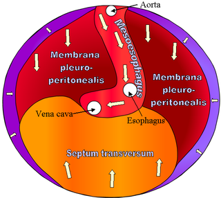
The septum transversum is a thick mass of cranial mesenchyme, formed in the embryo, that gives rise to parts of the thoracic diaphragm and the ventral mesentery of the foregut in the developed human being and other mammals.

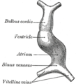
The sinus venosus is a large quadrangular cavity which precedes the atrium on the venous side of the chordate heart.
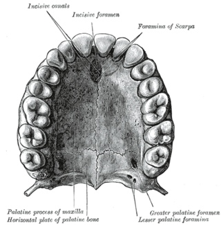
In the human mouth, the incisive foramen is the opening of the incisive canals on the hard palate immediately behind the incisor teeth. It gives passage to blood vessels and nerves. The incisive foramen is situated within the incisive fossa of the maxilla.

The vitelline veins are veins that drain blood from the yolk sac and the gut tube during gestation.
Endocardial cushions, or atrioventricular cushions, refer to a subset of cells in the development of the heart that play a vital role in the proper formation of the heart septa.

The infundibulum is a conical pouch formed from the upper and left angle of the right ventricle in the chordate heart, from which the pulmonary trunk arises. It develops from the bulbus cordis. Typically, the infundibulum refers to the corresponding internal structure, whereas the conus arteriosus refers to the external structure. Defects in infundibulum development can result in a heart condition known as tetralogy of Fallot.

The primitive ventricle or embryonic ventricle of the developing heart, together with the bulbus cordis that lies in front of it, gives rise to the left and right ventricles. The primitive ventricle provides the trabeculated parts of the walls, and the bulbus cordis the smooth parts.

A ventricular outflow tract is a portion of either the left ventricle or right ventricle of the heart through which blood passes in order to enter the great arteries.

The aorticopulmonary septum is developmentally formed from neural crest, specifically the cardiac neural crest, and actively separates the aorta and pulmonary arteries and fuses with the interventricular septum within the heart during heart development.
The truncus arteriosus is a structure that is present during embryonic development. It is an arterial trunk that originates from both ventricles of the heart that later divides into the aorta and the pulmonary trunk.

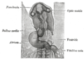
The ovarian ligament is a fibrous ligament that connects the ovary to the lateral surface of the uterus.
The heart is the first functional organ in a vertebrate embryo. There are 5 stages to heart development.

Human embryonic development or human embryogenesis is the development and formation of the human embryo. It is characterised by the processes of cell division and cellular differentiation of the embryo that occurs during the early stages of development. In biological terms, the development of the human body entails growth from a one-celled zygote to an adult human being. Fertilization occurs when the sperm cell successfully enters and fuses with an egg cell (ovum). The genetic material of the sperm and egg then combine to form the single cell zygote and the germinal stage of development commences. Embryonic development in the human, covers the first eight weeks of development; at the beginning of the ninth week the embryo is termed a fetus. The eight weeks has 23 stages.

The tubular heart or primitive heart tube is the earliest stage of heart development.
The aortic sac or aortic bulb is a dilated structure in mammalian embryos, lined by endothelial cells and is the most distal part of the truncus arteriosus. It is the primordial vascular channel from which the aortic arches arise and is homologous to the ventral aorta of gill-bearing vertebrates. The aortic sac eventually forms right and left horns, which subsequently give rise to the brachiocephalic trunk and the proximal segment of the arch of aorta, respectively.

Heart development, also known as cardiogenesis, refers to the prenatal development of the heart. This begins with the formation of two endocardial tubes which merge to form the tubular heart, also called the primitive heart tube. The heart is the first functional organ in vertebrate embryos.