Related Research Articles

Ascites is the abnormal build-up of fluid in the abdomen. Technically, it is more than 25 ml of fluid in the peritoneal cavity, although volumes greater than one liter may occur. Symptoms may include increased abdominal size, increased weight, abdominal discomfort, and shortness of breath. Complications can include spontaneous bacterial peritonitis.

A pleural effusion is accumulation of excessive fluid in the pleural space, the potential space that surrounds each lung. Under normal conditions, pleural fluid is secreted by the parietal pleural capillaries at a rate of 0.6 millilitre per kilogram weight per hour, and is cleared by lymphatic absorption leaving behind only 5–15 millilitres of fluid, which helps to maintain a functional vacuum between the parietal and visceral pleurae. Excess fluid within the pleural space can impair inspiration by upsetting the functional vacuum and hydrostatically increasing the resistance against lung expansion, resulting in a fully or partially collapsed lung.
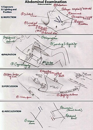
An abdominal examination is a portion of the physical examination which a physician or nurse uses to clinically observe the abdomen of a patient for signs of disease. The physical examination typically occurs after a thorough medical history is taken, that is, after the physician asks the patient the course of their symptoms. The abdominal examination is conventionally split into four different stages: first, inspection of the patient and the visible characteristics of their abdomen. Auscultation (listening) of the abdomen with a stethoscope. Palpation of the patient's abdomen. Finally, percussion (tapping) of the patient's abdomen and abdominal organs. Depending on the need to test for specific diseases such as ascites, special tests may be performed as a part of the physical examination. An abdominal examination may be performed because the physician suspects a disease of the organs inside the abdominal cavity, or simply as a part of a complete physical examination for other conditions. In a complete physical examination, the abdominal exam classically follows the respiratory examination and cardiovascular examination.
In anatomy, the precordium or praecordium is the portion of the body over the heart and lower chest.

A respiratory examination, or lung examination, is performed as part of a physical examination, in response to respiratory symptoms such as shortness of breath, cough, or chest pain, and is often carried out with a cardiac examination.
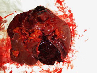
A splenic injury, which includes a ruptured spleen, is any injury to the spleen. The rupture of a normal spleen can be caused by trauma, such as a traffic collision.
Ewart's sign is a set of findings on physical examination in people with large collections of fluid around their heart.
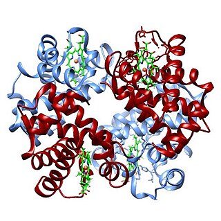
The oxygen–hemoglobin dissociation curve, also called the oxyhemoglobin dissociation curve or oxygen dissociation curve (ODC), is a curve that plots the proportion of hemoglobin in its saturated (oxygen-laden) form on the vertical axis against the prevailing oxygen tension on the horizontal axis. This curve is an important tool for understanding how our blood carries and releases oxygen. Specifically, the oxyhemoglobin dissociation curve relates oxygen saturation (SO2) and partial pressure of oxygen in the blood (PO2), and is determined by what is called "hemoglobin affinity for oxygen"; that is, how readily hemoglobin acquires and releases oxygen molecules into the fluid that surrounds it.
In medicine, shifting dullness refers to a sign elicited on physical examination for ascites.

Traube's (semilunar) space is an anatomic space of some clinical importance. It is a crescent-shaped space, encompassed by the lower edge of the left lung, the anterior border of the spleen, the left costal margin and the inferior margin of the left lobe of the liver. Thus, its surface markings are respectively the left sixth rib superiorly, the left mid axillary line laterally, and the left costal margin inferiorly.

Castell's sign is a medical sign assessed to evaluate splenomegaly and typically part of an abdominal examination. It is an alternative physical examination maneuver to percussion over Traube's space.
In medicine, the fluid wave test or fluid thrill test is a test for ascites. It is performed by having the patient push their hands down on the midline of the abdomen. The examiner then taps one flank, while feeling on the other flank for the tap. The pressure on the midline prevents vibrations through the abdominal wall while the fluid allows the tap to be felt on the other side. The result is considered positive if tap can be felt on the other side. However, even with the midline pressure, transmission through the skin must be excluded. A positive fluid wave test indicates that there is a free fluid (ascites) in the abdomen. When one side of the abdomen is pressed, the other side may also be painful due to the transfer of the fluid in it.
Sir Charles Alfred Ballance was an English surgeon who specialized in the fields of otology and neurotology.
In gastroenterology, the puddle sign is a physical examination maneuver that can be used to detect the presence of ascites. It is useful for detecting small amounts of ascites—as small as 120 mL; shifting dullness and bulging flanks typically require 500 mL.

Flanker is a position in the sport of rugby union. Each team of 15 players includes two flankers, who play in the forwards, and are generally classified as either blindside or openside flankers, numbers 6 and 7 respectively. The name comes from their position in a scrum in which they 'flank' each set of forwards. They compete for the ball – most commonly in rucks and mauls. Flankers also assist in pushing in a scrum, but are expected to detach from the scrum as soon as the ball is out to get to the play before the opposition's forwards. Flankers also participate in line-outs, either being lifted to contest or win possession, or to lift other players. Flankers are usually the key participants in the tackling process. The flankers, especially the openside, are often the fastest forwards on the team but still relied upon for tackling.
The cardiovascular examination is a portion of the physical examination that involves evaluation of the cardiovascular system. The exact contents of the examination will vary depending on the presenting complaint but a complete examination will involve the heart, lungs, belly and the blood vessels.
Lépine's sign is one of the medical signs of gallbladder disease. It is positive when effleurage with crooked third finger at the point of the gallbladder projection to anterior abdominal wall elicits pain. It is not to be confused with the following:

The human abdomen is divided into quadrants and regions by anatomists and physicians for the purposes of study, diagnosis, and treatment. The division into four quadrants allows the localisation of pain and tenderness, scars, lumps, and other items of interest, narrowing in on which organs and tissues may be involved. The quadrants are referred to as the left lower quadrant, left upper quadrant, right upper quadrant and right lower quadrant. These terms are not used in comparative anatomy, since most other animals do not stand erect.
In medical diagnosis Nixon's sign is an alternative to Castell's sign, useful in identifying splenomegaly.

Bernheim Syndrome is a presumed disorder whereby the right ventricle is severely compressed due to a shift in the ventricular septal wall of the heart leading to heart failure. It was first described by Hippolyte Bernheim in 1910. Today it is questioned whether or not Bernheim Syndrome is its own syndrome or a side effect of other cardiac conditions such as left ventricular heart failure whereby the left ventricle is substantially enlarged which encroaches on the space of the right ventricle.
References
- ↑ Rastogi, Vaibhav; Singh, Devina; Tekiner, Halil; Ye, Fan; Mazza, Joseph J.; Yale, Steven H. (March 2020). "Abdominal Physical Signs and Medical Eponyms: Part I. Percussion, 1871–1900". Clinical Medicine & Research. 18 (1): 42–47. doi: 10.3121/cmr.2018.1428 . ISSN 1539-4182.
- ↑ synd/608 at Who Named It?