Related Research Articles

Haematopoiesis is the formation of blood cellular components. All cellular blood components are derived from haematopoietic stem cells. In a healthy adult human, roughly ten billion to a hundred billion new blood cells are produced per day, in order to maintain steady state levels in the peripheral circulation.
A blood cell, also called a hematopoietic cell, hemocyte, or hematocyte, is a cell produced through hematopoiesis and found mainly in the blood. Major types of blood cells include red blood cells (erythrocytes), white blood cells (leukocytes), and platelets (thrombocytes). Together, these three kinds of blood cells add up to a total 45% of the blood tissue by volume, with the remaining 55% of the volume composed of plasma, the liquid component of blood.
Fibrinolysis is a process that prevents blood clots from growing and becoming problematic. Primary fibrinolysis is a normal body process, while secondary fibrinolysis is the breakdown of clots due to a medicine, a medical disorder, or some other cause.
Microangiopathic hemolytic anemia (MAHA) is a microangiopathic subgroup of hemolytic anemia caused by factors in the small blood vessels. It is identified by the finding of anemia and schistocytes on microscopy of the blood film.

A molar pregnancy, also known as a hydatidiform mole, is an abnormal form of pregnancy in which a non-viable fertilized egg implants in the uterus. It falls under the category of gestational trophoblastic diseases. During a molar pregnancy, the uterus contains a growing mass characterized by swollen chorionic villi, resembling clusters of grapes. The occurrence of a molar pregnancy can be attributed to the fertilized egg lacking an original maternal nucleus. As a result, the products of conception may or may not contain fetal tissue. These molar pregnancies are categorized into two types: partial moles and complete moles, where the term 'mole' simply denotes a clump of growing tissue or a ‘growth'.
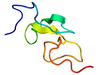
Epidermal growth factor (EGF) is a protein that stimulates cell growth and differentiation by binding to its receptor, EGFR. Human EGF is 6-kDa and has 53 amino acid residues and three intramolecular disulfide bonds.
Hematopoietic stem cells (HSCs) are the stem cells that give rise to other blood cells. This process is called haematopoiesis. In vertebrates, the very first definitive HSCs arise from the ventral endothelial wall of the embryonic aorta within the (midgestational) aorta-gonad-mesonephros region, through a process known as endothelial-to-hematopoietic transition. In adults, haematopoiesis occurs in the red bone marrow, in the core of most bones. The red bone marrow is derived from the layer of the embryo called the mesoderm.

A fatty streak is the first grossly visible lesion in the development of atherosclerosis. It appears as an irregular yellow-white discoloration on the luminal surface of an artery. It consists of aggregates of foam cells, which are lipoprotein-loaded macrophages, located in the intima, the innermost layer of the artery, beneath the endothelial cells that layer the lumina through which blood flows. Fatty streaks may also include T cells, aggregated platelets, and smooth muscle cells. Although fatty streaks can develop into atheromas, not all are destined to become advanced lesions.

A leiomyosarcoma, also known as LMS, is a rare malignant (cancerous) smooth muscle tumor. The origin of the word is from leio- + myo- + sarcoma which means malignant smooth muscle tumor. The stomach, bladder, uterus, blood vessels, and intestines are examples of hollow organs made up of smooth muscles where LMS can be located, however the uterus or abdomen are the most common sites.
Colony-stimulating factors (CSFs) are secreted glycoproteins that bind to receptor proteins on the surfaces of committed progenitors in the bone marrow, thereby activating intracellular signaling pathways that can cause the cells to proliferate and differentiate into a specific kind of blood cell.
In microbiology, colony-forming unit is a unit which estimates the number of microbial cells in a sample that are viable, able to multiply via binary fission under the controlled conditions. Counting with colony-forming units requires culturing the microbes and counts only viable cells, in contrast with microscopic examination which counts all cells, living or dead. The visual appearance of a colony in a cell culture requires significant growth, and when counting colonies, it is uncertain if the colony arose from one cell or a group of cells. Expressing results as colony-forming units reflects this uncertainty.
iC3b is a protein fragment that is part of the complement system, a component of the vertebrate immune system. iC3b is produced when complement factor I cleaves C3b. Complement receptors on white blood cells are able to bind iC3b, so iC3b functions as an opsonin. Unlike intact C3b, iC3b cannot associate with factor B, thus preventing amplification of the complement cascade through the alternative pathway. Complement factor I can further cleave iC3b into a protein fragment known as C3d.

Vinay Kumar is the Lowell T. Coggeshall Distinguished Service Professor of Pathology at the University of Chicago, where he was also the Chairman (2000-2016) of the Department of Pathology. He is a recipient of Life Time Achievement Award by National Board of Examinations.

CFU-GM, also known as granulocyte–macrophage progenitor (GMP), is a colony forming unit. It is derived from CFU-GEMM.

CFU-GEMM is a colony forming unit that generates myeloid cells. CFU-GEMM cells are the oligopotential progenitor cells for myeloid cells; they are thus also called common myeloid progenitor cells or myeloid stem cells. "GEMM" stands for granulocyte, erythrocyte, monocyte, megakaryocyte.

CFU-E stands for Colony Forming Unit-Erythroid. It arises from CFU-GEMM and gives rise to proerythroblasts.
CFU-Meg is a colony forming unit. Haematopoiesis in the bone marrow starts off from a haematopoietic stem cell (HSC) and this can differentiate into the myeloid and lymphoid cell lineages. In order to eventually produce a megakaryocyte, the haematopoietic stem cell must generate myeloid cells, so it becomes a common myeloid progenitor, CFU-GEMM. This in turn develops into CFU-Meg, which is the colony forming unit that leads to the production of megakaryocytes.
CFU-Eo is a colony forming unit that gives rise to eosinophils. Some sources prefer the term "CFU-Eos". It is also known as "hEoP".
Nonthrombocytopenic purpura is a type of purpura not associated with thrombocytopenia.
Abul K. Abbas is an American pathologist at University of California San Francisco where he is Distinguished Professor in Pathology and former chair of its Department of Pathology. He is senior editor of the pathology reference book Robbins and Cotran Pathologic Basis of Disease along with Vinay Kumar, as well as Basic Immunology, and Cellular & Molecular Immunology. He was editor for Immunity from 1993 to 1996, and continues to serve as a member of the editorial board. He was one of the inaugural co-editors of the Annual Review of Pathology: Mechanisms of Disease for issues from 2006 to 2020. He has published nearly 200 scientific papers.
References
- ↑ Tsuda T, Wong D, Dolovich J, Bienenstock J, Marshall J, Denburg JA (March 1991). "Synergistic effects of nerve growth factor and granulocyte-macrophage colony-stimulating factor on human basophilic cell differentiation". Blood. 77 (5): 971–9. doi: 10.1182/blood.V77.5.971.971 . PMID 1995103.
- ↑ Kumar V, Abbas AK, Fausto N, Robbins SL, Cotran RS (2005). Robbins and Cotran pathologic basis of disease . St. Louis, Mo: Elsevier Saunders. pp. 621. ISBN 978-0-7216-0187-8.
- ↑ Talpaz M, Kurzrock R (1999). Molecular Biology in Cancer Medicine (2nd ed.). London: Taylor & Francis Group. p. 81. ISBN 978-1-85317-676-0.
| | This cell biology article is a stub. You can help Wikipedia by expanding it. |