
Touton giant cells are a type of multinucleated giant cell seen in lesions with high lipid content such as fat necrosis, xanthoma, and xanthelasma and xanthogranulomas. They are also found in dermatofibroma. [1]
Touton giant cells are a type of multinucleated giant cell seen in lesions with high lipid content such as fat necrosis, xanthoma, and xanthelasma and xanthogranulomas. They are also found in dermatofibroma. [1]
Touton giant cells are named for Karl Touton, a German botanist and dermatologist. [2] Karl Touton first observed these cells in 1885 and named them "xanthelasmatic giant cells", a name which has since fallen out of favor. [3]
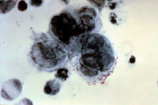
Touton giant cells, being multinucleated giant cells, can be distinguished by the presence of several nuclei in a distinct pattern. They contain a ring of nuclei surrounding a central homogeneous cytoplasm, while foamy cytoplasm surrounds the nuclei. [4] [5] The cytoplasm surrounded by the nuclei has been described as both amphophilic and eosinophilic, while the cytoplasm near the periphery of the cell is pale and foamy in appearance. [6]
Touton giant cells are formed by the fusion of macrophage-derived foam cells. It has been suggested that cytokines such as interferon gamma, interleukin-3, and M-CSF may be involved in the production of Touton giant cells. [7] [8]

Cytokines are a broad and loose category of small proteins important in cell signaling. Cytokines are peptides and cannot cross the lipid bilayer of cells to enter the cytoplasm. Cytokines have been shown to be involved in autocrine, paracrine and endocrine signaling as immunomodulating agents.

Phagocytes are cells that protect the body by ingesting harmful foreign particles, bacteria, and dead or dying cells. Their name comes from the Greek phagein, "to eat" or "devour", and "-cyte", the suffix in biology denoting "cell", from the Greek kutos, "hollow vessel". They are essential for fighting infections and for subsequent immunity. Phagocytes are important throughout the animal kingdom and are highly developed within vertebrates. One litre of human blood contains about six billion phagocytes. They were discovered in 1882 by Ilya Ilyich Mechnikov while he was studying starfish larvae. Mechnikov was awarded the 1908 Nobel Prize in Physiology or Medicine for his discovery. Phagocytes occur in many species; some amoebae behave like macrophage phagocytes, which suggests that phagocytes appeared early in the evolution of life.

A granuloma is an aggregation of macrophages that forms in response to chronic inflammation. This occurs when the immune system attempts to isolate foreign substances that it is otherwise unable to eliminate. Such substances include infectious organisms including bacteria and fungi, as well as other materials such as foreign objects, keratin, and suture fragments.

Cell-mediated immunity or cellular immunity is an immune response that does not involve antibodies. Rather, cell-mediated immunity is the activation of phagocytes, antigen-specific cytotoxic T-lymphocytes, and the release of various cytokines in response to an antigen.

An osteoclast is a type of bone cell that breaks down bone tissue. This function is critical in the maintenance, repair, and remodeling of bones of the vertebral skeleton. The osteoclast disassembles and digests the composite of hydrated protein and mineral at a molecular level by secreting acid and a collagenase, a process known as bone resorption. This process also helps regulate the level of blood calcium.

Interleukin 12 (IL-12) is an interleukin that is naturally produced by dendritic cells, macrophages, neutrophils, and human B-lymphoblastoid cells (NC-37) in response to antigenic stimulation. IL-12 belongs to the family of interleukin-12. IL-12 family is unique in comprising the only heterodimeric cytokines, which includes IL-12, IL-23, IL-27 and IL-35. Despite sharing many structural features and molecular partners, they mediate surprisingly diverse functional effects.

Interferon gamma (IFN-γ) is a dimerized soluble cytokine that is the only member of the type II class of interferons. The existence of this interferon, which early in its history was known as immune interferon, was described by E. F. Wheelock as a product of human leukocytes stimulated with phytohemagglutinin, and by others as a product of antigen-stimulated lymphocytes. It was also shown to be produced in human lymphocytes. or tuberculin-sensitized mouse peritoneal lymphocytes challenged with Mantoux test (PPD); the resulting supernatants were shown to inhibit growth of vesicular stomatitis virus. Those reports also contained the basic observation underlying the now widely employed IFN-γ release assay used to test for tuberculosis. In humans, the IFN-γ protein is encoded by the IFNG gene.

A giant cell is a mass formed by the union of several distinct cells, often forming a granuloma. Although there is typically a focus on the pathological aspects of multinucleate giant cells (MGCs), they also play many important physiological roles. Osteoclasts specifically are invaluable to healthy physiological functions and are key players in the skeletal system. Osteoclasts are frequently classified and discussed separately from other MGCs which are more closely linked with human pathologies.

Foam cells, also called lipid-laden macrophages, are a type of cell that contain cholesterol. These can form a plaque that can lead to atherosclerosis and trigger heart attacks and stroke.

Monoblasts are the committed progenitor cells that differentiated from a committed macrophage or dendritic cell precursor (MDP) in the process of hematopoiesis. They are the first developmental stage in the monocyte series leading to a macrophage. Their myeloid cell fate is induced by the concentration of cytokines they are surrounded by during development. These cytokines induce the activation of transcription factors which push completion of the monoblast's myeloid cell fate. Monoblasts are normally found in bone marrow and do not appear in the normal peripheral blood. They mature into monocytes which, in turn, develop into macrophages. They then are seen as macrophages in the normal peripheral blood and many different tissues of the body. Macrophages can produce a variety of effector molecules that initiate local, systemic inflammatory responses. These monoblast differentiated cells are equipped to fight off foreign invaders using pattern recognition receptors to detect antigen as part of the innate immune response.

Interleukin-18 (IL-18), also known as interferon-gamma inducing factor is a protein which in humans is encoded by the IL18 gene. The protein encoded by this gene is a proinflammatory cytokine. Many cell types, both hematopoietic cells and non-hematopoietic cells, have the potential to produce IL-18. It was first described in 1989 as a factor that induced interferon-γ (IFN-γ) production in mouse spleen cells. Originally, IL-18 production was recognized in Kupffer cells, liver-resident macrophages. However, IL-18 is constitutively expressed in non-hematopoietic cells, such as intestinal epithelial cells, keratinocytes, and endothelial cells. IL-18 can modulate both innate and adaptive immunity and its dysregulation can cause autoimmune or inflammatory diseases.

A foreign body reaction (FBR) is a typical tissue response to a foreign body within biological tissue. It usually includes the formation of a foreign body granuloma. Tissue-encapsulation of an implant is an example, as is inflammation around a splinter. Foreign body granuloma formation consists of protein adsorption, macrophages, multinucleated foreign body giant cells, fibroblasts, and angiogenesis. It has also been proposed that the mechanical property of the interface between an implant and its surrounding tissues is critical for the host response.
In dermatopathology, the Tzanck test, also Tzanck smear, is scraping of an ulcer base to look for Tzanck cells. It is sometimes also called the chickenpox skin test and the herpes skin test. It is a simple, low-cost, and rapid office based test.

According to a common point of view epithelioid cells are derivatives of activated macrophages resembling epithelial cells.
Multinucleate cells are eukaryotic cells that have more than one nucleus per cell, i.e., multiple nuclei share one common cytoplasm. Mitosis in multinucleate cells can occur either in a coordinated, synchronous manner where all nuclei divide simultaneously or asynchronously where individual nuclei divide independently in time and space. Certain organisms may have a multinuclear stage of their life cycle. For example, slime molds have a vegetative, multinucleate life stage called a plasmodium.
Type IV hypersensitivity, often called delayed-type hypersensitivity, is a type of hypersensitivity reaction that can take a day or more to develop. Unlike the other types, it is not humoral but rather is a type of cell-mediated response. This response involves the interaction of T cells, monocytes, and macrophages.

A foreign-body giant cell is a collection of fused macrophages which are generated in response to the presence of a large foreign body. This is particularly evident with catheters, parasites, or biomaterials that are inserted into the body for replacement or regeneration of diseased or damaged tissues. Foreign body giant cells are also produced to digest foreign material that is too large for phagocytosis. The inflammatory process that creates these cells often leads to a foreign body granuloma.
The pluripotency of biological compounds describes the ability of certain substances to produce several distinct biological responses. Pluripotent is also described as something that has no fixed developmental potential, as in being able to differentiate into different cell types in the case of pluripotent stem cells.
Karl Touton was a German dermatologist and amateur botanist.
Histopathology of dermatitis can be performed in uncertain cases of inflammatory skin condition that remain uncertain after history and physical examination.
| | This cell biology article is a stub. You can help Wikipedia by expanding it. |
| | This immunology article is a stub. You can help Wikipedia by expanding it. |