Cardioversion

Last updated
Cardioversion
Cardioversion.svg
Illustration of cardioversion
Specialty Cardiology
ICD-9-CM 99.6
MeSH D004554
MedlinePlus 007110

Cardioversion is a medical procedure by which an abnormally fast heart rate (tachycardia) or other cardiac arrhythmia is converted to a normal rhythm using electricity or drugs. Synchronized electrical cardioversion uses a therapeutic dose of electric current to the heart at a specific moment in the cardiac cycle, restoring the activity of the electrical conduction system of the heart. (Defibrillation uses a therapeutic dose of electric current to the heart at a random moment in the cardiac cycle, and is the most effective resuscitation measure for cardiac arrest associated with ventricular fibrillation and pulseless ventricular tachycardia. [1] ) Pharmacologic cardioversion, also called chemical cardioversion, uses antiarrhythmia medication instead of an electrical shock. [2]

Contents

'Burn' mark remaining one day after DC electrical cardioversion Electrical cardioversion.jpg
'Burn' mark remaining one day after DC electrical cardioversion

Electrical

To perform synchronized electrical cardioversion, two electrode pads are used (or, alternatively, the traditional hand-held "paddles"), each comprising a metallic plate which is faced with a saline based conductive gel. The pads are placed on the chest of the patient, or one is placed on the chest and one on the back. These are connected by cables to a machine which has the combined functions of an ECG display screen and the electrical function of a defibrillator. A synchronizing function (either manually operated or automatic) allows the cardioverter to deliver a reversion shock, by way of the pads, of a selected amount of electric current over a predefined number of milliseconds at the optimal moment in the cardiac cycle which corresponds to the R wave of the QRS complex on the ECG.

Timing the shock to the R wave prevents the delivery of the shock during the vulnerable period (or relative refractory period) of the cardiac cycle, which could induce ventricular fibrillation. If the patient is conscious, various drugs are often used to help sedate the patient and make the procedure more tolerable. However, if the patient is hemodynamically unstable or unconscious, the shock is given immediately upon confirmation of the arrhythmia. When synchronized electrical cardioversion is performed as an elective procedure, the shocks can be performed in conjunction with drug therapy until sinus rhythm is attained. After the procedure, the patient is monitored to ensure stability of the sinus rhythm.

Synchronized electrical cardioversion is used to treat hemodynamically unstable supraventricular (or narrow complex) tachycardias, including atrial fibrillation and atrial flutter. It is also used in the emergent treatment of wide complex tachycardias, including ventricular tachycardia, when a pulse is present. Pulseless ventricular tachycardia and ventricular fibrillation are treated with unsynchronized shocks referred to as defibrillation. Electrical therapy is inappropriate for sinus tachycardia, which should always be a part of the differential diagnosis.

Medication

Various antiarrhythmic agents can be used to return the heart to normal sinus rhythm. [3] Pharmacological cardioversion is an especially good option in patients with atrial fibrillation of recent onset. Drugs that are effective at maintaining normal rhythm after electric cardioversion can also be used for pharmacological cardioversion. Drugs like amiodarone, diltiazem, verapamil and metoprolol are frequently given before electrical cardioversion to decrease the heart rate, stabilize the patient and increase the chance that cardioversion is successful. There are various classes of agents that are most effective for pharmacological cardioversion.

Class I agents are sodium (Na) channel blockers (which slow conduction by blocking the Na+ channel) and are divided into 3 subclasses a, b and c. Class Ia slows phase 0 depolarization in the ventricles and increases the absolute refractory period. Procainamide, quinidine and disopyramide are Class Ia agents. Class 1b drugs lengthen phase 3 repolarization. They include lidocaine, mexiletine and phenytoin. Class Ic greatly slow phase 0 depolarization in the ventricles (however unlike 1a have no effect on the refractory period). Flecainide, moricizine and propafenone are Class Ic agents. [4]

Class II agents are beta blockers which inhibit SA and AV node depolarization and slow heart rate. They also decrease cardiac oxygen demand and can prevent cardiac remodeling. Not all beta blockers are the same; some are cardio selective (affecting only beta 1 receptors) while others are non-selective (affecting beta 1 and 2 receptors). Beta blockers that target the beta-1 receptor are called cardio selective because beta-1 is responsible for increasing heart rate; hence a beta blocker will slow the heart rate.

Class III agents (prolong repolarization by blocking outward K+ current): amiodarone and sotalol are effective class III agents. Ibutilide is another Class III agent but has a different mechanism of action (acts to promote influx of sodium through slow-sodium channels). It has been shown to be effective in acute cardioversion of recent-onset atrial fibrillation and atrial flutter.

Class IV drugs are calcium (Ca) channel blockers. They work by inhibiting the action potential of the SA and AV nodes.

If the patient is stable, adenosine may be used for restoration of sinus rhythm in patients with macro-reentrant supraventricular tachycardias. It causes a short-lived cessation of conduction through the atrio-ventricular node breaking the circus movement through the node and the macro-reentrant pathway restoring sinus rhythm.

Procedure

Preparation

Cardioversion for restoration of sinus rhythm from an atrial rhythm is largely a scheduled procedure. In addition to cardiology, anesthesiology is also usually involved to ensure comfort of the patient for the duration of the shock therapy. The presence of registered nurses, physician associates, or other medical personnel may also be helpful during the procedure.

Before starting the procedure, the patient's chest and back will be prepped for electrode placement. The skin should be free of any oily substances (e.g., lotions) and hair which may otherwise interfere with adhesion of the pads. [5] Once this is complete, the medical team will adhere the pads to the patient using a rolling motion to ensure the absence of air pockets. (see details on pad placement below). The anesthesiology team will then administer a general anesthetic (e.g., Propofol) in order to ensure patient comfort and amnesia during the procedure. Opioid analgesics (e.g., Fentanyl) may be combined with Propofol, although anesthesiology must weight the benefits against adverse effects including apnea. [6] Bite blocks and extremity restraints are then utilized to prevent self-injury during cardioversion. Once these medications are administered, the glabellar reflex or eyelash reflex may be used to determine the patient's level of consciousness.

The pads are connected to a machine that can interpret the patient's cardiac rate and rhythm and deliver a shock at the appropriate time. The machine should synchronize ('sync') with the R wave of the rhythm strip. Although uncommon, sometimes the machine will unintentionally sync to high amplitude T waves, so it is important to ensure that the machine is synced appropriately to R waves. [7] Interpretation of the patient's rhythm is imperative when using cardioversion to restore sinus rhythm from less emergent arrhythmias where a pulse is present (e.g., atrial flutter, atrial fibrillation). However, if a patient is confirmed to be in pulseless ventricular tachycardia "v-tach" or ventricular fibrillation "v-fib", then a shock is delivered immediately upon connection of the pads. In this application, electrical cardioversion is more properly termed defibrillation. [7]

LIFEPAK 20e Defibrillator and Monitor displaying synchronization with QRS complexes. (arrowheads) LIFEPAK 20e Defibrillator and Monitor displaying synchronization with QRS complexes. (arrowheads).jpg
LIFEPAK 20e Defibrillator and Monitor displaying synchronization with QRS complexes. (arrowheads)

Cardioversion

Once the machine is synced with the patient's cardiac rhythm, the machine must be charged. To determine the amount of energy (measured in joules "J") the patient requires, many factors are considered. As a rule of thumb, recent-onset atrial arrhythmias require less energy compared to persistent atrial arrhythmias. If the cardiologist suspects that the patient may be less respondent to cardioversion, a higher energy may be utilized. Once the machine is synced and charged, a shock can be delivered to the patient. [8]

  • Atrial Flutter and SVT: 50-100 J for biphasic devices; 100 J for monophasic devices
  • Atrial Fibrillation: 120-200 J for biphasic devices; 200 J for monophasic devices
  • Ventricular Tachycardia (with a pulse): 100 J for biphasic devices; 200 J for monophasic devices
  • Pulseless Ventricular Tachycardia and Ventricular Fibrillation: 120-200 J for biphasic devices; 360 J for monophasic devices [9] [10] [11]

After cardioversion

Following electrical cardioversion, the cardiologist will determine if sinus rhythm has been restored. To confirm sinus rhythm, a distinct P wave should be seen preceding each QRS complex. Additionally, each R-R interval should be evenly spaced. If sinus rhythm is restored, the pads may be disconnected, and any other medical equipment is removed from the patients (e.g., bite blocks, restraints, etc.). The patient will regain consciousness soon thereafter (the effects of Propofol generally last for only 3–8 minutes). However, if the arrhythmia is persistent, the machine may be re-charged to a higher energy level, and the cardioversion attempt may be repeated. It is recommended to wait 60 seconds between subsequent cardioversion attempts, but this amount of time may be adjusted based on the patient and/or provider.

Electrode pad placement

Pad placement for electrical cardioversion a cardiac arrhythmia may be either anterior-posterior or anterior-lateral. In an anterior-posterior setup one pad is placed on the chest and the other pad pad is placed on the back. In an anterior-lateral setup, one pad is placed on the chest and the other pad is placed along the left midaxillary line. Choosing the right pad placement can be an important aspect when measuring the success of electrical cardioversion. For example, the anterior-posterior pad positioning is commonly used when attempting to restore an atrial arrhythmia as the vector between the pads predominately runs through the atria. The anterior-lateral pad positioning may be used when attempting to restore pulseless ventricular tachycardia or ventricular fibrillation as there may not be enough time or strength to apply an electrode the patient's back.

Anterior-Posterior pad placement

The anterior pad should be placed inferior to the right clavicle while also being vertically centered over at the level of the right 4th intercostal space. The posterior pad should be placed just lateral to the left side of the spine and vertically centered at the level of T7. [12] [13] The inferior angle of the scapula can be used as a reference for the level of T7.

Anterior-Lateral pad placement

The anterior pad should be placed inferior to the right clavicle while also being vertically centered over at the level of the right 4th intercostal space. The lateral pad should be placed along the left midaxillary line at the level of the left 5th intercostal space. [12] [13] The left nipple can be used as a reference for the level of the left 4th intercostal space. From here, the midaxillary 5th intercostal space is identified by moving inferiorly one intercostal space and laterally towards the midaxillary line.

See also

Related Research Articles

<span class="mw-page-title-main">Cardiac arrest</span> Sudden stop in effective blood flow due to the failure of the heart to beat

Cardiac arrest, also known as sudden cardiac arrest, is when the heart suddenly and unexpectedly stops beating. As a result blood will not be pumped around the body in normal circulation, consciousness will be rapidly lost, and breathing will be abnormal or absent. Without immediate intervention such as cardiopulmonary resuscitation (CPR), and possibly defibrillation, death will occur within minutes.

<span class="mw-page-title-main">Defibrillation</span> Treatment for life-threatening cardiac arrhythmias

Defibrillation is a treatment for life-threatening cardiac arrhythmias, specifically ventricular fibrillation (V-Fib) and non-perfusing ventricular tachycardia (V-Tach). A defibrillator delivers a dose of electric current to the heart. Although not fully understood, this process depolarizes a large amount of the heart muscle, ending the arrhythmia. Subsequently, the body's natural pacemaker in the sinoatrial node of the heart is able to re-establish normal sinus rhythm. A heart which is in asystole (flatline) cannot be restarted by a defibrillator; it would be treated only by cardiopulmonary resuscitation (CPR) and medication, and then by cardioversion or defibrillation if it converts into a shockable rhythm.

<span class="mw-page-title-main">Tachycardia</span> Heart rate that exceeds the normal resting rate

Tachycardia, also called tachyarrhythmia, is a heart rate that exceeds the normal resting rate. In general, a resting heart rate over 100 beats per minute is accepted as tachycardia in adults. Heart rates above the resting rate may be normal or abnormal.

<span class="mw-page-title-main">Ventricular fibrillation</span> Rapid quivering of the ventricles of the heart

Ventricular fibrillation is an abnormal heart rhythm in which the ventricles of the heart quiver. It is due to disorganized electrical activity. Ventricular fibrillation results in cardiac arrest with loss of consciousness and no pulse. This is followed by sudden cardiac death in the absence of treatment. Ventricular fibrillation is initially found in about 10% of people with cardiac arrest.

<span class="mw-page-title-main">Asystole</span> Medical condition of the heart

Asystole is the absence of ventricular contractions in the context of a lethal heart arrhythmia. Asystole is the most serious form of cardiac arrest and is usually irreversible. Also referred to as cardiac flatline, asystole is the state of total cessation of electrical activity from the heart, which means no tissue contraction from the heart muscle and therefore no blood flow to the rest of the body.

<span class="mw-page-title-main">Automated external defibrillator</span> Portable electronic medical device

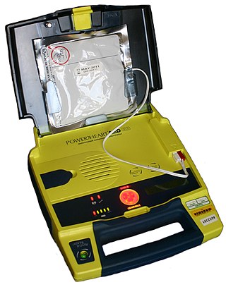
An automated external defibrillator or automatic electronic defibrillator (AED) is a portable electronic device that automatically diagnoses the life-threatening cardiac arrhythmias of ventricular fibrillation (VF) and pulseless ventricular tachycardia, and is able to treat them through defibrillation, the application of electricity which stops the arrhythmia, allowing the heart to re-establish an effective rhythm.

<span class="mw-page-title-main">Wolff–Parkinson–White syndrome</span> Medical condition

Wolff–Parkinson–White syndrome (WPWS) is a disorder due to a specific type of problem with the electrical system of the heart involving an accessory pathway able to conduct electrical current between the atria and the ventricles, thus bypassing the atrioventricular node. About 60% of people with the electrical problem developed symptoms, which may include an abnormally fast heartbeat, palpitations, shortness of breath, lightheadedness, or syncope. Rarely, cardiac arrest may occur. The most common type of irregular heartbeat that occurs is known as paroxysmal supraventricular tachycardia.

<span class="mw-page-title-main">Atrial flutter</span> Medical condition

Atrial flutter (AFL) is a common abnormal heart rhythm that starts in the atrial chambers of the heart. When it first occurs, it is usually associated with a fast heart rate and is classified as a type of supraventricular tachycardia. Atrial flutter is characterized by a sudden-onset (usually) regular abnormal heart rhythm on an electrocardiogram (ECG) in which the heart rate is fast. Symptoms may include a feeling of the heart beating too fast, too hard, or skipping beats, chest discomfort, difficulty breathing, a feeling as if one's stomach has dropped, a feeling of being light-headed, or loss of consciousness.

<span class="mw-page-title-main">Short QT syndrome</span> Medical condition

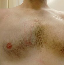
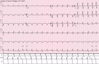
Short QT syndrome (SQT) is a very rare genetic disease of the electrical system of the heart, and is associated with an increased risk of abnormal heart rhythms and sudden cardiac death. The syndrome gets its name from a characteristic feature seen on an electrocardiogram (ECG) – a shortening of the QT interval. It is caused by mutations in genes encoding ion channels that shorten the cardiac action potential, and appears to be inherited in an autosomal dominant pattern. The condition is diagnosed using a 12-lead ECG. Short QT syndrome can be treated using an implantable cardioverter-defibrillator or medications including quinidine. Short QT syndrome was first described in 2000, and the first genetic mutation associated with the condition was identified in 2004.

<span class="mw-page-title-main">Ventricular tachycardia</span> Medical condition of the heart

Ventricular tachycardia is a cardiovascular disorder in which fast heart rate occurs in the ventricles of the heart. Although a few seconds of VT may not result in permanent problems, longer periods are dangerous; and multiple episodes over a short period of time are referred to as an electrical storm. Short periods may occur without symptoms, or present with lightheadedness, palpitations, or chest pain. Ventricular tachycardia may result in ventricular fibrillation (VF) and turn into cardiac arrest. This conversion of the VT into VF is called the degeneration of the VT. It is found initially in about 7% of people in cardiac arrest.

Precordial thump is a medical procedure used in the treatment of ventricular fibrillation or pulseless ventricular tachycardia under certain conditions. The procedure has a very low success rate, but may be used in those with witnessed, monitored onset of one of the "shockable" cardiac rhythms if a defibrillator is not immediately available. It should not delay cardiopulmonary resuscitation (CPR) and defibrillation, nor should it be used in those with unwitnessed out-of-hospital cardiac arrest.

<span class="mw-page-title-main">AV nodal reentrant tachycardia</span> Medical condition

AV-nodal reentrant tachycardia (AVNRT) is a type of abnormal fast heart rhythm. It is a type of supraventricular tachycardia (SVT), meaning that it originates from a location within the heart above the bundle of His. AV nodal reentrant tachycardia is the most common regular supraventricular tachycardia. It is more common in women than men. The main symptom is palpitations. Treatment may be with specific physical maneuvers, medications, or, rarely, synchronized cardioversion. Frequent attacks may require radiofrequency ablation, in which the abnormally conducting tissue in the heart is destroyed.

<span class="mw-page-title-main">Paroxysmal supraventricular tachycardia</span> Medical condition

Paroxysmal supraventricular tachycardia (PSVT) is a type of supraventricular tachycardia, named for its intermittent episodes of abrupt onset and termination. Often people have no symptoms. Otherwise symptoms may include palpitations, feeling lightheaded, sweating, shortness of breath, and chest pain.

Tachycardia-induced cardiomyopathy (TIC) is a disease where prolonged tachycardia or arrhythmia causes an impairment of the myocardium, which can result in heart failure. People with TIC may have symptoms associated with heart failure and/or symptoms related to the tachycardia or arrhythmia. Though atrial fibrillation is the most common cause of TIC, several tachycardias and arrhythmias have been associated with the disease.

Pediatric advanced life support (PALS) is a course offered by the American Heart Association (AHA) for health care providers who take care of children and infants in the emergency room, critical care and intensive care units in the hospital, and out of hospital. The course teaches healthcare providers how to assess injured and sick children and recognize and treat respiratory distress/failure, shock, cardiac arrest, and arrhythmias.

<span class="mw-page-title-main">Landiolol</span> Chemical compound

Landiolol (INN) is an ultra short-acting, β1-superselective intravenous adrenergic antagonist, which decreases the heart rate effectively with less negative effect on blood pressure or myocardial contractility. In comparison to other beta blockers, landiolol has the shortest elimination half-life, ultra-rapid onset of effect, and predictable effectiveness with inactive metabolites. The pure S-enantiomer structure of landiolol is believed to develop less hypotensive side effects in comparison to other β-blockers. This has a positive impact on the treatment of patients when reduction of heart rate without decrease in arterial blood pressure is desired. Landiolol was developed by modifying the chemical structure of esmolol to produce a compound with a higher rate of cardioselectivity and a greater potency without increasing its duration of action. It is sold as landiolol hydrochloride. Based on its positive benefit risk profile, landiolol has been granted the marketing authorization and introduced to the European markets under the brand names Rapibloc, Raploc, Runrapiq, Landibloc mid 2016. Landiolol is available in Japan under the brand names Onoact (50 mg) and Corbeta.

<span class="mw-page-title-main">Michel Haïssaguerre</span>

Michel Haïssaguerre is a French cardiologist and electrophysiologist. His investigations have been the basis for development of new markers and therapies for atrial and ventricular fibrillation.

<span class="mw-page-title-main">Atrial fibrillation</span> Irregular beating of the atria of the heart

Atrial fibrillation is an abnormal heart rhythm (arrhythmia) characterized by rapid and irregular beating of the atrial chambers of the heart. It often begins as short periods of abnormal beating, which become longer or continuous over time. It may also start as other forms of arrhythmia such as atrial flutter that then transform into AF.

<span class="mw-page-title-main">Arrhythmia</span> Group of medical conditions characterized by irregular heartbeat

Arrhythmias, also known as cardiac arrhythmias, heart arrhythmias, or dysrhythmias, are irregularities in the heartbeat, including when it is too fast or too slow. A resting heart rate that is too fast – above 100 beats per minute in adults – is called tachycardia, and a resting heart rate that is too slow – below 60 beats per minute – is called bradycardia. Some types of arrhythmias have no symptoms. Symptoms, when present, may include palpitations or feeling a pause between heartbeats. In more serious cases, there may be lightheadedness, passing out, shortness of breath, chest pain, or decreased level of consciousness. While most cases of arrhythmia are not serious, some predispose a person to complications such as stroke or heart failure. Others may result in sudden death.

<span class="mw-page-title-main">Automatic tachycardia</span> Medical condition

An automatic tachycardia is a cardiac arrhythmia which involves an area of the heart generating an abnormally fast rhythm, sometimes also called enhanced automaticity. These tachycardias, or fast heart rhythms, differ from reentrant tachycardias in which there is an abnormal electrical pathway which gives rise to the pathology. Most automatic tachycardias are supraventricular tachycardias (SVT). It is important to recognize an automatic tachycardia because the treatment will be different to that for a reentrant tachycardia. The most useful clue will be the presence of 'warm up' and 'cool down'. This means that whereas a reentrant tachycardia will both begin and end abruptly as cardiac conduction uses then ceases to use the accessory pathway, an automatic tachycardia will rise and fall gradually in rate as the automatic focus increases and decreases its automatic rate of electrical discharge.

References

  1. Marino PL (2014). Marino's the ICU book (Fourth ed.). Wolters Kluwer Health/Lippincott Williams & Wilkins. ISBN   978-1451121186.
  2. Shea JB, Maisel WH (November 2002). "Cardiology patient pages. Cardioversion". Circulation. 106 (22): e176–e178. doi: 10.1161/01.CIR.0000040586.24302.B9 . PMID   12451016.
  3. "Medications for Arrhythmia". American Heart Association. Retrieved 13 Sep 2020.
  4. "AED recyclen en inruilen" . Retrieved 15 April 2021.
  5. Sado DM, Deakin CD, Petley GW, Clewlow F (January 2004). "Comparison of the effects of removal of chest hair with not doing so before external defibrillation on transthoracic impedance". The American Journal of Cardiology. 93 (1): 98–100. doi:10.1016/j.amjcard.2003.09.020. PMID   14697478.
  6. Wafae BG, da Silva RM, Veloso HH (2019). "Propofol for sedation for direct current cardioversion". Annals of Cardiac Anaesthesia. 22 (2): 113–121. doi: 10.4103/aca.ACA_72_18 . PMC   6489399 . PMID   30971591.
  7. 1 2 Goyal A, Sciammarella JC, Chhabra L, Singhal M (2022). "Synchronized Electrical Cardioversion". StatPearls. Treasure Island (FL): StatPearls Publishing. PMID   29489237 . Retrieved 2022-08-12.
  8. Sirna SJ, Ferguson DW, Charbonnier F, Kerber RE (November 1988). "Factors affecting transthoracic impedance during electrical cardioversion". The American Journal of Cardiology. 62 (16): 1048–1052. doi:10.1016/0002-9149(88)90546-2. PMID   3189167.
  9. Fuster V, Rydén LE, Cannom DS, Crijns HJ, Curtis AB, Ellenbogen KA, et al. (August 2006). "ACC/AHA/ESC 2006 Guidelines for the Management of Patients with Atrial Fibrillation: a report of the American College of Cardiology/American Heart Association Task Force on Practice Guidelines and the European Society of Cardiology Committee for Practice Guidelines (Writing Committee to Revise the 2001 Guidelines for the Management of Patients With Atrial Fibrillation): developed in collaboration with the European Heart Rhythm Association and the Heart Rhythm Society". Circulation. 114 (7): e257–e354. doi: 10.1161/circulationaha.106.177292 . PMID   16908781.
  10. Zipes DP, Camm AJ, Borggrefe M, Buxton AE, Chaitman B, Fromer M, et al. (September 2006). "ACC/AHA/ESC 2006 Guidelines for Management of Patients With Ventricular Arrhythmias and the Prevention of Sudden Cardiac Death: a report of the American College of Cardiology/American Heart Association Task Force and the European Society of Cardiology Committee for Practice Guidelines (writing committee to develop Guidelines for Management of Patients With Ventricular Arrhythmias and the Prevention of Sudden Cardiac Death): developed in collaboration with the European Heart Rhythm Association and the Heart Rhythm Society". Circulation. 114 (10): e385–e484. doi: 10.1161/CIRCULATIONAHA.106.178233 . PMID   16935995.
  11. Link MS, Atkins DL, Passman RS, Halperin HR, Samson RA, White RD, et al. (November 2010). "Part 6: electrical therapies: automated external defibrillators, defibrillation, cardioversion, and pacing: 2010 American Heart Association Guidelines for Cardiopulmonary Resuscitation and Emergency Cardiovascular Care". Circulation. 122 (18 Suppl 3): S706–S719. doi: 10.1161/CIRCULATIONAHA.110.970954 . PMID   20956222.
  12. 1 2 Kirchhof P, Eckardt L, Loh P, Weber K, Fischer RJ, Seidl KH, et al. (October 2002). "Anterior-posterior versus anterior-lateral electrode positions for external cardioversion of atrial fibrillation: a randomised trial". Lancet. 360 (9342): 1275–1279. doi:10.1016/S0140-6736(02)11315-8. PMID   12414201. S2CID   25083798.
  13. 1 2 Botto GL, Politi A, Bonini W, Broffoni T, Bonatti R (December 1999). "External cardioversion of atrial fibrillation: role of paddle position on technical efficacy and energy requirements". Heart. 82 (6): 726–730. doi:10.1136/hrt.82.6.726. PMC   1729223 . PMID   10573502.