Classification of pneumonia

Last updated

Pneumonia
PneumonisWedge09.JPG
A chest X-ray showing a very prominent wedge-shape bacterial pneumonia in the right lung
Specialty Pulmonology   OOjs UI icon edit-ltr-progressive.svg

Pneumonia can be classified in several ways, most commonly by where it was acquired (hospital versus community), but may also by the area of lung affected or by the causative organism. [1] There is also a combined clinical classification, which combines factors such as age, risk factors for certain microorganisms, the presence of underlying lung disease or systemic disease and whether the person has recently been hospitalized.

Contents

By location acquired

Community-acquired

Community-acquired pneumonia (CAP) is infectious pneumonia in a person who has not recently been hospitalized. CAP is the most common type of pneumonia. The most common causes of CAP vary depending on a person's age, but they include Streptococcus pneumoniae , viruses, the atypical bacteria, and Haemophilus influenzae . Overall, Streptococcus pneumoniae is the most common cause of community-acquired pneumonia worldwide. Gram-negative bacteria cause CAP in certain at-risk populations. CAP is the fourth most common cause of death in the United Kingdom and the sixth in the United States. The term "walking pneumonia" has been used to describe a type of community-acquired pneumonia of less severity (because the sufferer can continue to "walk" rather than requiring hospitalization). [2] Walking pneumonia is usually caused by the atypical bacterium, Mycoplasma pneumoniae . [3]

Hospital-acquired

Hospital-acquired pneumonia, also called nosocomial pneumonia, is pneumonia acquired during or after hospitalization for another illness or procedure with onset at least 72 hrs after admission. The causes, microbiology, treatment and prognosis are different from those of community-acquired pneumonia. Up to 5% of patients admitted to a hospital for other causes subsequently develop pneumonia. Hospitalized patients may have many risk factors for pneumonia, including mechanical ventilation, prolonged malnutrition, underlying heart and lung diseases, decreased amounts of stomach acid, and immune disturbances. Additionally, the microorganisms a person is exposed to in a hospital are often different from those at home. Hospital-acquired microorganisms may include resistant bacteria such as MRSA, Pseudomonas , Enterobacter , and Serratia . Because individuals with hospital-acquired pneumonia usually have underlying illnesses and are exposed to more dangerous bacteria, it tends to be more deadly than community-acquired pneumonia. Ventilator-associated pneumonia (VAP) is a subset of hospital-acquired pneumonia. VAP is pneumonia which occurs after at least 48 hours of intubation and mechanical ventilation.

By cause

Pneumonia has historically been characterized as either typical or atypical depending on the presenting symptoms and thus the presumed underlying organism. [4] Attempting to make this distinction based on symptoms, however, has not been found to be accurate, and The American Thoracic Society does not recommend its use. [4]

Bronchiolitis obliterans organizing pneumonia

Bronchiolitis obliterans organizing pneumonia (BOOP) is caused by inflammation of the small airways of the lungs. It is also known as cryptogenic organizing pneumonitis (COP).
Eosinophilic pneumonia
Eosinophilic pneumonia is invasion of the lung by eosinophils, a particular kind of white blood cell. Eosinophilic pneumonia often occurs in response to infection with a parasite or after exposure to certain types of environmental factors.

Chemical pneumonia

Chemical pneumonia (usually called chemical pneumonitis) is caused by chemical toxicants such as pesticides, which may enter the body by inhalation or by skin contact. When the toxic substance is an oil, the pneumonia may be called lipoid pneumonia.

Aspiration pneumonia

Aspiration pneumonia (or aspiration pneumonitis) is caused by aspirating foreign objects which are usually oral or gastric contents, either while eating, or after reflux or vomiting which results in bronchopneumonia. The resulting lung inflammation is not an infection but can contribute to one, since the material aspirated may contain anaerobic bacteria or other unusual causes of pneumonia. Aspiration is a leading cause of death among hospital and nursing home patients, since they often cannot adequately protect their airways and may have otherwise impaired defenses.

Dust pneumonia

Dust pneumonia describes disorders caused by excessive exposure to dust storms, particularly during the Dust Bowl in the United States. With dust pneumonia, dust settles all the way into the alveoli of the lungs, stopping the cilia from moving and preventing the lungs from ever clearing themselves.

Necrotizing pneumonia

Necrotizing pneumonia (NP), also known as cavitary pneumonia or cavitatory necrosis, is a rare but severe complication of lung parenchymal infection. [5] [6] [7] In necrotizing pneumonia, there is a substantial liquefaction following death of the lung tissue, which may lead to gangrene formation in the lung. [8] [9] In most cases patients with NP have fever, cough and bad breath, and those with more indolent infections have weight loss. [10] Often patients clinically present with acute respiratory failure. [10] The most common pathogens responsible for NP are Streptococcus pneumonia , Staphylococcus aureus , Klebsiella pneumoniae . [11]

Opportunistic pneumonia

People with weakened immune defense, such as HIV/AIDS patients, are highly susceptible to opportunistic infections affecting the lungs. [12] Most common pathogens are Pneumocystis jiroveci , Mycobacterium avium-intracellulare complex, Streptococcus pneumoniae , Haemophilus species. Less frequent pathogens are Cryptococcus neoformans , Histoplasma capsulatum , Coccidioides immitis , cytomegalovirus (CMV), and Toxoplasma gondii . [13]
Chemotherapy-induced immunodeficiency may lead to severe lung infections. [14] Pathogens commonly associated with lung infectioins are bacteria (like Pseudomonas aeruginosa , Stenotrophomonas maltophilia , and Nocardia species), viruses (eg, respiratory syncytial virus, parainfluenza virus, influenza virus A and influenza B, and cytomegalovirus), and fungi (eg, Aspergillus, Fusarium, and Mucorales species, and Pneumocystis jirovecii). [14]

Double pneumonia (bilateral pneumonia)

This is a historical term for acute lung injury (ALI) or acute respiratory distress syndrome (ARDS). [15] However, the term was and, especially by lay people, still is used to denote pneumonia affecting both lungs. Accordingly, the term 'double pneumonia' is more likely to be used to describe bilateral pneumonia than it is ALI or ARDS.

Severe acute respiratory syndrome

Severe acute respiratory syndrome (SARS) is a highly contagious and deadly type of pneumonia which first occurred in November 2002 after initial outbreaks in China caused by SARS-CoV, which had almost disappeared by the month of May 2004 (it was later called SARS-CoV-1 to distinguish it from other similar viruses). The second outbreak, SARS-CoV-2, started in December 2019 from Wuhan, China and was declared pandemic by the WHO on 11 March 2020. SARS is caused by the SARS coronavirus, a previously unknown pathogen.

By area of lung affected

Initial descriptions of pneumonia focused on the anatomic or pathologic appearance of the lung, either by direct inspection at autopsy or by its appearance under a microscope.

The discovery of x-rays made it possible to determine the anatomic type of pneumonia without direct examination of the lungs at autopsy and led to the development of a radiological classification. Early investigators distinguished between typical lobar pneumonia and atypical (e.g. Chlamydophila) or viral pneumonia using the location, distribution, and appearance of the opacities they saw on chest x-rays. Certain x-ray findings can be used to help predict the course of illness, although it is not possible to clearly determine the microbiologic cause of a pneumonia with x-rays alone.

With the advent of modern microbiology, classification based upon the causative microorganism became possible. Determining which microorganism is causing an individual's pneumonia is an important step in deciding treatment type and length. Sputum cultures, blood cultures, tests on respiratory secretions, and specific blood tests are used to determine the microbiologic classification. Because such laboratory testing typically takes several days, microbiologic classification is usually not possible at the time of initial diagnosis.

Clinical

Traditionally, clinicians have classified pneumonia by clinical characteristics, dividing them into "acute" (less than three weeks duration) and "chronic" pneumonias. This is useful because chronic pneumonias tend to be either non-infectious, or mycobacterial, fungal, or mixed bacterial infections caused by airway obstruction. Acute pneumonias are further divided into the classic bacterial bronchopneumonias (such as Streptococcus pneumoniae ), the atypical pneumonias (such as the interstitial pneumonitis of Mycoplasma pneumoniae or Chlamydia pneumoniae ), and the aspiration pneumonia syndromes.[ citation needed ]

Chronic pneumonias, on the other hand, mainly include those of Nocardia , Actinomyces and Blastomyces dermatitidis , as well as the granulomatous pneumonias ( Mycobacterium tuberculosis and atypical mycobacteria, Histoplasma capsulatum and Coccidioides immitis ). [17]

The combined clinical classification, now the most commonly used classification scheme, attempts to identify a person's risk factors when he or she first comes to medical attention. The advantage of this classification scheme over previous systems is that it can help guide the selection of appropriate initial treatments even before the microbiologic cause of the pneumonia is known. There are two broad categories of pneumonia in this scheme: community-acquired pneumonia and hospital-acquired pneumonia. A recently[ when? ] introduced type of healthcare-associated pneumonia (in patients living outside the hospital who have recently been in close contact with the health care system) lies between these two categories.[ citation needed ]

Related Research Articles

<i>Streptococcus</i> Genus of bacteria

Streptococcus is a genus of gram-positive coccus or spherical bacteria that belongs to the family Streptococcaceae, within the order Lactobacillales, in the phylum Bacillota. Cell division in streptococci occurs along a single axis, so as they grow, they tend to form pairs or chains that may appear bent or twisted. This differs from staphylococci, which divide along multiple axes, thereby generating irregular, grape-like clusters of cells. Most streptococci are oxidase-negative and catalase-negative, and many are facultative anaerobes.

<span class="mw-page-title-main">Pneumonia</span> Inflammation of the alveoli of the lungs

Pneumonia is an inflammatory condition of the lung primarily affecting the small air sacs known as alveoli. Symptoms typically include some combination of productive or dry cough, chest pain, fever, and difficulty breathing. The severity of the condition is variable.

Atypical pneumonia, also known as walking pneumonia, is any type of pneumonia not caused by one of the pathogens most commonly associated with the disease. Its clinical presentation contrasts to that of "typical" pneumonia. A variety of microorganisms can cause it. When it develops independently from another disease, it is called primary atypical pneumonia (PAP).

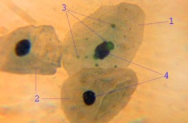
Mycoplasma pneumoniae is a very small bacterium in the class Mollicutes. It is a human pathogen that causes the disease mycoplasma pneumonia, a form of atypical bacterial pneumonia related to cold agglutinin disease. M. pneumoniae is characterized by the absence of a peptidoglycan cell wall and resulting resistance to many antibacterial agents. The persistence of M. pneumoniae infections even after treatment is associated with its ability to mimic host cell surface composition.

<i>Streptococcus pneumoniae</i> Species of bacterium

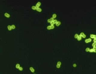
Streptococcus pneumoniae, or pneumococcus, is a Gram-positive, spherical bacteria, alpha-hemolytic member of the genus Streptococcus. They are usually found in pairs (diplococci) and do not form spores and are non motile. As a significant human pathogenic bacterium S. pneumoniae was recognized as a major cause of pneumonia in the late 19th century, and is the subject of many humoral immunity studies.

<span class="mw-page-title-main">Lower respiratory tract infection</span> Medical term

Lower respiratory tract infection (LRTI) is a term often used as a synonym for pneumonia but can also be applied to other types of infection including lung abscess and acute bronchitis. Symptoms include shortness of breath, weakness, fever, coughing and fatigue. A routine chest X-ray is not always necessary for people who have symptoms of a lower respiratory tract infection.

<span class="mw-page-title-main">Bacterial pneumonia</span> Disease of the lungs

Bacterial pneumonia is a type of pneumonia caused by bacterial infection.

<span class="mw-page-title-main">Aspiration pneumonia</span> Medical condition

Aspiration pneumonia is a type of lung infection that is due to a relatively large amount of material from the stomach or mouth entering the lungs. Signs and symptoms often include fever and cough of relatively rapid onset. Complications may include lung abscess, acute respiratory distress syndrome, empyema, and parapneumonic effusion. Some include chemical induced inflammation of the lungs as a subtype, which occurs from acidic but non-infectious stomach contents entering the lungs.

<span class="mw-page-title-main">Bronchopneumonia</span> Human disease

Bronchopneumonia is a subtype of pneumonia. It is the acute inflammation of the bronchi, accompanied by inflamed patches in the nearby lobules of the lungs.

<i>Chlamydia pneumoniae</i> Species of bacterium

Chlamydia pneumoniae is a species of Chlamydia, an obligate intracellular bacterium that infects humans and is a major cause of pneumonia. It was known as the Taiwan acute respiratory agent (TWAR) from the names of the two original isolates – Taiwan (TW-183) and an acute respiratory isolate designated AR-39. Briefly, it was known as Chlamydophila pneumoniae, and that name is used as an alternate in some sources. In some cases, to avoid confusion, both names are given.

<span class="mw-page-title-main">Opportunistic infection</span> Infection caused by pathogens that take advantage of an opportunity not normally available

An opportunistic infection is an infection caused by pathogens that take advantage of an opportunity not normally available. These opportunities can stem from a variety of sources, such as a weakened immune system, an altered microbiome, or breached integumentary barriers. Many of these pathogens do not necessarily cause disease in a healthy host that has a non-compromised immune system, and can, in some cases, act as commensals until the balance of the immune system is disrupted. Opportunistic infections can also be attributed to pathogens which cause mild illness in healthy individuals but lead to more serious illness when given the opportunity to take advantage of an immunocompromised host.

<span class="mw-page-title-main">Lung abscess</span> Medical condition

Lung abscess is a type of liquefactive necrosis of the lung tissue and formation of cavities containing necrotic debris or fluid caused by microbial infection.

Community-acquired pneumonia (CAP) refers to pneumonia contracted by a person outside of the healthcare system. In contrast, hospital-acquired pneumonia (HAP) is seen in patients who have recently visited a hospital or who live in long-term care facilities. CAP is common, affecting people of all ages, and its symptoms occur as a result of oxygen-absorbing areas of the lung (alveoli) filling with fluid. This inhibits lung function, causing dyspnea, fever, chest pains and cough.

Ventilator-associated pneumonia (VAP) is a type of lung infection that occurs in people who are on mechanical ventilation breathing machines in hospitals. As such, VAP typically affects critically ill persons that are in an intensive care unit (ICU) and have been on a mechanical ventilator for at least 48 hours. VAP is a major source of increased illness and death. Persons with VAP have increased lengths of ICU hospitalization and have up to a 20–30% death rate. The diagnosis of VAP varies among hospitals and providers but usually requires a new infiltrate on chest x-ray plus two or more other factors. These factors include temperatures of >38 °C or <36 °C, a white blood cell count of >12 × 109/ml, purulent secretions from the airways in the lung, and/or reduction in gas exchange.

<span class="mw-page-title-main">Lobar pneumonia</span> Medical condition

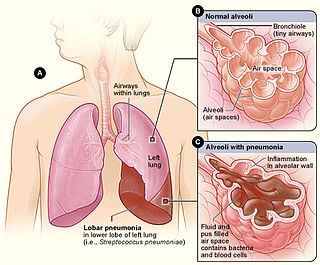
Lobar pneumonia is a form of pneumonia characterized by inflammatory exudate within the intra-alveolar space resulting in consolidation that affects a large and continuous area of the lobe of a lung.

Pneumococcal pneumonia is a type of bacterial pneumonia that is caused by Streptococcus pneumoniae (pneumococcus). It is the most common bacterial pneumonia found in adults, the most common type of community-acquired pneumonia, and one of the common types of pneumococcal infection. The estimated number of Americans with pneumococcal pneumonia is 900,000 annually, with almost 400,000 cases hospitalized and fatalities accounting for 5-7% of these cases.

<span class="mw-page-title-main">Hospital-acquired pneumonia</span>

Hospital-acquired pneumonia (HAP) or nosocomial pneumonia refers to any pneumonia contracted by a patient in a hospital at least 48–72 hours after being admitted. It is thus distinguished from community-acquired pneumonia. It is usually caused by a bacterial infection, rather than a virus.

<span class="mw-page-title-main">Acute exacerbation of chronic obstructive pulmonary disease</span> Medical condition

An acute exacerbation of chronic obstructive pulmonary disease, or acute exacerbations of chronic bronchitis (AECB), is a sudden worsening of chronic obstructive pulmonary disease (COPD) symptoms including shortness of breath, quantity and color of phlegm that typically lasts for several days.

<span class="mw-page-title-main">Ground-glass opacity</span> Radiologic sign on radiographs and computed tomography scans

Ground-glass opacity (GGO) is a finding seen on chest x-ray (radiograph) or computed tomography (CT) imaging of the lungs. It is typically defined as an area of hazy opacification (x-ray) or increased attenuation (CT) due to air displacement by fluid, airway collapse, fibrosis, or a neoplastic process. When a substance other than air fills an area of the lung it increases that area's density. On both x-ray and CT, this appears more grey or hazy as opposed to the normally dark-appearing lungs. Although it can sometimes be seen in normal lungs, common pathologic causes include infections, interstitial lung disease, and pulmonary edema.

Necrotizing pneumonia (NP), also known as cavitary pneumonia or cavitatory necrosis, is a rare but severe complication of lung parenchymal infection. In necrotizing pneumonia, there is a substantial liquefaction following death of the lung tissue, which may lead to gangrene formation in the lung. In most cases patients with NP have fever, cough and bad breath, and those with more indolent infections have weight loss. Often patients clinically present with acute respiratory failure. The most common pathogens responsible for NP are Streptococcus pneumoniae, Staphylococcus aureus, Klebsiella pneumoniae. Diagnosis is usually done by chest imaging, e.g. chest X-ray, CT scan. Among these CT scan is the most sensitive test which shows loss of lung architecture and multiple small thin walled cavities. Often cultures from bronchoalveolar lavage and blood may be done for identification of the causative organism(s). It is primarily managed by supportive care along with appropriate antibiotics. However, if patient develops severe complications like sepsis or fails to medical therapy, surgical resection is a reasonable option for saving life.

References

  1. Dunn L (2005). "Pneumonia: classification, diagnosis and nursing management". Nurs Stand. 19 (42): 50–4. doi:10.7748/ns2005.06.19.42.50.c3901. PMID   16013205.
  2. "UpToDate Inc".
  3. Krause DC, Balish MF (February 2004). "Cellular engineering in a minimal microbe: structure and assembly of the terminal organelle of Mycoplasma pneumoniae". Mol. Microbiol. 51 (4): 917–24. doi: 10.1046/j.1365-2958.2003.03899.x . PMID   14763969.
  4. 1 2 Ebby, Orin (December 2005). "Community-Acquired Pneumonia: From Common Pathogens To Emerging Resistance". Emergency Medicine Practice. 7 (12).
  5. Sawicki, G. S.; Lu, F. L.; Valim, C.; Cleveland, R. H.; Colin, A. A. (5 March 2008). "Necrotising pneumonia is an increasingly detected complication of pneumonia in children". European Respiratory Journal. 31 (6): 1285–1291. doi: 10.1183/09031936.00099807 . ISSN   0903-1936. PMID   18216055.
  6. Tsai, Yueh-Feng; Ku, Yee-Huang (2012). "Necrotizing pneumonia". Current Opinion in Pulmonary Medicine. 18 (3): 246–252. doi:10.1097/MCP.0b013e3283521022. ISSN   1070-5287. PMID   22388585.
  7. Masters, I. Brent; Isles, Alan F.; Grimwood, Keith (25 July 2017). "Necrotizing pneumonia: an emerging problem in children?". Pneumonia. 9 (1): 11. doi: 10.1186/s41479-017-0035-0 . ISSN   2200-6133. PMC   5525269 . PMID   28770121.
  8. Scotta, Marcelo C.; Marostica, Paulo J.C.; Stein, Renato T. (2019). "Pneumonia in Children". Kendig's Disorders of the Respiratory Tract in Children. Elsevier. p. 435.e4. doi:10.1016/b978-0-323-44887-1.00025-0. ISBN   978-0-323-44887-1. S2CID   81700501.
  9. Widysanto, Allen; Liem, Maranatha; Puspita, Karina Dian; Pradhana, Cindy Meidy Leony (2020). "Management of necrotizing pneumonia with bronchopleural fistula caused by multidrug‐resistant Acinetobacter baumannii". Respirology Case Reports. 8 (8): e00662. doi: 10.1002/rcr2.662 . PMC   7507560 . PMID   32999723.
  10. 1 2 Reimel, Beth Ann; Krishnadasen, Baiya; Cuschieri, Joseph; Klein, Matthew B; Gross, Joel; Karmy-Jones, Riyad (1 January 2000). "Surgical management of acute necrotizing lung infections". Canadian Respiratory Journal. 13 (7): 369–373. doi: 10.1155/2006/760390 . PMC   2683290 . PMID   17036090.
  11. Krutikov, Maria; Rahman, Ananna; Tiberi, Simon (2019). "Necrotizing pneumonia (aetiology, clinical features and management)". Current Opinion in Pulmonary Medicine. 25 (3): 225–232. doi:10.1097/mcp.0000000000000571. ISSN   1070-5287. PMID   30844921. S2CID   73507080.
  12. Conces, Dewey J. (1 January 1993). "Opportunistic pneumonia". Current Problems in Diagnostic Radiology. 22 (1): 5–35. doi:10.1016/0363-0188(93)90005-E. ISSN   0363-0188. PMID   8428506 . Retrieved 23 February 2021.
  13. Huang, Laurence; Crothers, Kristina A. (2009). "HIV-associated Opportunistic Pneumonias". Respirology. 14 (4): 474–485. doi: 10.1111/j.1440-1843.2009.01534.x . PMC   2835537 . PMID   19645867.
  14. 1 2 Vento, Sandro; Cainelli, Francesca; Temesgen, Zelalem (1 October 2008). "Lung infections after cancer chemotherapy". The Lancet Oncology. 9 (10): 982–992. doi:10.1016/S1470-2045(08)70255-9. ISSN   1470-2045. PMID   19071255 . Retrieved 23 February 2021.
  15. Girard TD, Bernard GR (March 2007). "Mechanical ventilation in ARDS: a state-of-the-art review". Chest. 131 (3): 921–9. doi:10.1378/chest.06-1515. PMID   17356115.
  16. Feldman C, Kallenbach JM, Levy H, Thorburn JR, Hurwitz MD, Koornhof HJ (1991). "Comparison of bacteraemic community-acquired lobar pneumonia due to Streptococcus pneumoniae and Klebsiella pneumoniae in an intensive care unit". Respiration. 58 (5–6): 265–70. doi:10.1159/000195943. PMID   1792415.
  17. Table 13-7 in: Mitchell, Richard Sheppard; Kumar, Vinay; Abbas, Abul K.; Fausto, Nelson (2007). Robbins Basic Pathology (8th ed.). Philadelphia: Saunders. ISBN   978-1-4160-2973-1.