
The abducens nerve is the sixth cranial nerve (CNVI), in humans, that controls the movement of the lateral rectus muscle, responsible for outward gaze. It is a somatic efferent nerve.

In the human skull, the zygomatic bone is a paired irregular bone which articulates with the maxilla, the temporal bone, the sphenoid bone and the frontal bone. It is situated at the upper and lateral part of the face and forms the prominence of the cheek, part of the lateral wall and floor of the orbit, and parts of the temporal fossa and the infratemporal fossa. It presents a malar and a temporal surface; four processes, and four borders.

In human anatomy, the forehead is an area of the head bounded by three features, two of the skull and one of the scalp. The top of the forehead is marked by the hairline, the edge of the area where hair on the scalp grows. The bottom of the forehead is marked by the supraorbital ridge, the bone feature of the skull above the eyes. The two sides of the forehead are marked by the temporal ridge, a bone feature that links the supraorbital ridge to the coronal suture line and beyond. However, your eyebrows do not form part of your forehead.

The supraorbital nerve is one of two branches of the frontal nerve, itself a branch of the ophthalmic nerve. The other branch of the frontal nerve is the supratrochlear nerve.

In anatomy, the orbit is the cavity or socket of the skull in which the eye and its appendages are situated. "Orbit" can refer to the bony socket, or it can also be used to imply the contents. In the adult human, the volume of the orbit is 30 millilitres, of which the eye occupies 6.5 ml. The orbital contents comprise the eye, the orbital and retrobulbar fascia, extraocular muscles, cranial nerves II, III, IV, V, and VI, blood vessels, fat, the lacrimal gland with its sac and duct, the eyelids, medial and lateral palpebral ligaments, check ligaments, the suspensory ligament, septum, ciliary ganglion and short ciliary nerves.

The superior oblique muscle, or obliquus oculi superior, is a fusiform muscle originating in the upper, medial side of the orbit which abducts, depresses and internally rotates the eye. It is the only extraocular muscle innervated by the trochlear nerve.

The digastric muscle is a small muscle located under the jaw. The term "digastric muscle" refers to this specific muscle. However, other muscles that have two separate muscle bellies include the ligament of Treitz, omohyoid, occipitofrontalis.

The platysma is a superficial muscle of the human neck that overlaps the sternocleidomastoid.

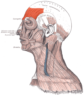
The corrugator supercilii is a small, narrow, pyramidal muscle close to the eye. It is located at the medial end of the eyebrow, beneath the frontalis and just above orbicularis oculi muscle.

The nasalis is a sphincter-like muscle of the nose whose function is to compress the nasal cartilages. It is the muscle responsible for "flaring" of the nostrils. Some people can use it to close the nostrils to prevent entry of water when underwater.

The orbicularis oculi is a muscle in the face that closes the eyelids. It arises from the nasal part of the frontal bone, from the frontal process of the maxilla in front of the lacrimal groove, and from the anterior surface and borders of a short fibrous band, the medial palpebral ligament.
The frontalis muscle is a muscle which covers parts of the forehead of the skull. Some sources consider the frontalis muscle to be a distinct muscle. However, Terminologia Anatomica currently classifies it as part of the occipitofrontalis muscle along with the occipitalis muscle.

The nasociliary nerve is a branch of the ophthalmic nerve, itself a branch of the trigeminal nerve. It is intermediate in size between the other two branches of the ophthalmic nerve, the frontal nerve and lacrimal nerve.

The supratrochlear nerve is a branch of the frontal nerve, itself a branch of the ophthalmic nerve. It provides sensory innervation to the skin of the forehead and upper eyelid.

The infraorbital artery is an artery in the head that branches off the maxillary artery, emerging through the infraorbital foramen, just under the orbit of the eye.

The marginal mandibular branch of the facial nerve passes forward beneath the platysma and depressor anguli oris, supplying the muscles of the lower lip and chin, and communicating with the mental branch of the inferior alveolar nerve.
The buccal branches of the facial nerve, are of larger size than the rest of the branches, pass horizontally forward to be distributed below the orbit and around the mouth.
The temporal branches of the facial nerve crosses the zygomatic arch to the temporal region, supplying the auriculares anterior and superior, and joining with the zygomaticotemporal branch of the maxillary nerve, and with the auriculotemporal branch of the mandibular nerve.

The facial muscles are a group of striated skeletal muscles supplied by the facial nerve that, among other things, control facial expression. These muscles are also called mimetic muscles.

The following outline is provided as an overview of and topical guide to human anatomy:
