The facial nerve, also known as the seventh cranial nerve, cranial nerve VII, or simply CN VII, is a cranial nerve that emerges from the pons of the brainstem, controls the muscles of facial expression, and functions in the conveyance of taste sensations from the anterior two-thirds of the tongue. The nerve typically travels from the pons through the facial canal in the temporal bone and exits the skull at the stylomastoid foramen. It arises from the brainstem from an area posterior to the cranial nerve VI and anterior to cranial nerve VIII.

In neuroanatomy, the mandibular nerve (V3) is the largest of the three divisions of the trigeminal nerve, the fifth cranial nerve (CN V). Unlike the other divisions of the trigeminal nerve (ophthalmic nerve, maxillary nerve) which contain only afferent fibers, the mandibular nerve contains both afferent and efferent fibers. These nerve fibers innervate structures of the lower jaw and face, such as the tongue, lower lip, and chin. The mandibular nerve also innervates the muscles of mastication.

The parotid gland is a major salivary gland in many animals. In humans, the two parotid glands are present on either side of the mouth and in front of both ears. They are the largest of the salivary glands. Each parotid is wrapped around the mandibular ramus, and secretes serous saliva through the parotid duct into the mouth, to facilitate mastication and swallowing and to begin the digestion of starches. There are also two other types of salivary glands; they are submandibular and sublingual glands. Sometimes accessory parotid glands are found close to the main parotid glands.

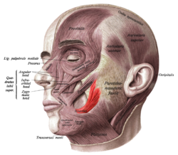
In anatomy, the masseter is one of the muscles of mastication. Found only in mammals, it is particularly powerful in herbivores to facilitate chewing of plant matter. The most obvious muscle of mastication is the masseter muscle, since it is the most superficial and one of the strongest.

The zygomaticus major muscle is a muscle of the face. It arises from either zygomatic arch (cheekbone); it inserts at the corner of the mouth. It is innervated by branches of the facial nerve.

The zygomaticus minor muscle is a muscle of facial expression. It originates from the zygomatic bone, lateral to the rest of the levator labii superioris muscle, and inserts into the outer part of the upper lip. It draws the upper lip backward, upward, and outward and is used in smiling. It is innervated by the facial nerve (VII).

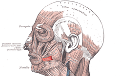
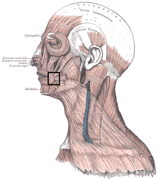
The depressor anguli oris muscle is a facial muscle. It originates from the mandible and inserts into the angle of the mouth. It is associated with frowning, as it depresses the corner of the mouth.

The facial artery is a branch of the external carotid artery that supplies structures of the superficial face.

The occipitofrontalis muscle is a muscle which covers parts of the skull. It consists of two parts or bellies: the occipital belly, near the occipital bone, and the frontal belly, near the frontal bone. It is supplied by the supraorbital artery, the supratrochlear artery, and the occipital artery. It is innervated by the facial nerve. In humans, the occipitofrontalis helps to create facial expressions.
In facial anatomy, the modiolus is a dense, compact, mobile, fibromuscular tissue mass of facial muscles formed by the interlacing of a number of muscles just lateral to the angle of the mouth opposite the second upper premolar tooth.

The sural nerve(L4-S1) is generally considered a pure cutaneous nerve of the posterolateral leg to the lateral ankle. The sural nerve originates from a combination of either the sural communicating branch and medial sural cutaneous nerve, or the lateral sural cutaneous nerve. This group of nerves is termed the sural nerve complex. There are eight documented variations of the sural nerve complex. Once formed the sural nerve takes its course midline posterior to posterolateral around the lateral malleolus. The sural nerve terminates as the lateral dorsal cutaneous nerve.

The infratemporal fossa is an irregularly shaped cavity that is a part of the skull. It is situated below and medial to the zygomatic arch. It is not fully enclosed by bone in all directions. It contains superficial muscles, including the lower part of the temporalis muscle, the lateral pterygoid muscle, and the medial pterygoid muscle. It also contains important blood vessels such as the middle meningeal artery, the pterygoid plexus, and the retromandibular vein, and nerves such as the mandibular nerve (CN V3) and its branches.
The masseteric nerve is a nerve of the face. It is a branch of the mandibular nerve (CN V3). It passes through the mandibular notch to reach masseter muscle. It provides motor innervation the masseter muscle, and sensory innervation to the temporomandibular joint.
The masseteric fascia and parotideomasseteric fascia are fascias of the head varyingly described depending upon the source consulted. They may or may not be described as one and the same structure.

The marginal mandibular branch of the facial nerve arises from the facial nerve in the parotid gland at the parotid plexus. It passes anterior-ward deep to the platysma and depressor anguli oris muscles. It provides motor innervation to muscles of the lower lip and chin: the depressor labii inferioris muscle, depressor anguli oris muscle, and mentalis muscle. It communicates with the mental branch of the inferior alveolar nerve.
The facial muscles are a group of striated skeletal muscles supplied by the facial nerve that, among other things, control facial expression. These muscles are also called mimetic muscles. They are only found in mammals, although they derive from neural crest cells found in all vertebrates. They are the only muscles that attach to the dermis.
Jaw reduction or mandible angle reduction is a type of surgery to narrow the lower one-third of the face—particularly the contribution from the mandible and its muscular attachments. There are several techniques for treatment—including surgical and non-surgical methods. A square lower jaw can be considered a masculine trait, especially in Asian countries. As a result, whereas square lower jaws are often considered a positive trait in men, a wide mandible can be perceived as discordant or masculine on women, or sometimes in certain men, particularly when there is asymmetry.

Smile surgery or smile reconstruction is a surgical procedure that restores the smile for people with facial nerve paralysis. Facial nerve paralysis is a relatively common condition with a yearly incidence of 0.25% leading to function loss of the mimic muscles. The facial nerve gives off several branches in the face. If one or more facial nerve branches are paralysed, the corresponding mimetic muscles lose their ability to contract. This may lead to several symptoms such as incomplete eye closure with or without exposure keratitis, oral incompetence, poor articulation, dental caries, drooling, and a low self-esteem. This is because the different branches innervate the frontalis muscle, orbicularis oculi and oris muscles, lip elevators and depressors, and the platysma. The elevators of the upper lip and corner of the mouth are innervated by the zygomatic and buccal branches. When these branches are paralysed, there is an inability to create a symmetric smile.

The submasseterric space is a fascial space of the head and neck. It is a potential space in the face over the angle of the jaw, and is paired on each side. It is located between the lateral aspect of the mandible and the medial aspect of the masseter muscle and its investing fascia. The term is derived from sub- meaning "under" in Latin and masseteric which refers to the masseter muscle. The submasseteric space is one of the four compartments of the masticator space. Sometimes the submasseteric space is described as a series of spaces, created because the masseter muscle has multiple insertions that cover most of the lateral surface of the ramus of the mandible.
The parotid fascia is a tough fascia enclosing the parotid gland. It has a superficial layer and a deep layer.