Descending neuron

Last updated

A descending neuron is a neuron that conveys signals from the brain to neural circuits in the spinal cord (vertebrates) or ventral nerve cord (invertebrates). As the sole conduits of information between the brain and the body, descending neurons play a key role in behavior. Their activity can initiate, maintain, modulate, and terminate behaviors such as locomotion. Because the number of descending neurons is several orders of magnitude smaller than the number of neurons in either the brain or spinal cord/ventral nerve cord, this class of cells represents a critical bottleneck in the flow of information from sensory systems to motor circuits.

Contents

Anatomy

Descending neurons have their somas and dendrites (primary input zones) in the brain. Their axons traverse the neck in connectives, or tracts, and output onto neurons in the spinal cord (vertebrates) or ventral nerve cord (invertebrates).

Schematic of major descending pathways in mammals. The corticospinal and corticobulbar tracts are pyramidal tracts controlling voluntary movement. The tectospinal, rubrospinal, vestibulospinal, and reticulospinal tracts are extrapyramidal tracts controlling involuntary movement. Descending pathways in mammals.svg
Schematic of major descending pathways in mammals. The corticospinal and corticobulbar tracts are pyramidal tracts controlling voluntary movement. The tectospinal, rubrospinal, vestibulospinal, and reticulospinal tracts are extrapyramidal tracts controlling involuntary movement.

Mammals possess hundreds of thousands of descending neurons. [1] [2] They can be divided functionally into two major pathways: pyramidal tracts, which originate in the motor cortex, and extrapyramidal tracts, which originate in the brainstem (see schematic). An example of the former is the corticospinal tract, which is responsible for voluntary movement of the body. An example of the latter is the reticulospinal tract, which contributes to the unconscious regulation of locomotion and posture. Reticulospinal neurons originate in the medullary reticular formation, where they receive information from upstream locomotor centers, such as the mesencephalic locomotor region and the basal ganglia. [3]

Side-view schematic of major descending pathways in Drosophila melanogaster. In the ventral nerve chord, the major pathways target the dorsal wing, neck, and haltere neuropils, the ventral leg neuropils, and the intermediate tectulum, an integrative region. Adapted from Namiki et al. (2018) . Descending pathways in Drosophila.svg
Side-view schematic of major descending pathways in Drosophila melanogaster. In the ventral nerve chord, the major pathways target the dorsal wing, neck, and haltere neuropils, the ventral leg neuropils, and the intermediate tectulum, an integrative region. Adapted from Namiki et al. (2018) .

Insects possess only several hundreds of descending neurons. [5] [6] [7] [8] Work in the fruit fly Drosophila melanogaster suggests that they are organized into three broad pathways (see schematic). [8] Two direct pathways link specific regions in the brain to motor circuits in the ventral nerve cord controlling the legs and wings, respectively. A third pathway couples a broad array of brain regions to a large integrative region in the ventral nerve cord that may control both sets of appendages.


Function

Descending neurons play an important role in initiating, maintaining, modulating, and terminating behaviors. Several descending neurons involved in controlling specific behaviors have been identified in both vertebrates and invertebrates. These include descending neurons that can initiate and terminate locomotion, [9] [10] [11] [12] modulate locomotion speed [10] [13] [14] and direction, [15] [16] [17] [18] [19] and help coordinate limbs. [20]

While some descending neurons are sufficient to elicit specific behaviors, [21] [22] [19] most behaviors are likely not controlled by single, command-like descending neurons, but instead by the combined activity of different descending neurons. [23] [24]

Some descending pathways form direct connections with motor neurons and premotor interneurons, [25] including central pattern generators. [26] But how exactly descending signals are integrated in circuits in the spinal cord (vertebrates) or ventral nerve cord (invertebrates) during behavior is not well understood. [3] [27]

See also

Related Research Articles

<span class="mw-page-title-main">Motor neuron</span> Nerve cell sending impulse to muscle

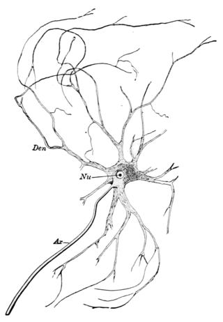
A motor neuron is a neuron whose cell body is located in the motor cortex, brainstem or the spinal cord, and whose axon (fiber) projects to the spinal cord or outside of the spinal cord to directly or indirectly control effector organs, mainly muscles and glands. There are two types of motor neuron – upper motor neurons and lower motor neurons. Axons from upper motor neurons synapse onto interneurons in the spinal cord and occasionally directly onto lower motor neurons. The axons from the lower motor neurons are efferent nerve fibers that carry signals from the spinal cord to the effectors. Types of lower motor neurons are alpha motor neurons, beta motor neurons, and gamma motor neurons.

<span class="mw-page-title-main">Brainstem</span> Posterior part of the brain, adjoining and structurally continuous

The brainstem is the stalk-like part of the brain that interconnects the cerebrum and diencephalon with the spinal cord. In the human brain, the brainstem is composed of the midbrain, the pons, and the medulla oblongata. The midbrain is continuous with the thalamus of the diencephalon through the tentorial notch.

<span class="mw-page-title-main">Motor nerve</span> Nerve located in the central nervous system

A motor nerve is a nerve that transmits motor signals from the central nervous system (CNS) to the muscles of the body. This is different from the motor neuron, which includes a cell body and branching of dendrites, while the nerve is made up of a bundle of axons. Motor nerves act as efferent nerves which carry information out from the CNS to muscles, as opposed to afferent nerves, which transfer signals from sensory receptors in the periphery to the CNS. Efferent nerves can also connect to glands or other organs/issues instead of muscles. In addition, there are nerves that serve as both sensory and motor nerves called mixed nerves.

In biology, a reflex, or reflex action, is an involuntary, unplanned sequence or action and nearly instantaneous response to a stimulus.

<span class="mw-page-title-main">Extrapyramidal system</span> Connection between brain and spinal cord

In anatomy, the extrapyramidal system is a part of the motor system network causing involuntary actions. The system is called extrapyramidal to distinguish it from the tracts of the motor cortex that reach their targets by traveling through the pyramids of the medulla. The pyramidal tracts may directly innervate motor neurons of the spinal cord or brainstem, whereas the extrapyramidal system centers on the modulation and regulation of anterior (ventral) horn cells.

<span class="mw-page-title-main">Periaqueductal gray</span> Nucleus surrounding the cerebral aqueduct

The periaqueductal gray is a brain region that plays a critical role in autonomic function, motivated behavior and behavioural responses to threatening stimuli. PAG is also the primary control center for descending pain modulation. It has enkephalin-producing cells that suppress pain.

<span class="mw-page-title-main">Reticular formation</span> Spinal trigeminal nucleus

The reticular formation is a set of interconnected nuclei that are located throughout the brainstem. It is not anatomically well defined, because it includes neurons located in different parts of the brain. The neurons of the reticular formation make up a complex set of networks in the core of the brainstem that extend from the upper part of the midbrain to the lower part of the medulla oblongata. The reticular formation includes ascending pathways to the cortex in the ascending reticular activating system (ARAS) and descending pathways to the spinal cord via the reticulospinal tracts.

Central pattern generators (CPGs) are self-organizing biological neural circuits that produce rhythmic outputs in the absence of rhythmic input. They are the source of the tightly-coupled patterns of neural activity that drive rhythmic and stereotyped motor behaviors like walking, swimming, breathing, or chewing. The ability to function without input from higher brain areas still requires modulatory inputs, and their outputs are not fixed. Flexibility in response to sensory input is a fundamental quality of CPG-driven behavior. To be classified as a rhythmic generator, a CPG requires:

  1. "two or more processes that interact such that each process sequentially increases and decreases, and
  2. that, as a result of this interaction, the system repeatedly returns to its starting condition."
<span class="mw-page-title-main">Scratch reflex</span> Response to activation of sensory neurons

The scratch reflex is a response to activation of sensory neurons whose peripheral terminals are located on the surface of the body. Some sensory neurons can be activated by stimulation with an external object such as a parasite on the body surface. Alternatively, some sensory neurons can respond to a chemical stimulus that produces an itch sensation. During a scratch reflex, a nearby limb reaches toward and rubs against the site on the body surface that has been stimulated. The scratch reflex has been extensively studied to understand the functioning of neural networks in vertebrates. Despite decades of research, key aspects of the scratch reflex are still unknown, such as the neural mechanisms by which the reflex is terminated.

The zona incerta (ZI) is a horizontally elongated region of gray matter in the subthalamus below the thalamus. Its connections project extensively over the brain from the cerebral cortex down into the spinal cord.

<span class="mw-page-title-main">Escape reflex</span>

Escape reflex, or escape behavior, is any kind of escape response found in an animal when it is presented with an unwanted stimulus. It is a simple reflectory reaction in response to stimuli indicative of danger, that initiates an escape motion of an animal. The escape response has been found to be processed in the telencephalon.

<span class="mw-page-title-main">Ventral nerve cord</span>

The ventral nerve cord is a major structure of the invertebrate central nervous system. It is the functional equivalent of the vertebrate spinal cord. The ventral nerve cord coordinates neural signaling from the brain to the body and vice versa, integrating sensory input and locomotor output. Because arthropods have an open circulatory system, decapitated insects can still walk, groom, and mate—illustrating that the circuitry of the ventral nerve cord is sufficient to perform complex motor programs without brain input.

<span class="mw-page-title-main">Spinal cord</span> Long, tubular central nervous system structure in the vertebral column

The spinal cord is a long, thin, tubular structure made up of nervous tissue that extends from the medulla oblongata in the brainstem to the lumbar region of the vertebral column (backbone) of vertebrate animals. The center of the spinal cord is hollow and contains a structure called central canal, which contains cerebrospinal fluid. The spinal cord is also covered by meninges and enclosed by the neural arches. Together, the brain and spinal cord make up the central nervous system (CNS).

<span class="mw-page-title-main">Primary motor cortex</span> Brain region

The primary motor cortex is a brain region that in humans is located in the dorsal portion of the frontal lobe. It is the primary region of the motor system and works in association with other motor areas including premotor cortex, the supplementary motor area, posterior parietal cortex, and several subcortical brain regions, to plan and execute voluntary movements. Primary motor cortex is defined anatomically as the region of cortex that contains large neurons known as Betz cells, which, along with other cortical neurons, send long axons down the spinal cord to synapse onto the interneuron circuitry of the spinal cord and also directly onto the alpha motor neurons in the spinal cord which connect to the muscles.

Pigment dispersing factor (pdf) is a gene that encodes the protein PDF, which is part of a large family of neuropeptides. Its hormonal product, pigment dispersing hormone (PDH), was named for the diurnal pigment movement effect it has in crustacean retinal cells upon its initial discovery in the central nervous system of arthropods. The movement and aggregation of pigments in retina cells and extra-retinal cells is hypothesized to be under a split hormonal control mechanism. One hormonal set is responsible for concentrating chromatophoral pigment by responding to changes in the organism's exposure time to darkness. Another hormonal set is responsible for dispersion and responds to the light cycle. However, insect pdf genes do not function in such pigment migration since they lack the chromatophore.

<span class="mw-page-title-main">Neural substrate of locomotor central pattern generators in mammals</span>

Central pattern generators are biological neural networks organized to produce any rhythmic output without requiring a rhythmic input. In mammals, locomotor CPGs are organized in the lumbar and cervical segments of the spinal cord, and are used to control rhythmic muscle output in the arms and legs. Certain areas of the brain initiate the descending neural pathways that ultimately control and modulate the CPG signals. In addition to this direct control, there exist different feedback loops that coordinate the limbs for efficient locomotion and allow for the switching of gaits under appropriate circumstances.

The parabrachial nuclei, also known as the parabrachial complex, are a group of nuclei in the dorsolateral pons that surrounds the superior cerebellar peduncle as it enters the brainstem from the cerebellum. They are named from the Latin term for the superior cerebellar peduncle, the brachium conjunctivum. In the human brain, the expansion of the superior cerebellar peduncle expands the parabrachial nuclei, which form a thin strip of grey matter over most of the peduncle. The parabrachial nuclei are typically divided along the lines suggested by Baxter and Olszewski in humans, into a medial parabrachial nucleus and lateral parabrachial nucleus. These have in turn been subdivided into a dozen subnuclei: the superior, dorsal, ventral, internal, external and extreme lateral subnuclei; the lateral crescent and subparabrachial nucleus along the ventrolateral margin of the lateral parabrachial complex; and the medial and external medial subnuclei

<span class="mw-page-title-main">Spinal interneuron</span> Interneuron relaying signals between sensory and motor neurons in the spinal cord

A spinal interneuron, found in the spinal cord, relays signals between (afferent) sensory neurons, and (efferent) motor neurons. Different classes of spinal interneurons are involved in the process of sensory-motor integration. Most interneurons are found in the grey column, a region of grey matter in the spinal cord.

<span class="mw-page-title-main">Mesencephalic locomotor region</span>

The mesencephalic locomotor region (MLR) is a functionally defined area of the midbrain that is associated with the initiation and control of locomotor movements in vertebrate species.

<span class="mw-page-title-main">Ole Kiehn</span> Danish-Swedish neuroscientist

Ole Kiehn is a Danish-Swedish neuroscientist. He is Professor of Integrative Neuroscience at the Department of Neuroscience, University of Copenhagen, Denmark, and professor of neurophysiology at Karolinska Institute, Sweden.

References

  1. Lemon, Roger N. (July 21, 2008). "Descending Pathways in Motor Control". Annual Review of Neuroscience . 31: 195–218. doi:10.1146/annurev.neuro.31.060407.125547. ISSN   0147-006X. OCLC   57214750. PMID   18558853.
  2. Liang, Huazheng; Paxinos, George; Watson, Charles (October 9, 2010). "Projections from the brain to the spinal cord in the mouse". Brain Structure and Function . 215 (3–4): 159–186. doi:10.1007/s00429-010-0281-x. hdl: 20.500.11937/30100 . ISSN   1863-2653. LCCN   2007243247. OCLC   804279700. PMID   20936329. S2CID   1880945.
  3. 1 2 Leiras, Roberto; Cregg, Jared M.; Kiehn, Ole (July 8, 2022). "Brainstem Circuits for Locomotion". Annual Review of Neuroscience. 45 (1): annurev–neuro–082321-025137. doi:10.1146/annurev-neuro-082321-025137. ISSN   0147-006X. PMID   34985919. S2CID   245771230.
  4. Namiki, Shigehiro; Dickinson, Michael H; Wong, Allan M; Korff, Wyatt; Card, Gwyneth M (June 26, 2018). Scott, Kristin (ed.). "The functional organization of descending sensory-motor pathways in Drosophila". eLife. 7: e34272. doi: 10.7554/eLife.34272 . ISSN   2050-084X. PMC   6019073 . PMID   29943730.
  5. Okada, Ryuichi; Sakura, Midori; Mizunami, Makoto (March 31, 2003). "Distribution of dendrites of descending neurons and its implications for the basic organization of the cockroach brain". The Journal of Comparative Neurology. 458 (2): 158–174. doi:10.1002/cne.10580. ISSN   0021-9967. PMID   12596256. S2CID   14396370.
  6. Gal, Ram; Libersat, Frederic (September 2006). "New vistas on the initiation and maintenance of insect motor behaviors revealed by specific lesions of the head ganglia". Journal of Comparative Physiology A. 192 (9): 1003–1020. doi:10.1007/s00359-006-0135-4. ISSN   0340-7594. PMID   16733727. S2CID   28032937.
  7. Hsu, Cynthia T.; Bhandawat, Vikas (April 2016). "Organization of descending neurons in Drosophila melanogaster". Scientific Reports. 6 (1): 20259. Bibcode:2016NatSR...620259H. doi:10.1038/srep20259. ISSN   2045-2322. PMC   4738306 . PMID   26837716.
  8. 1 2 Namiki, Shigehiro; Dickinson, Michael H; Wong, Allan M; Korff, Wyatt; Card, Gwyneth M (June 26, 2018). "The functional organization of descending sensory-motor pathways in Drosophila". eLife. 7: e34272. doi: 10.7554/eLife.34272 . ISSN   2050-084X. PMC   6019073 . PMID   29943730.
  9. Bidaye, Salil S.; Laturney, Meghan; Chang, Amy K.; Liu, Yuejiang; Bockemühl, Till; Büschges, Ansgar; Scott, Kristin (November 11, 2020). "Two Brain Pathways Initiate Distinct Forward Walking Programs in Drosophila". Neuron. 108 (3): 469–485.e8. doi:10.1016/j.neuron.2020.07.032. ISSN   0896-6273. PMC   9435592 . PMID   32822613. S2CID   221198570.
  10. 1 2 Capelli, Paolo; Pivetta, Chiara; Soledad Esposito, Maria; Arber, Silvia (November 2017). "Locomotor speed control circuits in the caudal brainstem". Nature. 551 (7680): 373–377. Bibcode:2017Natur.551..373C. doi:10.1038/nature24064. ISSN   1476-4687. PMID   29059682. S2CID   205260887.
  11. Zacarias, Ricardo; Namiki, Shigehiro; Card, Gwyneth M.; Vasconcelos, Maria Luisa; Moita, Marta A. (September 12, 2018). "Speed dependent descending control of freezing behavior in Drosophila melanogaster". Nature Communications. 9 (1): 3697. Bibcode:2018NatCo...9.3697Z. doi:10.1038/s41467-018-05875-1. ISSN   2041-1723. PMC   6135764 . PMID   30209268.
  12. Bouvier, Julien; Caggiano, Vittorio; Leiras, Roberto; Caldeira, Vanessa; Bellardita, Carmelo; Balueva, Kira; Fuchs, Andrea; Kiehn, Ole (November 19, 2015). "Descending Command Neurons in the Brainstem that Halt Locomotion". Cell. 163 (5): 1191–1203. doi:10.1016/j.cell.2015.10.074. ISSN   1097-4172. PMC   4899047 . PMID   26590422.
  13. Severi, Kristen E.; Portugues, Ruben; Marques, João C.; O’Malley, Donald M.; Orger, Michael B.; Engert, Florian (August 6, 2014). "Neural Control and Modulation of Swimming Speed in the Larval Zebrafish". Neuron. 83 (3): 692–707. doi:10.1016/j.neuron.2014.06.032. ISSN   0896-6273. PMC   4126853 . PMID   25066084.
  14. Namiki, Shigehiro; Ros, Ivo G.; Morrow, Carmen; Rowell, William J.; Card, Gwyneth M.; Korff, Wyatt; Dickinson, Michael H. (March 14, 2022). "A population of descending neurons that regulates the flight motor of Drosophila". Current Biology. 32 (5): 1189–1196.e6. doi:10.1016/j.cub.2022.01.008. ISSN   1879-0445. PMC   9206711 . PMID   35090590. S2CID   236961767.
  15. Cregg, Jared M.; Leiras, Roberto; Montalant, Alexia; Wanken, Paulina; Wickersham, Ian R.; Kiehn, Ole (June 2020). "Brainstem neurons that command mammalian locomotor asymmetries". Nature Neuroscience. 23 (6): 730–740. doi:10.1038/s41593-020-0633-7. ISSN   1546-1726. PMC   7610510 . PMID   32393896.
  16. Orger, Michael B.; Kampff, Adam R.; Severi, Kristen E.; Bollmann, Johann H.; Engert, Florian (March 2008). "Control of visually guided behavior by distinct populations of spinal projection neurons". Nature Neuroscience. 11 (3): 327–333. doi:10.1038/nn2048. ISSN   1546-1726. PMC   2894808 . PMID   18264094.
  17. Schnell, Bettina; Ros, Ivo G.; Dickinson, Michael H. (April 24, 2017). "A Descending Neuron Correlated with the Rapid Steering Maneuvers of Flying Drosophila". Current Biology. 27 (8): 1200–1205. doi:10.1016/j.cub.2017.03.004. ISSN   0960-9822. PMC   6309624 . PMID   28392112. S2CID   5052663.
  18. Rayshubskiy, Aleksandr; Holtz, Stephen L.; D’Alessandro, Isabel; Li, Anna A.; Vanderbeck, Quinn X.; Haber, Isabel S.; Gibb, Peter W.; Wilson, Rachel I. (July 18, 2020). "Neural circuit mechanisms for steering control in walking Drosophila": 2020.04.04.024703. doi:10.1101/2020.04.04.024703.{{cite journal}}: Cite journal requires |journal= (help)
  19. 1 2 Bidaye, Salil S.; Machacek, Christian; Wu, Yang; Dickson, Barry J. (April 4, 2014). "Neuronal Control of Drosophila Walking Direction". Science. 344 (6179): 97–101. Bibcode:2014Sci...344...97B. doi:10.1126/science.1249964. ISSN   0036-8075. PMID   24700860. S2CID   27815021.
  20. Ruder, Ludwig; Takeoka, Aya; Arber, Silvia (December 7, 2016). "Long-Distance Descending Spinal Neurons Ensure Quadrupedal Locomotor Stability". Neuron. 92 (5): 1063–1078. doi: 10.1016/j.neuron.2016.10.032 . ISSN   0896-6273. PMID   27866798. S2CID   2203590.
  21. Korn, Henri; Faber, Donald S. (July 7, 2005). "The Mauthner Cell Half a Century Later: A Neurobiological Model for Decision-Making?". Neuron. 47 (1): 13–28. doi: 10.1016/j.neuron.2005.05.019 . ISSN   0896-6273. PMID   15996545. S2CID   2851487.
  22. Hampel, Stefanie; Franconville, Romain; Simpson, Julie H; Seeds, Andrew M (September 7, 2015). Borst, Alexander (ed.). "A neural command circuit for grooming movement control". eLife. 4: e08758. doi: 10.7554/eLife.08758 . ISSN   2050-084X. PMC   4599031 . PMID   26344548.
  23. Cande, Jessica; Namiki, Shigehiro; Qiu, Jirui; Korff, Wyatt; Card, Gwyneth M; Shaevitz, Joshua W; Stern, David L; Berman, Gordon J (June 26, 2018). "Optogenetic dissection of descending behavioral control in Drosophila". eLife. 7: e34275. doi: 10.7554/eLife.34275 . ISSN   2050-084X. PMC   6031430 . PMID   29943729.
  24. Namiki, Shigehiro; Ros, Ivo G.; Morrow, Carmen; Rowell, William J.; Card, Gwyneth M.; Korff, Wyatt; Dickinson, Michael H. (January 31, 2022). "A population of descending neurons that regulates the flight motor of Drosophila". Current Biology. 32 (5): 1189–1196.e6. doi: 10.1016/j.cub.2022.01.008 . ISSN   0960-9822. PMC   9206711 . PMID   35090590. S2CID   236961767.
  25. Lemon, Roger N. (July 1, 2008). "Descending Pathways in Motor Control". Annual Review of Neuroscience. 31 (1): 195–218. doi:10.1146/annurev.neuro.31.060407.125547. ISSN   0147-006X. PMID   18558853.
  26. Jordan, Larry M.; Liu, Jun; Hedlund, Peter B.; Akay, Turgay; Pearson, Keir G. (January 1, 2008). "Descending command systems for the initiation of locomotion in mammals". Brain Research Reviews. Networks in Motion. 57 (1): 183–191. doi:10.1016/j.brainresrev.2007.07.019. ISSN   0165-0173. PMID   17928060. S2CID   39052299.
  27. Bidaye, Salil S.; Bockemühl, Till; Büschges, Ansgar (February 1, 2018). "Six-legged walking in insects: how CPGs, peripheral feedback, and descending signals generate coordinated and adaptive motor rhythms". Journal of Neurophysiology. 119 (2): 459–475. doi: 10.1152/jn.00658.2017 . ISSN   1522-1598. PMID   29070634.