Diaphragm pacing

Last updated

Diaphragm pacing
Gray806.png
Electrical stimulation of the phrenic nerve has been known to stimulate respiration for centuries.
Other namesphrenic nerve pacing

Diaphragm pacing (and even earlier as electrophrenic respiration [1] [2] ) is the rhythmic application of electrical impulses to the diaphragm to provide artificial ventilatory support for respiratory failure or sleep apnea. [3] [4] Historically, this has been accomplished through the electrical stimulation of a phrenic nerve by an implanted receiver/electrode, [5] though today an alternative option of attaching percutaneous wires to the diaphragm exists. [6]

Contents

History

The idea of stimulating the diaphragm through the phrenic nerve was first firmly postulated by German physician Christoph Wilhelm Hufeland, who in 1783 proposed that such a technique could be applied as a treatment for asphyxia. [7] [8] :545–549 French neurologist Duchenne de Boulogne made a similar proposal in 1855, though neither of them tested it. [9] It was not until a year later that Hugo Wilhelm von Ziemssen demonstrated diaphragm pacing on a 27-year-old woman asphyxiated on charcoal fumes by rhythmically faradizing her phrenic nerves, saving her life. [8] [10] :49 Duchenne would later in 1872 declare the technique the "best means of imitating natural respiration". [11] However, advances in mechanical ventilation by the likes of George Poe in the early twentieth century [12] ended up being initially favored over phrenic nerve stimulation.

Harvard researchers Sarnoff et al. revisited diaphragm pacing via the phrenic nerve in 1948, publishing their experimental results on dogs. [1] In a separate publication a few days before, the same group also revealed they had an opportunity to use the technique "on a five-year-old boy with complete respiratory paralysis following rupture of a cerebral aneurysm". Referring to the process as "electrophrenic respiration", Sarnoff was able to artificially respirate the young boy for 52 hours. [13] The technology behind diaphragm pacing was advanced further in 1968 with the publication of doctors John P. Judson and William W. L. Glenn's research on the use of radio-frequency transmission to at whim "adjust the amplitude of stimulation and to control the rate of stimulation externally". [14] Teaming up with Avery Laboratories, Glenn brought his prototype device to the commercial market in the early 1970s. [15] The Avery Breathing Pacemaker received pre-market approval from the FDA in 1987 for "chronic ventilatory support because of upper motor neuron respiratory muscle paralysis" in patients of all ages. [16] In the 1980s, "sequential multipole stimulation" was developed in Tampere, Finland. This technology was commercialized as the Atrostim PNS system and became commercially available in Europe in 1990. [17]

By the early 1990s, long-term evaluations of the technology were being published, with some researchers such as Bach and O'Connor stating that phrenic nerve pacing is a valid option "for the properly screened patient but that expense, failure rate, morbidity, and mortality remain excessive and that alternative methods of ventilatory support should be explored". [18] Others such as Brouillette and Marzocchi suggested that advances in encapsulation and electrode technologies could improve system longevity and reduce damage to diaphragm muscle. [19] Additionally, new surgical techniques such as a thoracoscopic approach began to appear in the late 1990s. [20]

In the mid-2000s, U.S. company Synapse Biomedical began researching a new diaphragm pacing system that would not have to attach to the phrenic nerve but instead depended on "four electrodes implanted in the muscle of the diaphragm to electronically stimulate contraction". The marketed NeuRx device received several FDA approvals under a Humanitarian Device Exemption (HDE), one in 2008 and another in 2011. [21]

Methodology and devices

The basic principle behind a diaphragm pacing device (the U.S. Food and Drug Administration identifies the device as a "diaphragmatic/phrenic nerve stimulator" [22] ) involves passing an electric current through electrodes that are attached internally. The diaphragm contracts, expanding the chest cavity, and causing air to be sucked into the lungs (inspiration). When not stimulated, the diaphragm relaxes and air moves out of the lungs (expiration).

According to the United States Medicare system, phrenic nerve stimulators are indicated for "selected patients with partial or complete respiratory insufficiency" and "can be effective only if the patient has an intact phrenic nerve and diaphragm". [23] Common patient diagnoses for phrenic nerve pacing include patients with spinal cord injury, central sleep apnea, congenital central hypoventilation syndrome (i.e., Ondine's curse), and diaphragm paralysis. [21] [23]

There are currently three commercially distributed diaphragm pacing devices: Synapse Biomedical, Inc.'s NeuRx (US), Avery Biomedical Devices, Inc.'s Mark IV Breathing Pacemaker (US), [21] and Atrotech OY's Atrostim PNS (Finland). [24] The Synapse and Avery devices are distributed worldwide and approved for use in the United States. [21] The Atrotech device is not available in the U.S. As of December 2019, FDA Premarket Approval was given to Avery's Spirit Transmitter Device, replacing the Mark IV transmitter. [25]

In May 2020, Canadian company Lungpacer Medical received approval for emergency use by the USFDA amid the COVID-19 pandemic. [26]

Surgical procedure

In the case of the Atrostim and Mark IV devices, several surgical techniques may be used. Surgery is typically performed by placing an electrode around the phrenic nerve, either in the neck (i.e., cervically; an older technique), or in the chest (i.e., thoracically; more modern). This electrode is connected to a radiofrequency receiver which is implanted just under the skin. An external transmitter sends radio signals to the device by an antenna that is worn over the receiver. [27] For the cervical surgical technique, the phrenic nerve is approached via a small (~5 cm) incision slightly above, and midline to, the clavic. The phrenic nerve is then isolated under the scalenus anticus muscle. For the thoracic surgical technique, a small (~5 cm) incisions over the 2nd or 3rd intercostal space. The electrodes are placed around the phrenic nerves alongside the pericardium. The use of a thorascope allows for this technique to be performed in a minimally-invasive manner. [27]

In the case of the NeuRx device, a series of four incisions are made in the abdominal skin. Several tools such as a laparoscope and probe are used to find the best four locations on the diaphragm to attach four electrodes, which have connections outside the body. A fifth electrode is placed just under the skin in the same area. All these connect to the device. [28]

Research

Research into the efficacy of diaphragmatic pacing (DP) for ventilatory support has had mixed results, that depend largely on the disease process of the patient population being studied. [29]

Numerous studies have shown that DP can be a very effective tool for patients with spinal cord injuries (SCI). [30] [31] [32] [29] One retrospective long-term multivariate analysis of patients with cervical SCI injuries, found that DP produced better survival rates and updated self-reported quality of life ratings than did the use of mechanical ventilation (MV) . [31]

According to a number of high profile studies, diaphragm pacing should not be used to treat respiratory failure in patients with Amyotrophic Lateral Sclerosis (ALS). [33] [34] Studies have demonstrated a significantly higher mortality rate in patients with activated DP implants, than those who received the traditional MV for treatment in later stages of ALS. There have been virtually no clinical trials published on the efficacy of DP in patients with ALS since around 2016, due to the results of the DiPALS study, that showed a mortality rate of 76% of patients with the implant compared to 51% of patients who received MV alone. [34] The USA has allowed the implementation of DP for patients with ALS using a “Compassionate Authorization”, however Europe has stopped all authorization for DP in patients with ALS. [35]

Related Research Articles

<span class="mw-page-title-main">Phrenic nerve</span> Nerve controlling the diaphragm

The phrenic nerve is a mixed motor/sensory nerve that originates from the C3-C5 spinal nerves in the neck. The nerve is important for breathing because it provides exclusive motor control of the diaphragm, the primary muscle of respiration. In humans, the right and left phrenic nerves are primarily supplied by the C4 spinal nerve, but there is also a contribution from the C3 and C5 spinal nerves. From its origin in the neck, the nerve travels downward into the chest to pass between the heart and lungs towards the diaphragm.

<span class="mw-page-title-main">Thoracic diaphragm</span> Sheet of internal skeletal muscle

The thoracic diaphragm, or simply the diaphragm, is a sheet of internal skeletal muscle in humans and other mammals that extends across the bottom of the thoracic cavity. The diaphragm is the most important muscle of respiration, and separates the thoracic cavity, containing the heart and lungs, from the abdominal cavity: as the diaphragm contracts, the volume of the thoracic cavity increases, creating a negative pressure there, which draws air into the lungs. Its high oxygen consumption is noted by the many mitochondria and capillaries present; more than in any other skeletal muscle.

The control of ventilation is the physiological mechanisms involved in the control of breathing, which is the movement of air into and out of the lungs. Ventilation facilitates respiration. Respiration refers to the utilization of oxygen and balancing of carbon dioxide by the body as a whole, or by individual cells in cellular respiration.

<span class="mw-page-title-main">Central hypoventilation syndrome</span> Medical condition

Central hypoventilation syndrome (CHS) is a sleep-related breathing disorder that causes ineffective breathing, apnea, or respiratory arrest during sleep. CHS can either be congenital (CCHS) or acquired (ACHS) later in life. The condition can be fatal if untreated. CCHS was once known as Ondine's curse.

<span class="mw-page-title-main">Functional electrical stimulation</span> Technique that uses low-energy electrical pulses

Functional electrical stimulation (FES) is a technique that uses low-energy electrical pulses to artificially generate body movements in individuals who have been paralyzed due to injury to the central nervous system. More specifically, FES can be used to generate muscle contraction in otherwise paralyzed limbs to produce functions such as grasping, walking, bladder voiding and standing. This technology was originally used to develop neuroprostheses that were implemented to permanently substitute impaired functions in individuals with spinal cord injury (SCI), head injury, stroke and other neurological disorders. In other words, a person would use the device each time he or she wanted to generate a desired function. FES is sometimes also referred to as neuromuscular electrical stimulation (NMES).

<span class="mw-page-title-main">Artificial ventilation</span> Assisted breathing to support life

Artificial ventilation is a means of assisting or stimulating respiration, a metabolic process referring to the overall exchange of gases in the body by pulmonary ventilation, external respiration, and internal respiration. It may take the form of manually providing air for a person who is not breathing or is not making sufficient respiratory effort, or it may be mechanical ventilation involving the use of a mechanical ventilator to move air in and out of the lungs when an individual is unable to breathe on their own, for example during surgery with general anesthesia or when an individual is in a coma or trauma.

A side stitch is an intense stabbing abdominal pain under the lower edge of the ribcage that occurs during exercise. It is also called a side ache, side cramp, muscle stitch, or simply stitch, and the medical term is exercise-related transient abdominal pain (ETAP). It sometimes extends to shoulder tip pain, and commonly occurs during running, swimming, and horseback riding. Approximately two-thirds of runners will experience at least one episode of a stitch each year. The precise cause is unclear, although it most likely involves irritation of the abdominal lining, and the condition is more likely after consuming a meal or a sugary beverage. If the pain is present only when exercising and is completely absent at rest, in an otherwise healthy person, it does not require investigation. Typical treatment strategies involve deep breathing and/or manual pressure on the affected area.

<span class="mw-page-title-main">Implant (medicine)</span> Device surgically placed within the body for medical purposes

An implant is a medical device manufactured to replace a missing biological structure, support a damaged biological structure, or enhance an existing biological structure. For example, an implant may be a rod, used to strengthen weak bones. Medical implants are human-made devices, in contrast to a transplant, which is a transplanted biomedical tissue. The surface of implants that contact the body might be made of a biomedical material such as titanium, silicone, or apatite depending on what is the most functional. In 2018, for example, American Elements developed a nickel alloy powder for 3D printing robust, long-lasting, and biocompatible medical implants. In some cases implants contain electronics, e.g. artificial pacemaker and cochlear implants. Some implants are bioactive, such as subcutaneous drug delivery devices in the form of implantable pills or drug-eluting stents.

Neuroprosthetics is a discipline related to neuroscience and biomedical engineering concerned with developing neural prostheses. They are sometimes contrasted with a brain–computer interface, which connects the brain to a computer rather than a device meant to replace missing biological functionality.

<span class="mw-page-title-main">Pericardiacophrenic artery</span>

The pericardiacophrenic artery is a long slender branch of the internal thoracic artery.

<span class="mw-page-title-main">Spinal cord stimulator</span> SCS TREATMENT

A spinal cord stimulator (SCS) or dorsal column stimulator (DCS) is a type of implantable neuromodulation device that is used to send electrical signals to select areas of the spinal cord for the treatment of certain pain conditions. SCS is a consideration for people who have a pain condition that has not responded to more conservative therapy. There are also spinal cord stimulators under research and development that could enable patients with spinal cord injury to walk again via epidural electrical stimulation (EES).

Sacral nerve stimulation, also termed sacral neuromodulation, is a type of medical electrical stimulation therapy.

Diaphragmatic paradox or paradoxical diaphragm phenomenon is an abnormal medical sign observed during respiration, in which the diaphragm moves opposite to the normal directions of its movements. The diaphragm normally moves downwards during inspiration and upwards during expiration. But in diaphragmatic paradox, it moves upwards during inspiration and downwards during expiration.

Neurally adjusted ventilatory assist (NAVA) is a mode of mechanical ventilation. NAVA delivers assistance in proportion to and in synchrony with the patient's respiratory efforts, as reflected by an electrical signal. This signal represents the electrical activity of the diaphragm, the body's principal breathing muscle.

Central sleep apnea (CSA) or central sleep apnea syndrome (CSAS) is a sleep-related disorder in which the effort to breathe is diminished or absent, typically for 10 to 30 seconds either intermittently or in cycles, and is usually associated with a reduction in blood oxygen saturation. CSA is usually due to an instability in the body's feedback mechanisms that control respiration. Central sleep apnea can also be an indicator of Arnold–Chiari malformation.

Neurostimulation is the purposeful modulation of the nervous system's activity using invasive or non-invasive means. Neurostimulation usually refers to the electromagnetic approaches to neuromodulation.

Neuromodulation is "the alteration of nerve activity through targeted delivery of a stimulus, such as electrical stimulation or chemical agents, to specific neurological sites in the body". It is carried out to normalize – or modulate – nervous tissue function. Neuromodulation is an evolving therapy that can involve a range of electromagnetic stimuli such as a magnetic field (rTMS), an electric current, or a drug instilled directly in the subdural space. Emerging applications involve targeted introduction of genes or gene regulators and light (optogenetics), and by 2014, these had been at minimum demonstrated in mammalian models, or first-in-human data had been acquired. The most clinical experience has been with electrical stimulation.

Stentrode is a small stent-mounted electrode array permanently implanted into a blood vessel in the brain, without the need for open brain surgery. It is in clinical trials as a brain–computer interface (BCI) for people with paralyzed or missing limbs, who will use their neural signals or thoughts to control external devices, which currently include computer operating systems. The device may ultimately be used to control powered exoskeletons, robotic prosthesis, computers or other devices.

<span class="mw-page-title-main">Intermittent hypoxia</span>

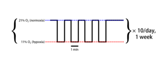
Intermittent hypoxia (also known as episodic hypoxia) is an intervention in which a person or animal undergoes alternating periods of normoxia and hypoxia. Normoxia is defined as exposure to oxygen levels normally found in Earth's atmosphere (~21% O2) and hypoxia as any oxygen levels lower than those of normoxia. Normally, exposure to hypoxia is negatively associated to physiological changes to the body, such as altitude sickness. However, when used in moderation, intermittent hypoxia may be used clinically as a means to alleviate various pathological conditions.

Neural dust is a hypothetical class of nanometer-sized devices operated as wirelessly powered nerve sensors; it is a type of brain–computer interface. The sensors may be used to study, monitor, or control the nerves and muscles and to remotely monitor neural activity. In practice, a medical treatment could introduce thousands of neural dust devices into human brains. The term is derived from "smart dust", as the sensors used as neural dust may also be defined by this concept.

References

  1. 1 2 Sarnoff, S.J.; Whittenberger, J.L.; Hardenbergh, E. (1948). "Electrophrenic respiration. Mechanism of the inhibition of spontaneous respiration". American Journal of Physiology. 155 (2): 203–207. doi:10.1152/ajplegacy.1948.155.2.203. PMID   18107083.
  2. Marshall, L.B., ed. (1951). "Electrophrenic Respiration". United States Navy Medical News Letter. 18 (4): 10–12.
  3. Bhimji, S. (16 December 2015). Mosenifar, Z. (ed.). "Overview - Indications and Contraindications". Medscape - Diaphragm Pacing. WebMD LLC. Retrieved 19 February 2016.
  4. Khanna, V.K. (2015). "Chapter 19: Diaphragmatic/Phrenic Nerve Stimulation". Implantable Medical Electronics: Prosthetics, Drug Delivery, and Health Monitoring. Springer International Publishing AG Switzerland. p. 453. ISBN   9783319254487 . Retrieved 19 February 2016.
  5. Chen, M.L.; Tablizo, M.A.; Kun, S.; Keens, T.G. (2005). "Diaphragm pacers as a treatment for congenital central hypoventilation syndrome". Expert Review of Medical Devices. 2 (5): 577–585. doi:10.1586/17434440.2.5.577. PMID   16293069. S2CID   12142444.
  6. "Use and Care of the NeuRx Diaphragm Pacing System" (PDF). Synapse Biomedical, Inc. Retrieved 19 February 2016.
  7. Hufeland, C.W. (1783). Usum uis electriciae in asphyxia experimentis illustratum. Dissertatio inauguralis medica sistens.
  8. 1 2 Althaus, Julius (1870). A Treatise on Medical Electricity, Theoretical and Practical: And Its Use in the Treatment of Paralysis, Neuralgia and Other Diseases (2nd ed.). London: Longmans, Green, and Co. p. 676. Retrieved 19 February 2016.
  9. Duchenne, G.B.A. (1855). De l'electrisation localisée et de son application a la physiologie, a la pathologie et a la thérapeutique. Paris: Baillière. Retrieved 19 February 2016.
  10. von Ziemssen, H.W. (1857). Die Electricität in der Medicin Studien. Berlin: Hirschwald. p. 106. Retrieved 19 February 2016.
  11. Duchenne, G.B.A. (1872). De l'électrisation localisée et de son application à la pathologie et à la thérapeutique par courants induits et par courants galvaniques interrompus et continus. Paris: Baillière. Retrieved 19 February 2016.
  12. "Smother Small Dog To See it Revived". The New York Times . 29 May 1908. Retrieved 19 February 2016 via WikiMedia Commons.
  13. Sarnoff, S.J.; Hardenbergh, E.; Whittenberger, J.L. (1948). "Electrophrenic Respiration". Science. 108 (2809): 482. Bibcode:1948Sci...108..482S. doi:10.1126/science.108.2809.482. PMID   17733839.
  14. Judson, J.P.; Glenn, W.W.L. (1968). "Radio-Frequency electrophrenic respiration: Long-term application to a patient with primary hypoventilation". JAMA. 203 (12): 1033–1037. doi:10.1001/jama.1968.03140120031007. PMID   5694362.
  15. "History of Pacing". Avery Biomedical Devices, Inc. Retrieved 19 February 2016.
  16. Wang, Diep (June 2015). "Diaphragm Pacing without Tracheostomy in Congenital Central Hypoventilation Syndrome Patients". Respiration. 89 (6). Retrieved 2 December 2019.
  17. "Phrenic Nerve Stimulation". Atrotech. Retrieved 2 December 2019.
  18. Bach, J.R.; O'Connor, K. (1991). "Electrophrenic ventilation: A different perspective". The Journal of the American Paraplegia Society. 14 (1): 9–17. doi:10.1080/01952307.1991.11735829. PMID   2022962.
  19. Brouillette, R.T.; Marzocchi, M. (1994). "Diaphragm pacing: clinical and experimental results". Biology of the Neonate. 65 (3–4): 265–271. doi:10.1159/000244063. PMID   8038293.
  20. Shaul, D.B.; Danielson, P.D.; McComb, J.G.; Keens, T.G. (2002). "Thoracoscopic placement of phrenic nerve electrodes for diaphragmatic pacing in children". Journal of Pediatric Surgery. 37 (7): 974–978. doi:10.1053/jpsu.2002.33821. PMID   12077752.
  21. 1 2 3 4 "Diaphragmatic/Phrenic Nerve Stimulation and Diaphragm Pacing Systems". Policy # MED.00100. Anthem Insurance Companies, Inc. 5 February 2015. Retrieved 19 February 2016.
  22. "PART 882 -- NEUROLOGICAL DEVICES". CFR - Code of Federal Regulations Title 21. U.S. Food and Drug Administration. 21 August 2015. Retrieved 19 February 2016.
  23. 1 2 "Chapter 1, Part 2, Section 160.19: Phrenic Nerve Stimulator" (PDF). Medicare National Coverage Determinations Manual. Centers for Medicare and Medicaid Services. 27 March 2015. Retrieved 19 February 2016.
  24. "Phrenic Nerve Stimulation". Atrotech OY. Retrieved 19 February 2016.
  25. "Premarket Approval". fda.gov. US Food and Drug Administration. Retrieved 2 December 2019.
  26. SFU professor's invention to help recovery of ventilator patients approved for emergency use in U.S. - Meera Bains, Canadian Broadcasting Corporation, 8 May 2020
  27. 1 2 Bhimji, S. (16 December 2015). Mosenifar, Z. (ed.). "Technique - Insertion of Pacemaker". Medscape - Diaphragm Pacing. WebMD LLC. Retrieved 19 February 2016.
  28. "Surgery: What to Expect" (PDF). NeuRx Diaphragm Pacing System Patient/Caregiver Information and Instruction Manual. Synapse Biomedical, Inc. 2011. p. 18. Retrieved 19 February 2016.
  29. 1 2 Woo, A-la; Tchoe, Ha-jin; Shin, Hae-won; Shin, Chae-min; Lim, Chae-Man (2020). "Assisted Breathing with a Diaphragm Pacing System: A Systematic Review". Yonsei Medical Journal. 61 (12): 1024. doi: 10.3349/ymj.2020.61.12.1024 . ISSN   0513-5796. PMC   7700882 .
  30. Wijkstra, Peter J.; van der Aa, Hans; Hofker, H. Sijbrand; Curto, Francesco; Giacomini, Matteo; Stagni, Giuliana; Dura Agullo, Maria Asuncion; Curià Casanoves, Francesc Xavier; Benito-Penalva, Jesús; Martinez-Barenys, Carlos; Vidal, Joan (2022). "Diaphragm Pacing in Patients with Spinal Cord Injury: A European Experience". Respiration. 101 (1): 18–24. doi: 10.1159/000517401 . ISSN   0025-7931.
  31. 1 2 Romero, F J; Gambarrutta, C; Garcia-Forcada, A; Marín, M A; Diaz de la Lastra, E; Paz, F; Fernandez-Dorado, M T; Mazaira, J (16 July 2021). "Long-term evaluation of phrenic nerve pacing for respiratory failure due to high cervical spinal cord injury". Spinal Cord. 50 (12): 895–898. doi: 10.1038/sc.2012.74 . ISSN   1362-4393.
  32. Posluszny, Joseph A.; Onders, Raymond; Kerwin, Andrew J.; Weinstein, Michael S.; Stein, Deborah M.; Knight, Jennifer; Lottenberg, Lawrence; Cheatham, Michael L.; Khansarinia, Saeid; Dayal, Saraswati; Byers, Patricia M.; Diebel, Lawrence (1 February 2014). "Multicenter review of diaphragm pacing in spinal cord injury: Successful not only in weaning from ventilators but also in bridging to independent respiration". Journal of Trauma and Acute Care Surgery. 76 (2): 303–310. doi:10.1097/TA.0000000000000112. ISSN   2163-0755.
  33. Gonzalez-Bermejo, Jésus; Morélot-Panzini, Capucine; Tanguy, Marie-Laure; Meininger, Vincent; Pradat, Pierre-François; Lenglet, Timothée; Bruneteau, Gaëlle; Forestier, Nadine Le; Couratier, Philippe; Guy, Nathalie; Desnuelle, Claude; Prigent, Hélène; Perrin, Christophe; Attali, Valérie; Fargeot, Catherine (11 October 2016). "Early diaphragm pacing in patients with amyotrophic lateral sclerosis (RespiStimALS): a randomised controlled triple-blind trial". The Lancet Neurology. 15 (12): 1217–1227. doi:10.1016/S1474-4422(16)30233-2.
  34. 1 2 "Safety and efficacy of diaphragm pacing in patients with respiratory insufficiency due to amyotrophic lateral sclerosis (DiPALS): a multicentre, open-label, randomised controlled trial". The Lancet Neurology. 14 (9): 883–892. 30 July 2015. doi: 10.1016/S1474-4422(15)00152-0 . hdl: 10026.1/3556 .
  35. Le Pimpec-Barthes, Francoise; Legras, Antoine; Arame, Alex; Pricopi, Ciprian; Boucherie, Jean-Claude; Badia, Alain; Panzini, Capucine Morelot (8 April 2016). "Diaphragm pacing: the state of the art". Journal of Thoracic Disease. 8 (S4): S376–S386. doi: 10.21037/jtd.2016.03.97 . PMC   4856845 .

Further reading