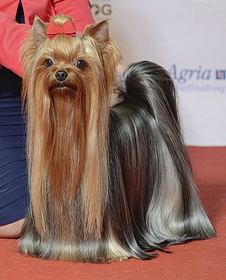
The Yorkshire Terrier, also known as a Yorkie, is a British breed of toy dog of terrier type. It is among the smallest of the terriers and indeed of all dog breeds, with a weight of no more than 3.2 kg (7 lb). It originated in the nineteenth century in the English county of Yorkshire, for which it is named. The coat is tan on the head and dark steel-grey on the body; no other colour is accepted by either The Kennel Club or the Fédération Cynologique Internationale.

A labradoodle is a crossbreed dog created by crossing a Labrador Retriever and a Standard or Miniature Poodle. Labradoodles were intended to be a good choice for people with canine dander allergies.

Eye surgery, also known as ophthalmic surgery or ocular surgery, is surgery performed on the eye or its adnexa. Eye surgery is part of ophthalmology and is performed by an ophthalmologist or eye surgeon. The eye is a fragile organ, and requires due care before, during, and after a surgical procedure to minimize or prevent further damage. An eye surgeon is responsible for selecting the appropriate surgical procedure for the patient, and for taking the necessary safety precautions. Mentions of eye surgery can be found in several ancient texts dating back as early as 1800 BC, with cataract treatment starting in the fifth century BC. It continues to be a widely practiced class of surgery, with various techniques having been developed for treating eye problems.

The Portuguese Water Dog originated from the Algarve region of Portugal. From there the breed expanded to all around Portugal's coast, where they were taught to herd fish into fishermen's nets, retrieve lost tackle or broken nets, and act as couriers from ship to ship, or ship to shore. Portuguese Water Dogs rode in fishing trawlers as they worked their way from the Atlantic waters of Portugal to the waters off the coast of Iceland fishing for cod.

An eyelid is a thin fold of skin that covers and protects an eye. The levator palpebrae superioris muscle retracts the eyelid, exposing the cornea to the outside, giving vision. This can be either voluntarily or involuntarily. "Palpebral" means relating to the eyelids. Its key function is to regularly spread the tears and other secretions on the eye surface to keep it moist, since the cornea must be continuously moist. They keep the eyes from drying out when asleep. Moreover, the blink reflex protects the eye from foreign bodies. A set of specialized hairs known as lashes grow from the upper and lower eyelid margins to further protect the eye from dust and debris.

Dry eye syndrome, also known as keratoconjunctivitis sicca, is the condition of having dry eyes. Symptoms include dryness in the eye, irritation, redness, discharge, blurred vision, and easily fatigued eyes. Symptoms range from mild and occasional to severe and continuous. Dry eye syndrome can lead to blurred vision, instability of the tear film, increased risk of damage to the ocular surface such as scarring of the cornea, and changes in the eye including the neurosensory system.

Cherry eye is a disorder of the nictitating membrane (NM), also called the third eyelid, present in the eyes of dogs and cats. Cherry eye is most often seen in young dogs under the age of two. Common misnomers include adenitis, hyperplasia, adenoma of the gland of the third eyelid; however, cherry eye is not caused by hyperplasia, neoplasia, or primary inflammation. In many species, the third eyelid plays an essential role in vision by supplying oxygen and nutrients to the eye via tear production. Normally, the gland can turn inside-out without detachment. Cherry eye results from a defect in the retinaculum which is responsible for anchoring the gland to the periorbita. This defect causes the gland to prolapse and protrude from the eye as a red fleshy mass. Problems arise as sensitive tissue dries out and is subjected to external trauma Exposure of the tissue often results in secondary inflammation, swelling, or infection. If left untreated, this condition can lead to dry eye syndrome and other complications.
Trichiasis is a medical term for abnormally positioned eyelashes that grow back toward the eye, touching the cornea or conjunctiva. This can be caused by infection, inflammation, autoimmune conditions, congenital defects, eyelid agenesis and trauma such as burns or eyelid injury.
Exophthalmos is a bulging of the eye anteriorly out of the orbit. Exophthalmos can be either bilateral or unilateral. Complete or partial dislocation from the orbit is also possible from trauma or swelling of surrounding tissue resulting from trauma.

Entropion is a medical condition in which the eyelid folds inward. It is very uncomfortable, as the eyelashes continuously rub against the cornea causing irritation. Entropion is usually caused by genetic factors. This is different from when an extra fold of skin on the lower eyelid causes lashes to turn in towards the eye (epiblepharon). In epiblepharons, the eyelid margin itself is in the correct position, but the extra fold of skin causes the lashes to be misdirected. Entropion can also create secondary pain of the eye. The upper or lower eyelid can be involved, and one or both eyes may be affected. When entropion occurs in both eyes, this is known as "bilateral entropion". Repeated cases of trachoma infection may cause scarring of the inner eyelid, which may cause entropion. In human cases, this condition is most common to people over 60 years of age.

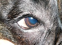
A corneal ulcer, or ulcerative keratitis, is an inflammatory condition of the cornea involving loss of its outer layer. It is very common in dogs and is sometimes seen in cats. In veterinary medicine, the term corneal ulcer is a generic name for any condition involving the loss of the outer layer of the cornea, and as such is used to describe conditions with both inflammatory and traumatic causes.
Follicular dysplasia is a genetic disease of dogs causing alopecia, also called hair loss. It is caused by hair follicles that are misfunctioning due to structural abnormality. There are several types, some affecting only certain breeds. Diagnosis is achieved through a biopsy, and treatment is rarely successful. Certain breeds, such as the Mexican Hairless Dog and Chinese Crested Dog, are bred specifically for alopecia.
Progressive retinal atrophy (PRA) is a group of genetic diseases seen in certain breeds of dogs and, more rarely, cats. Similar to retinitis pigmentosa in humans, it is characterized by the bilateral degeneration of the retina, causing progressive vision loss culminating in blindness. The condition in nearly all breeds is inherited as an autosomal recessive trait, with the exception of the Siberian Husky (inherited as an X chromosome linked trait) and the Bullmastiff (inherited as an autosomal dominant trait). There is no treatment.

Veterinary surgery is surgery performed on non-human animals by veterinarians, whereby the procedures fall into three broad categories: orthopaedics, soft tissue surgery, and neurosurgery. Advanced surgical procedures such as joint replacement, fracture repair, stabilization of cranial cruciate ligament deficiency, oncologic (cancer) surgery, herniated disc treatment, complicated gastrointestinal or urogenital procedures, kidney transplant, skin grafts, complicated wound management, and minimally invasive procedures are performed by veterinary surgeons. Most general practice veterinarians perform routine surgeries such as neuters and minor mass excisions; some also perform additional procedures.

Corneal ulcer, also called keratitis, is an inflammatory or, more seriously, infective condition of the cornea involving disruption of its epithelial layer with involvement of the corneal stroma. It is a common condition in humans particularly in the tropics and in farming. In developing countries, children afflicted by vitamin A deficiency are at high risk for corneal ulcer and may become blind in both eyes persisting throughout life. In ophthalmology, a corneal ulcer usually refers to having an infection, while the term corneal abrasion refers more to a scratch injury.

Corneal dystrophies are a group of diseases that affect the cornea in dogs.

Canine glaucoma refers to a group of diseases in dogs that affect the optic nerve and involve a loss of retinal ganglion cells in a characteristic pattern. An intraocular pressure greater than 22 mmHg (2.9 kPa) is a significant risk factor for the development of glaucoma. Untreated glaucoma in dogs leads to permanent damage of the optic nerve and resultant visual field loss, which can progress to blindness.

Brachycephalic airway obstructive syndrome (BAOS) is a pathological condition affecting short nosed dogs and cats which can lead to severe respiratory distress. There are four different anatomical abnormalities that contribute to the disease, all of which occur more commonly in brachycephalic breeds: an elongated soft palate, stenotic nares, a hypoplastic trachea, and everted laryngeal saccules. Because all of these components make it more difficult to breathe in situations of exercise, stress, or heat, an animal with these abnormalities may be unable to take deep or fast enough breaths to blow off carbon dioxide. This leads to distress and further increases respiratory rate and heart rate, creating a vicious cycle that can quickly lead to a life-threatening situation.
Feline corneal sequestrum is the development of dark areas of dead tissue in the cornea of domestic cats. This disease is painful to the cat, although it develops slowly over a longer period of time. Cats will usually demonstrate teary eye(s), squinting or closing of the eye(s), and covering of the third eyelid.