Articles related to anatomy include:

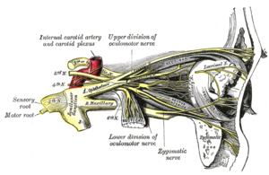
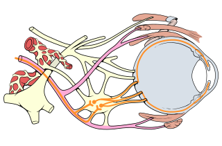
The oculomotor nerve, also known as the third cranial nerve, cranial nerve III, or simply CN III, is a cranial nerve that enters the orbit through the superior orbital fissure and innervates extraocular muscles that enable most movements of the eye and that raise the eyelid. The nerve also contains fibers that innervate the intrinsic eye muscles that enable pupillary constriction and accommodation. The oculomotor nerve is derived from the basal plate of the embryonic midbrain. Cranial nerves IV and VI also participate in control of eye movement.
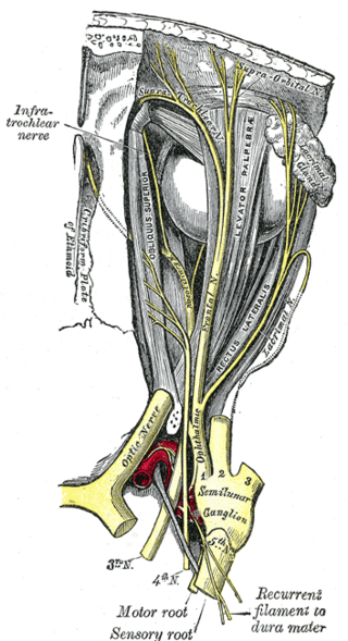
The supraorbital nerve is one of two branches of the frontal nerve, itself a branch of the ophthalmic nerve. The other branch of the frontal nerve is the supratrochlear nerve.
Blinking is a bodily function; it is a semi-autonomic rapid closing of the eyelid. A single blink is determined by the forceful closing of the eyelid or inactivation of the levator palpebrae superioris and the activation of the palpebral portion of the orbicularis oculi, not the full open and close. It is an essential function of the eye that helps spread tears across and remove irritants from the surface of the cornea and conjunctiva.

An eyelid is a thin fold of skin that covers and protects an eye. The levator palpebrae superioris muscle retracts the eyelid, exposing the cornea to the outside, giving vision. This can be either voluntarily or involuntarily. "Palpebral" means relating to the eyelids. Its key function is to regularly spread the tears and other secretions on the eye surface to keep it moist, since the cornea must be continuously moist. They keep the eyes from drying out when asleep. Moreover, the blink reflex protects the eye from foreign bodies. A set of specialized hairs known as lashes grow from the upper and lower eyelid margins to further protect the eye from dust and debris.
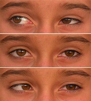
Duane syndrome is a congenital rare type of strabismus most commonly characterized by the inability of the eye to move outward. The syndrome was first described by ophthalmologists Jakob Stilling (1887) and Siegmund Türk (1896), and subsequently named after Alexander Duane, who discussed the disorder in more detail in 1905.

The levator palpebrae superioris is the muscle in the orbit that elevates the upper eyelid.

The extraocular muscles, or extrinsic ocular muscles, are the seven extrinsic muscles of the human eye. Six of the extraocular muscles, the four recti muscles, and the superior and inferior oblique muscles, control movement of the eye and the other muscle, the levator palpebrae superioris, controls eyelid elevation. The actions of the six muscles responsible for eye movement depend on the position of the eye at the time of muscle contraction.

The orbicularis oculi is a muscle in the face that closes the eyelids. It arises from the nasal part of the frontal bone, from the frontal process of the maxilla in front of the lacrimal groove, and from the anterior surface and borders of a short fibrous band, the medial palpebral ligament.
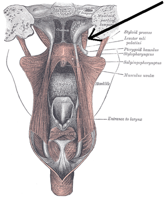
The tensor veli palatini muscle is a thin, triangular muscle of the head that tenses the soft palate and opens the Eustachian tube to equalise pressure in the middle ear.

The superior tarsal muscle is a smooth muscle adjoining the levator palpebrae superioris muscle that helps to raise the upper eyelid.

Ptosis, also known as blepharoptosis, is a drooping or falling of the upper eyelid. This condition is sometimes called "lazy eye," but that term normally refers to the condition amblyopia. If severe enough and left untreated, the drooping eyelid can cause other conditions, such as amblyopia or astigmatism, so it is especially important to treat the disorder in children before it can interfere with vision development.
Blepharochalasis is an inflammation of the eyelid that is characterized by exacerbations and remissions of eyelid edema, which results in a stretching and subsequent atrophy of the eyelid tissue, leading to the formation of redundant folds over the lid margins. It typically affects only the upper eyelids, and may be unilateral as well as bilateral.

Weber's syndrome, also known as midbrain stroke syndrome or superior alternating hemiplegia, is a form of stroke that affects the medial portion of the midbrain. It involves oculomotor fascicles in the interpeduncular cisterns and cerebral peduncle so it characterizes the presence of an ipsilateral lower motor neuron type oculomotor nerve palsy and contralateral hemiparesis or hemiplegia.
Oculomotor nerve palsy or oculomotor neuropathy is an eye condition resulting from damage to the third cranial nerve or a branch thereof. As the name suggests, the oculomotor nerve supplies the majority of the muscles controlling eye movements. Damage to this nerve will result in an inability to move the eye normally. The nerve also supplies the upper eyelid muscle and is accompanied by parasympathetic fibers innervating the muscles responsible for pupil constriction. The limitations of eye movement resulting from the condition are generally so severe that patients are often unable to maintain normal eye alignment when gazing straight ahead, leading to strabismus and, as a consequence, double vision (diplopia).
Synkinesis is a neurological symptom in which a voluntary muscle movement causes the simultaneous involuntary contraction of other muscles. An example might be smiling inducing an involuntary contraction of the eye muscles, causing a person to squint when smiling. Facial and extraocular muscles are affected most often; in rare cases, a person's hands might perform mirror movements.
External pterygoid can refer to:
Bienfang's test is a clinical test used in the diagnosis of ocular myasthenia gravis. It is used in conjunction with other examination techniques such as Cogan's lid twitch test or enhancement of blepharoptosis from prolonged upward gaze. It is a simple, quick, and non-invasive test for ocular myasthenia gravis that can be performed not only by ophthalmologists or neurologists, but also by other physicians evaluating patients with ptosis, diplopia, or other symptoms of myasthenia gravis.
In ophthalmology, apraxia of lid opening (ALO) is an inability to initiate voluntary opening of the eyelid following a period of eyelid closure, with normal function at other times. Manual lifting of the eyelid often resolves the problem and the lid is able to stay open.

The ciliary ganglion is a parasympathetic ganglion located just behind the eye in the posterior orbit. Three types of axons enter the ciliary ganglion but only the preganglionic parasympathetic axons synapse there. The entering axons are arranged into three roots of the ciliary ganglion, which join enter the posterior surface of the ganglion.