Ebstein's anomaly

Last updated
Ebstein's anomaly
Ebstein4.jpg
Pathological specimen and ultrasound image of a heart with Ebstein's anomaly: Abbreviations: RA: right atrium; ARV: atrialized right ventricle; FRV: functional right ventricle; AL: anterior leaflet; SL: septal leaflet; LA: left atrium; LV: left ventricle; asterisk: grade II tethering of the tricuspid septal leaflet
Specialty Cardiology   OOjs UI icon edit-ltr-progressive.svg

Ebstein's anomaly is a congenital heart defect in which the septal and posterior leaflets of the tricuspid valve are displaced downwards towards the apex of the right ventricle of the heart. [1] EA has great anatomical heterogeneity that generates a wide spectrum of clinical features at presentation and is complicated by the fact that the lesion is often accompanied by other congenital cardiac lesions. [2] It is classified as a critical congenital heart defect [3] accounting for less than 1% of all congenital heart defects presenting in around 1 per 200,000 live births. [4] Ebstein's anomaly usually presents with a systolic murmur (sometimes diastolic) and frequently with a gallop rhythm. [5]

Contents

Signs and symptoms

The annulus of the valve is still in the normal position. The valve leaflets, however, are to a varying degree, attached to the walls and septum of the right ventricle. A subsequent "atrialization" of a portion of the morphologic right ventricle (which is then contiguous with the right atrium) is seen. This causes the right atrium to be large and the anatomic right ventricle to be small in size.[ citation needed ]

A diagram showing the downward displacement of the tricuspid valve from its normal position in the fibrous ring down into the right ventricle. EbsteinAnomaly.svg
A diagram showing the downward displacement of the tricuspid valve from its normal position in the fibrous ring down into the right ventricle.

While Ebstein's anomaly is defined as the congenital displacement of the tricuspid valve towards the apex of the right ventricle, it is often associated with other abnormalities.[ citation needed ]

Anatomic abnormalities

Typically, anatomic abnormalities of the tricuspid valve exist, with enlargement of the anterior leaflet of the valve. The other leaflets are described as being plastered to the endocardium.[ citation needed ] Tethering the underlying ventricular wall is the most common for the posterior and septal leaflets, and sail-like anterior leaflets may be tethered to the RV free wall also.[ citation needed ]

ECGs recorded during sinus rhythm and AVRT in a 9-year-old girl with Ebstein's anomaly and a Mahaim accessory pathway. ECGs in Ebstein's anomoly with Mahaim accessory pathway.png
ECGs recorded during sinus rhythm and AVRT in a 9-year-old girl with Ebstein's anomaly and a Mahaim accessory pathway.

About 50% of individuals with Ebstein's anomaly have an associated shunt between the right and left atria, either an atrial septal defect or a patent foramen ovale. [6]

Electrophysiologic abnormalities

About 50% of individuals with Ebstein's anomaly have an accessory pathway with evidence of Wolff-Parkinson-White syndrome, secondary to the atrialized right ventricular tissue. This can lead to abnormal heart rhythms including atrioventricular re-entrant tachycardia.[ citation needed ]

A 10-lead ECG of a woman with Ebstein's anomaly: The ECG shows signs of right atrial enlargement, best seen in V1. Other P waves are broad and tall, these are termed "Himalayan" P waves. Also, a right bundle-branch block pattern and a first-degree atrioventricular block (prolonged PR-interval) due to intra-atrial conduction delay are seen. No evidence of a Kent-bundle is seen in this patient. The T wave inversion in V1-4 and a marked Q wave in III occur; these changes are characteristic for Ebstein's anomaly and do not reflect ischemic ECG changes in this patient. Ebstein's anomaly ECG.png
A 10-lead ECG of a woman with Ebstein's anomaly: The ECG shows signs of right atrial enlargement, best seen in V1. Other P waves are broad and tall, these are termed "Himalayan" P waves. Also, a right bundle-branch block pattern and a first-degree atrioventricular block (prolonged PR-interval) due to intra-atrial conduction delay are seen. No evidence of a Kent-bundle is seen in this patient. The T wave inversion in V1-4 and a marked Q wave in III occur; these changes are characteristic for Ebstein's anomaly and do not reflect ischemic ECG changes in this patient.

Other abnormalities that can be seen on the ECG include:

  1. signs of right atrial enlargement or tall and broad 'Himalayan' P waves
  2. first degree atrioventricular block manifesting as a prolonged PR-interval [7]
  3. low amplitude QRS complexes in the right precordial leads
  4. atypical right bundle branch block
  5. T wave inversion in V1-V4 and Q waves in V1-V4 and II, III and aVF. [8]

Risk factors

An enlargement of the aorta may occur; an increased risk of abnormality is seen in babies of women taking lithium during the first trimester of pregnancy [9] (though some have questioned this) [10] and in those with Wolff-Parkinson-White syndrome.

Diagnosis

An echocardiogram is the most common and specific way to diagnose Ebstein’s anomaly because it effectively shows all 4 chambers of the heart, which displays the distance between the hinge point of the septal leaflet of the tricuspid valve and the anterior leaflet of the mitral valved (displacement index) to determine if the value is greater than 8mm/m2. [11]

Treatment

Medication

Ebstein's cardio physiology typically presents as an (antidromic) AV reentrant tachycardia with associated pre-excitation. In this setting, the preferred medication treatment agent is procainamide. Since AV-blockade may promote conduction over the accessory pathway, drugs such as beta blockers, calcium channel blockers, and digoxin are contraindicated[ citation needed ].

If atrial fibrillation with pre-excitation occurs, treatment options include procainamide, flecainide, propafenone, dofetilide, and ibutilide, since these medications slow conduction in the accessory pathway causing the tachycardia and should be administered before considering electrical cardioversion. Intravenous amiodarone may also convert atrial fibrillation and/or slow the ventricular response.[ citation needed ]

Surgery

The Canadian Cardiovascular Society (CCS) recommends surgical intervention for these indications: [12]

The CCS further recommends patients who require operation for Ebstein's anomaly should be operated on by congenital heart surgeons who have substantial specific experience and success with this operation. Every effort should be made to preserve the native tricuspid valve. [12]

History

Ebstein's anomaly was named after Wilhelm Ebstein, [13] [14] who in 1866 described the heart of the 19-year-old patient Joseph Prescher. Joseph Prescher was cyanotic with dyspnea, palpitations, jugular venous distension, and cardiomegaly. At autopsy, “Ebstein described an enlarged and fenestrated anterior leaflet of the tricuspid valve.” In addition, “the posterior and septal leaflets were hypoplastic, thickened, and adherent to the right ventricle. There was also a thinned and dilated atrialized portion of the right ventricle, an enlarged right atrium, and a patent foramen ovale.” [15]

Related Research Articles

<span class="mw-page-title-main">Heart valve</span> A flap of tissue that prevent backflow of blood around the heart

A heart valve is a biological one-way valve that allows blood to flow in one direction through the chambers of the heart. Four valves are usually present in a mammalian heart and together they determine the pathway of blood flow through the heart. A heart valve opens or closes according to differential blood pressure on each side.

<span class="mw-page-title-main">Heart sounds</span> Noise generated by the beating heart

Heart sounds are the noises generated by the beating heart and the resultant flow of blood through it. Specifically, the sounds reflect the turbulence created when the heart valves snap shut. In cardiac auscultation, an examiner may use a stethoscope to listen for these unique and distinct sounds that provide important auditory data regarding the condition of the heart.

<span class="mw-page-title-main">Mitral valve</span> Valve in the heart connecting the left atrium and left ventricle

The mitral valve, also known as the bicuspid valve or left atrioventricular valve, is one of the four heart valves. It has two cusps or flaps and lies between the left atrium and the left ventricle of the heart. The heart valves are all one-way valves allowing blood flow in just one direction. The mitral valve and the tricuspid valve are known as the atrioventricular valves because they lie between the atria and the ventricles.

<span class="mw-page-title-main">Tricuspid valve</span> One-way valve present between right auricle and right ventricle

The tricuspid valve, or right atrioventricular valve, is on the right dorsal side of the mammalian heart, at the superior portion of the right ventricle. The function of the valve is to allow blood to flow from the right atrium to the right ventricle during diastole, and to close to prevent backflow (regurgitation) from the right ventricle into the right atrium during right ventricular contraction (systole).

<span class="mw-page-title-main">Papillary muscle</span> Heart ventricle muscles

The papillary muscles are muscles located in the ventricles of the heart. They attach to the cusps of the atrioventricular valves via the chordae tendineae and contract to prevent inversion or prolapse of these valves on systole.

<span class="mw-page-title-main">Mitral valve prolapse</span> Medical condition

Mitral valve prolapse (MVP) is a valvular heart disease characterized by the displacement of an abnormally thickened mitral valve leaflet into the left atrium during systole. It is the primary form of myxomatous degeneration of the valve. There are various types of MVP, broadly classified as classic and nonclassic. In severe cases of classic MVP, complications include mitral regurgitation, infective endocarditis, congestive heart failure, and, in rare circumstances, cardiac arrest.

<span class="mw-page-title-main">Atrioventricular node</span> Part of the electrical conduction system of the heart

The atrioventricular node or AV node electrically connects the heart's atria and ventricles to coordinate beating in the top of the heart; it is part of the electrical conduction system of the heart. The AV node lies at the lower back section of the interatrial septum near the opening of the coronary sinus, and conducts the normal electrical impulse from the atria to the ventricles. The AV node is quite compact.

<span class="mw-page-title-main">Ostium primum atrial septal defect</span> Medical condition

The ostium primum atrial septal defect is a defect in the atrial septum at the level of the tricuspid and mitral valves. This is sometimes known as an endocardial cushion defect because it often involves the endocardial cushion, which is the portion of the heart where the atrial septum meets the ventricular septum and the mitral valve meets the tricuspid valve.

<span class="mw-page-title-main">Bicuspid aortic valve</span> Medical condition

Bicuspid aortic valve (BAV) is a form of heart disease in which two of the leaflets of the aortic valve fuse during development in the womb resulting in a two-leaflet (bicuspid) valve instead of the normal three-leaflet (tricuspid) valve. BAV is the most common cause of heart disease present at birth and affects approximately 1.3% of adults. Normally, the mitral valve is the only bicuspid valve and this is situated between the heart's left atrium and left ventricle. Heart valves play a crucial role in ensuring the unidirectional flow of blood from the atrium to the ventricles, or from the ventricle to the aorta or pulmonary trunk. BAV is normally inherited.

<span class="mw-page-title-main">Ventricular septal defect</span> Medical condition

A ventricular septal defect (VSD) is a defect in the ventricular septum, the wall dividing the left and right ventricles of the heart. The extent of the opening may vary from pin size to complete absence of the ventricular septum, creating one common ventricle. The ventricular septum consists of an inferior muscular and superior membranous portion and is extensively innervated with conducting cardiomyocytes.

<span class="mw-page-title-main">Mitral regurgitation</span> Form of valvular heart disease

Mitral regurgitation(MR), also known as mitral insufficiency or mitral incompetence, is a form of valvular heart disease in which the mitral valve is insufficient and does not close properly when the heart pumps out blood. It is the abnormal leaking of blood backwards – regurgitation from the left ventricle, through the mitral valve, into the left atrium, when the left ventricle contracts. Mitral regurgitation is the most common form of valvular heart disease.

<span class="mw-page-title-main">Atrium (heart)</span> Part of the human heart

The atrium is one of the two upper chambers in the heart that receives blood from the circulatory system. The blood in the atria is pumped into the heart ventricles through the atrioventricular mitral and tricuspid heart valves.

<span class="mw-page-title-main">Transposition of the great vessels</span> Group of congenital heart defects

Transposition of the great vessels (TGV) is a group of congenital heart defects involving an abnormal spatial arrangement of any of the great vessels: superior and/or inferior venae cavae, pulmonary artery, pulmonary veins, and aorta. Congenital heart diseases involving only the primary arteries belong to a sub-group called transposition of the great arteries (TGA), which is considered the most common congenital heart lesion that presents in neonates.

A transthoracic echocardiogram (TTE) is the most common type of echocardiogram, which is a still or moving image of the internal parts of the heart using ultrasound. In this case, the probe is placed on the chest or abdomen of the subject to get various views of the heart. It is used as a non-invasive assessment of the overall health of the heart, including a patient's heart valves and degree of heart muscle contraction. The images are displayed on a monitor for real-time viewing and then recorded.

<span class="mw-page-title-main">Tricuspid atresia</span> Medical condition

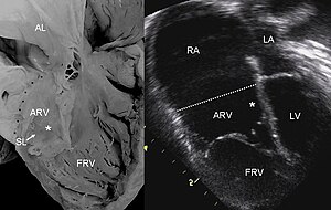
Tricuspid atresia is a form of congenital heart disease whereby there is a complete absence of the tricuspid valve. Therefore, there is an absence of right atrioventricular connection. This leads to a hypoplastic (undersized) or absent right ventricle. This defect is contracted during prenatal development, when the heart does not finish developing. It causes the systemic circulation to be filled with relatively deoxygenated blood. The causes of tricuspid atresia are unknown.

<span class="mw-page-title-main">Valvular heart disease</span> Disease in the valves of the heart

Valvular heart disease is any cardiovascular disease process involving one or more of the four valves of the heart. These conditions occur largely as a consequence of aging, but may also be the result of congenital (inborn) abnormalities or specific disease or physiologic processes including rheumatic heart disease and pregnancy.

<span class="mw-page-title-main">Atrioventricular septal defect</span> Medical condition

Atrioventricular septal defect (AVSD) or atrioventricular canal defect (AVCD), also known as "common atrioventricular canal" or "endocardial cushion defect" (ECD), is characterized by a deficiency of the atrioventricular septum of the heart that creates connections between all four of its chambers. It is a very specific combination of 3 defects:

<span class="mw-page-title-main">Tricuspid regurgitation</span> Type of valvular heart disease

Tricuspid regurgitation (TR), also called tricuspid insufficiency, is a type of valvular heart disease in which the tricuspid valve of the heart, located between the right atrium and right ventricle, does not close completely when the right ventricle contracts (systole). TR allows the blood to flow backwards from the right ventricle to the right atrium, which increases the volume and pressure of the blood both in the right atrium and the right ventricle, which may increase central venous volume and pressure if the backward flow is sufficiently severe.

The following outline is provided as an overview of and topical guide to cardiology, the branch of medicine dealing with disorders of the human heart. The field includes medical diagnosis and treatment of congenital heart defects, coronary artery disease, heart failure, valvular heart disease and electrophysiology. Physicians who specialize in cardiology are called cardiologists.

References

  1. Boston, Umar S.; Bayle, Ken; Kumar, T. K. Susheel; Knott-Craig, Christopher J. (2020). "107. Neonatal Ebstein's Anomaly". In Raja, Shahzad G. (ed.). Cardiac Surgery: A Complete Guide. Switzerland: Springer. pp. 971–980. ISBN   978-3-030-24176-6.
  2. https://www.sciencedirect.com/science/article/pii/S0735109723055080?via%3Dihub
  3. "Facts About Critical Congenital Heart Defects | NCBDDD | CDC". www.cdc.gov. 2017-06-27. Retrieved 2017-10-12.
  4. Jost, Christine H. Attenhofer; Connolly, Heidi M.; Dearani, Joseph A.; Edwards, William D.; Danielson, Gordon K. (2007-01-16). "Ebstein's Anomaly". Circulation. 115 (2): 277–285. doi: 10.1161/CIRCULATIONAHA.106.619338 . ISSN   0009-7322. PMID   17228014.
  5. Engle, Mary Allen; Payne, Torrence P. B.; Bruins, Caroline; Taussig, Helen B. (June 1950). "Ebstein's Anomaly of the Tricuspid Valve: Report of Three Cases and Analysis of Clinical Syndrome". Circulation. 1 (6): 1246–1260. doi:10.1161/01.CIR.1.6.1246. ISSN   0009-7322.
  6. Mohan, Jagdish C. (2014). A Practical Approach to Clinical Echocardiography. Jaypee Brothers Medical Pub. p. 119. ISBN   978-93-5152-140-2.
  7. "Atrioventricular Block". The Lecturio Medical Concept Library. Retrieved 3 July 2021.
  8. Khairy P, Marelli AJ (December 2007). "Clinical use of electrocardiography in adults with congenital heart disease". Circulation. 116 (23): 2734–46. doi: 10.1161/CIRCULATIONAHA.107.691568 . PMID   18056539.
  9. Attenhofer Jost CH, Connolly HM, Dearani JA, Edwards WD, Danielson GK (2007). "Ebstein's anomaly". Circulation. 115 (2): 277–85. doi: 10.1161/CIRCULATIONAHA.106.619338 . PMID   17228014.
  10. Yacobi S, Ornoy A (2008). "Is lithium a real teratogen? What can we conclude from the prospective versus retrospective studies? A review". Isr J Psychiatry Relat Sci. 45 (2): 95–106. PMID   18982835. Archived from the original on September 30, 2020.
  11. Alsaied, Tarek; Christopher, Adam B.; Da Silva, Jose; Gupta, Aditi; Morell, Victor O.; Lanford, Lizabeth; Weinberg, Jacqueline G.; Feingold, Brian; Seery, Thomas; Hoskoppal, Arvind; Goldstein, Bryan H.; Johnson, Jennifer A.; Olivieri, Laura J.; De Fonseca Da Silva, Luciana (23 September 2022). "Multimodality Imaging in Ebstein Anomaly". Pediatric Cardiology. doi:10.1007/s00246-022-03011-x.
  12. 1 2 Silversides, C. K.; Salehian, O.; Oechslin, E.; Schwerzmann, M.; Vonder Muhll, I.; Khairy, P.; Horlick, E.; Landzberg, M.; Meijboom, F.; Warnes, C.; Therrien, J. (2010). "Canadian Cardiovascular Society 2009 Consensus Conference on the management of adults with congenital heart disease: Complex congenital cardiac lesions". The Canadian Journal of Cardiology. 26 (3): e98–117. doi:10.1016/S0828-282X(10)70356-1. PMC   2851473 . PMID   20352139.
  13. synd/435 at Who Named It?
  14. W. Ebstein. Über einen sehr seltenen Fall von Insufficienz der Valvula tricuspidalis, bedingt durch eine angeborene hochgradige Missbildung derselben. Archiv für Anatomie, Physiologie und wissenschaftliche Medicin, Leipzig, 1866, 238-254.
  15. Attenhofer Jost, C. H., Connolly, H. M., Dearani, J. A., Edwards, W. D., & Danielson, G. K. (2007). Ebstein’s anomaly. Circulation, 115(2), 277–285. https://doi.org/10.1161/circulationaha.106.619338