Persistent truncus arteriosus

Last updated
Persistent truncus arteriosus
Other namesTruncus Arteriosus, Truncus Arteriosus Communis, Patent truncus arteriosus, or Common arterial trunk
Truncus arteriosus.jpg
Illustration of truncus arteriosus
Specialty Medical genetics   OOjs UI icon edit-ltr-progressive.svg

Persistent truncus arteriosus (PTA), [1] often referred to simply as truncus arteriosus, [2] is a rare form of congenital heart disease that presents at birth. In this condition, the embryological structure known as the truncus arteriosus fails to properly divide into the pulmonary trunk and aorta. This results in one arterial trunk arising from the heart and providing mixed blood to the coronary arteries, pulmonary arteries, and systemic circulation. [3] For the International Classification of Diseases (ICD-11), the International Paediatric and Congenital Cardiac Code (IPCCC) was developed to standardize the nomenclature of congenital heart disease. Under this system, English is now the official language, and persistent truncus arteriosus should properly be termed common arterial trunk. [2]

Contents

Causes

Most of the time, this defect occurs spontaneously. Genetic disorders and teratogens (viruses, metabolic imbalance, and industrial or pharmacological agents) have been associated as possible causes. Up to 50% (varies in studies) of cases are associated with chromosome 22q11 deletions (DiGeorge Syndrome). The neural crest, specifically a population known as the cardiac neural crest, directly contributes to the aorticopulmonary septum. [4] [5]

Microablation of the cardiac neural crest in developing chick embryos and genetic anomalies affecting this population of cells in rodents results in persistent truncus arteriosus. [6] [7] [8]

Numerous perturbations affecting the cardiac neural crest have been associated with persistent truncus arteriosus, some of which include growth factors (fibroblast growth factor 8 and bone morphogenetic protein), transcription factors (T-box, Pax, Nkx2-5, GATA-6, and Forkhead), and gap junction proteins (Connexin). The cardiac neural crest also contributes the smooth muscle of the great arteries.[ citation needed ]

Pathophysiology

TruncusArteriosus.svg

Anatomical changes associated with this disorder includes:[ citation needed ]

Diagnosis

The diagnosis is based on:[ citation needed ]

Classification

A well-known classification is the fourfold system developed by Collett and Edwards in 1949. [9] Collett/Edwards Types I, II, and III are distinguished by the branching pattern of the pulmonary arteries: [10] [11]

Another well-known classification was defined by Stella and Richard Van Praagh in 1965. [11] [12] In this classification scheme, the preceding letter ("A" or "B") refers to the presence or absence, respectively, of a ventricular septal defect. Type B common arterial trunk is extremely rare; so below, only Type A is considered:[ citation needed ]

As both of the above schemes involve four numerals, they can be easily confused. For this reason, the Collette & Edwards scheme usually uses roman numerals while the Van Praagh system uses arabic numerals and the preceding "A". Ambiguity as to the system being used can lead to misunderstanding.

The classification in the International Paediatric and Congenital Cardiac Code (IPCCC) attempts to eliminate this source of confusion with the following nomenclature scheme, which removes the use of numbered types: [2]

Treatment

Treatment is with neonatal surgical repair, with the objective of restoring a normal pattern of blood flow. [13] The surgery is open heart, and the patient will be placed on cardiopulmonary bypass to allow the surgeon to work on a still heart. The heart is opened and the ventricular septal defect is closed with a patch. The pulmonary arteries are then detached from the common artery (truncus arteriosus) and connected to the right ventricle using a tube (a conduit or tunnel). The common artery, now separated from the pulmonary circulation, functions as the aorta with the truncal valve operating as the aortic valve. Most babies survive this surgical repair, but may require further surgery as they grow up. For example, the conduit does not grow with the child and may need to be replaced as the child grows. Furthermore, the truncal valve is often abnormal and may require future surgery to improve its function. There have been cases where the condition has been diagnosed at birth and surgical intervention is an option. A number of these cases have survived well into adulthood. [14]

Epidemiology

Persistent truncus arteriosus is a rare cardiac abnormality that has a prevalence of less than 1%. [3] [15]

Additional images

See also

Related Research Articles

<span class="mw-page-title-main">Aorta</span> Largest artery in the human body

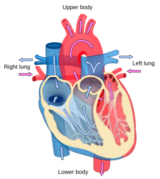
The aorta is the main and largest artery in the human body, originating from the left ventricle of the heart, branching upwards immediately after, and extending down to the abdomen, where it splits at the aortic bifurcation into two smaller arteries. The aorta distributes oxygenated blood to all parts of the body through the systemic circulation.

<span class="mw-page-title-main">Cardiology</span> Branch of medicine dealing with the heart

Cardiology is the study of the heart. Cardiology is a branch of medicine that deals with disorders of the heart and the cardiovascular system. The field includes medical diagnosis and treatment of congenital heart defects, coronary artery disease, heart failure, valvular heart disease, and electrophysiology. Physicians who specialize in this field of medicine are called cardiologists, a specialty of internal medicine. Pediatric cardiologists are pediatricians who specialize in cardiology. Physicians who specialize in cardiac surgery are called cardiothoracic surgeons or cardiac surgeons, a specialty of general surgery.

<span class="mw-page-title-main">Tetralogy of Fallot</span> Type of congenital heart defect

Tetralogy of Fallot (TOF), formerly known as Steno-Fallot tetralogy, is a congenital heart defect characterized by four specific cardiac defects. Classically, the four defects are:

<span class="mw-page-title-main">Pulmonary artery</span> Artery in pulmonary circulation carrying deoxygenated blood from heart to lungs

A pulmonary artery is an artery in the pulmonary circulation that carries deoxygenated blood from the right side of the heart to the lungs. The largest pulmonary artery is the main pulmonary artery or pulmonary trunk from the heart, and the smallest ones are the arterioles, which lead to the capillaries that surround the pulmonary alveoli.

dextro-Transposition of the great arteries Medical condition

dextro-Transposition of the great arteries is a potentially life-threatening birth defect in the large arteries of the heart. The primary arteries are transposed.

<span class="mw-page-title-main">Ductus arteriosus</span> Blood vessel connecting the pulmonary artery to the proximal descending aorta

The ductus arteriosus, also called the ductus Botalli, named after the Italian physiologist Leonardo Botallo, is a blood vessel in the developing fetus connecting the trunk of the pulmonary artery to the proximal descending aorta. It allows most of the blood from the right ventricle to bypass the fetus's fluid-filled non-functioning lungs. Upon closure at birth, it becomes the ligamentum arteriosum.

<span class="mw-page-title-main">Congenital heart defect</span> Defect in the structure of the heart that is present at birth

A congenital heart defect (CHD), also known as a congenital heart anomaly, congenital cardiovascular malformation, and congenital heart disease, is a defect in the structure of the heart or great vessels that is present at birth. A congenital heart defect is classed as a cardiovascular disease. Signs and symptoms depend on the specific type of defect. Symptoms can vary from none to life-threatening. When present, symptoms are variable and may include rapid breathing, bluish skin (cyanosis), poor weight gain, and feeling tired. CHD does not cause chest pain. Most congenital heart defects are not associated with other diseases. A complication of CHD is heart failure.

<span class="mw-page-title-main">Transposition of the great vessels</span> Group of congenital heart defects

Transposition of the great vessels (TGV) is a group of congenital heart defects involving an abnormal spatial arrangement of any of the great vessels: superior and/or inferior venae cavae, pulmonary artery, pulmonary veins, and aorta. Congenital heart diseases involving only the primary arteries belong to a sub-group called transposition of the great arteries (TGA), which is considered the most common congenital heart lesion that presents in neonates.

<span class="mw-page-title-main">Interventricular septum</span> Wall of tissue separating ventricles of human heart

The interventricular septum is the stout wall separating the ventricles, the lower chambers of the heart, from one another.

A right-to-left shunt is a cardiac shunt which allows blood to flow from the right heart to the left heart. This terminology is used both for the abnormal state in humans and for normal physiological shunts in reptiles.

<span class="mw-page-title-main">Arterial switch operation</span> Open heart surgical procedure

Arterial switch operation (ASO) or arterial switch, is an open heart surgical procedure used to correct dextro-transposition of the great arteries (d-TGA).

Interrupted aortic arch is a very rare heart defect in which the aorta is not completely developed. There is a gap between the ascending and descending thoracic aorta. In a sense it is the complete form of a coarctation of the aorta. Almost all patients also have other cardiac anomalies, including a ventricular septal defect (VSD), aorto-pulmonary window, and truncus arteriosus. There are three types of interrupted aortic arch, with type B being the most common. Interrupted aortic arch is often associated with DiGeorge syndrome.

<span class="mw-page-title-main">Aorticopulmonary septum</span>

The aorticopulmonary septum is developmentally formed from neural crest, specifically the cardiac neural crest, and actively separates the aorta and pulmonary arteries and fuses with the interventricular septum within the heart during heart development.

<span class="mw-page-title-main">Truncus arteriosus</span>

The truncus arteriosus is a structure that is present during embryonic development. It is an arterial trunk that originates from both ventricles of the heart that later divides into the aorta and the pulmonary trunk.

The heart is the first functional organ in a vertebrate embryo. There are 5 stages to heart development.

Aortopulmonary septal defect is a rare congenital heart disorder accounting for only 0.1-0.3% of congenital heart defects worldwide. It is characterized by a communication between the aortic and pulmonary arteries, with preservation of two normal semilunar valves. It is the result of an incomplete separation of the aorticopulmonary trunk that normally occurs in early fetal development with formation of the spiral septum. Aortopulmonary septal defects occur in isolation in about half of cases, the remainder are associated with more complex heart abnormalities.

Neural crest cells are multipotent cells required for the development of cells, tissues and organ systems. A subpopulation of neural crest cells are the cardiac neural crest complex. This complex refers to the cells found amongst the midotic placode and somite 3 destined to undergo epithelial-mesenchymal transformation and migration to the heart via pharyngeal arches 3, 4 and 6.

<span class="mw-page-title-main">Heart development</span> Prenatal development of the heart

Heart development, also known as cardiogenesis, refers to the prenatal development of the heart. This begins with the formation of two endocardial tubes which merge to form the tubular heart, also called the primitive heart tube. The heart is the first functional organ in vertebrate embryos.

Stella Van Praagh was a pediatric cardiologist and pathologist at Children's Hospital Boston. She was internationally known for her contributions to the pathology of congenital heart disease.

References

  1. Ruan, Wen; Loh, Yee Jim; Guo, Kenneth Wei Qiang; Tan, Ju Le (2016). "Surgical correction of persistent truncus arteriosus on a 33-year-old male with unilateral pulmonary hypertension from migration of pulmonary artery band". Journal of Cardiothoracic Surgery. 11: 39. doi: 10.1186/s13019-016-0435-x . PMC   4812612 . PMID   27025216. S2CID   13742890.
  2. 1 2 3 Franklin, Rodney; Béland, Marie; Colan, Steven; Walters, Henry; Aiello, Vera; Anderson, Robert; Bailliard, Frédérique; Boris, Jeffrey; Cohen, Meryl; Gaynor, J William; Guleserian, Kristine; Houyel, Lucile; Jacobs, Marshall; Juraszek, Amy; Krogmann, Otto; Kurosawa, Hiromi; Lopez, Leo; Maruszewski, Bohdan; St Louis, James; Seslar, Stephen; Srivastava, Shubhika; Stellin, Giovanni; Tchervenkov, Christo; Weinberg, Paul; Jacobs, Jeffrey (Dec 2017). "Nomenclature for congenital and paediatric cardiac disease: the International Pediatric and Congenital Cardiac Code (IPCCC) and the Eleventh Iteration of the International Classification of Diseases (ICD-11)". Cardiology in the Young. 27 (10): 1872–1938. doi: 10.1017/S1047951117002244 . PMID   29286277.
  3. 1 2 Barata IA (August 2013). "Cardiac emergencies". Emergency Medicine Clinics of North America. 31 (3): 677–704. doi:10.1016/j.emc.2013.04.007. PMID   23915599.
  4. Kirby ML, Gale TF, Stewart DE (June 1983). "Neural crest cells contribute to normal aorticopulmonary septation". Science. 220 (4601): 1059–61. Bibcode:1983Sci...220.1059K. doi:10.1126/science.6844926. PMID   6844926.
  5. Jiang X, Rowitch DH, Soriano P, McMahon AP, Sucov HM (April 2000). "Fate of the mammalian cardiac neural crest". Development. 127 (8): 1607–16. doi:10.1242/dev.127.8.1607. PMID   10725237.
  6. Hutson MR, Kirby ML (February 2003). "Neural crest and cardiovascular development: a 20-year perspective". Birth Defects Research. Part C, Embryo Today. 69 (1): 2–13. doi: 10.1002/bdrc.10002 . PMID   12768653.
  7. Waller BR, McQuinn T, Phelps AL, Markwald RR, Lo CW, Thompson RP, Wessels A (November 2000). "Conotruncal anomalies in the trisomy 16 mouse: an immunohistochemical analysis with emphasis on the involvement of the neural crest". The Anatomical Record. 260 (3): 279–93. doi: 10.1002/1097-0185(20001101)260:3<279::AID-AR65>3.0.CO;2-2 . PMID   11066038. S2CID   45359927.
  8. Franz T (1989). "Persistent truncus arteriosus in the Splotch mutant mouse". Anatomy and Embryology. 180 (5): 457–64. doi:10.1007/BF00305120. PMID   2619088. S2CID   24116967.
  9. Collett RW, Edwards JE: Persistent truncus arteriosus: a classification according to anatomic types. Surg Clin North Am 1949; 29: 1245-70.
  10. "Persistent Truncus Arteriosus: Congenital Cardiovascular Anomalies: Merck Manual Professional" . Retrieved 2021-02-11.
  11. 1 2 3 "eMedicine - Truncus Arteriosus : Article by Doff McElhinney, MD" . Retrieved 2007-11-04.
  12. Van Praagh R, Van Praagh S (September 1965). "The anatomy of common aorticopulmonary trunk (truncus arteriosus communis) and its embryologic implications. A study of 57 necropsy cases". The American Journal of Cardiology. 16 (3): 406–25. doi: 10.1016/0002-9149(65)90732-0 . PMID   5828135.
  13. Rodefeld MD, Hanley FL (2002). "Neonatal truncus arteriosus repair: surgical techniques and clinical management". Seminars in Thoracic and Cardiovascular Surgery. Pediatric Cardiac Surgery Annual. 5 (1): 212–7. doi:10.1053/pcsu.2002.31497. PMID   11994881.
  14. Haskal ZJ (2005). "SIR 2005 Annual Meeting Film Panel Case: hemoptysis and bronchial artery embolization in an adult with uncorrected truncus arteriosus and Eisenmenger syndrome". Journal of Vascular and Interventional Radiology. 16 (5): 635–8. doi:10.1097/01.RVI.0000161372.87971.84. PMID   15872317.
  15. Parker SE, Mai CT, Canfield MA, Rickard R, Wang Y, Meyer RE, Anderson P, Mason CA, Collins JS, Kirby RS, Correa A (December 2010). "Updated National Birth Prevalence estimates for selected birth defects in the United States, 2004-2006". Birth Defects Research. Part A, Clinical and Molecular Teratology. 88 (12): 1008–16. doi:10.1002/bdra.20735. PMID   20878909.