Related Research Articles

Paramagnetism is a form of magnetism whereby some materials are weakly attracted by an externally applied magnetic field, and form internal, induced magnetic fields in the direction of the applied magnetic field. In contrast with this behavior, diamagnetic materials are repelled by magnetic fields and form induced magnetic fields in the direction opposite to that of the applied magnetic field. Paramagnetic materials include most chemical elements and some compounds; they have a relative magnetic permeability slightly greater than 1 and hence are attracted to magnetic fields. The magnetic moment induced by the applied field is linear in the field strength and rather weak. It typically requires a sensitive analytical balance to detect the effect and modern measurements on paramagnetic materials are often conducted with a SQUID magnetometer.
Site-directed spin labeling (SDSL) is a technique for investigating the structure and local dynamics of proteins using electron spin resonance. The theory of SDSL is based on the specific reaction of spin labels with amino acids. A spin label's built-in protein structure can be detected by EPR spectroscopy. SDSL is also a useful tool in examinations of the protein folding process.
Microwave spectroscopy is the spectroscopy method that employs microwaves, i.e. electromagnetic radiation at GHz frequencies, for the study of matter.

Singlet oxygen, systematically named dioxygen(singlet) and dioxidene, is a gaseous inorganic chemical with the formula O=O, which is in a quantum state where all electrons are spin paired. It is kinetically unstable at ambient temperature, but the rate of decay is slow.

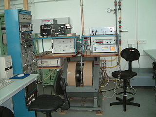
Electron paramagnetic resonance (EPR) or electron spin resonance (ESR) spectroscopy is a method for studying materials with unpaired electrons. The basic concepts of EPR are analogous to those of nuclear magnetic resonance (NMR), but the spins excited are those of the electrons instead of the atomic nuclei. EPR spectroscopy is particularly useful for studying metal complexes and organic radicals. EPR was first observed in Kazan State University by Soviet physicist Yevgeny Zavoisky in 1944, and was developed independently at the same time by Brebis Bleaney at the University of Oxford.
Ferromagnetic resonance, or FMR, is coupling between an electromagnetic wave and the magnetization of a medium through which it passes. This coupling induces a significant loss of power of the wave. The power is absorbed by the precessing magnetization of the material and lost as heat. For this coupling to occur, the frequency of the incident wave must be equal to the precession frequency of the magnetization and the polarization of the wave must match the orientation of the magnetization.

Spin trapping is an analytical technique employed in chemistry and biology for the detection and identification of short-lived free radicals through the use of electron paramagnetic resonance (EPR) spectroscopy. EPR spectroscopy detects paramagnetic species such as the unpaired electrons of free radicals. However, when the half-life of radicals is too short to detect with EPR, compounds known as spin traps are used to react covalently with the radical products and form more stable adduct that will also have paramagnetic resonance spectra detectable by EPR spectroscopy. The use of radical-addition reactions to detect short-lived radicals was developed by several independent groups by 1968.

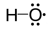
In chemistry, a radical is an atom, molecule, or ion that has at least one unpaired valence electron. With some exceptions, these unpaired electrons make radicals highly chemically reactive. Many radicals spontaneously dimerize. Most organic radicals have short lifetimes.

Iron oxide nanoparticles are iron oxide particles with diameters between about 1 and 100 nanometers. The two main forms are magnetite and its oxidized form maghemite. They have attracted extensive interest due to their superparamagnetic properties and their potential applications in many fields.
Electron nuclear double resonance (ENDOR) is a magnetic resonance technique for elucidating the molecular and electronic structure of paramagnetic species. The technique was first introduced to resolve interactions in electron paramagnetic resonance (EPR) spectra. It is currently practiced in a variety of modalities, mainly in the areas of biophysics and heterogeneous catalysis.
Acoustic paramagnetic resonance (APR) is a phenomenon of resonant absorption of sound by a system of magnetic particles placed in an external magnetic field. It occurs when the energy of the sound wave quantum becomes equal to the splitting of the energy levels of the particles, the splitting being induced by the magnetic field. APR is a variation of electron paramagnetic resonance (EPR) where the acoustic rather than electromagnetic waves are absorbed by the studied sample. APR was theoretically predicted in 1952, independently by Semen Altshuler and Alfred Kastler, and was experimentally observed by W. G. Proctor and W. H. Tanttila in 1955.
William Dale Phillips (1925-1993) was a chemist, nuclear magnetic resonance spectroscopist, federal science policy advisor and member of the National Academy of Sciences. He was born October 10, 1925, in Kansas City, Missouri and died in St. Louis, Missouri, on December 15, 1993.
Magnetochemistry is concerned with the magnetic properties of chemical compounds. Magnetic properties arise from the spin and orbital angular momentum of the electrons contained in a compound. Compounds are diamagnetic when they contain no unpaired electrons. Molecular compounds that contain one or more unpaired electrons are paramagnetic. The magnitude of the paramagnetism is expressed as an effective magnetic moment, μeff. For first-row transition metals the magnitude of μeff is, to a first approximation, a simple function of the number of unpaired electrons, the spin-only formula. In general, spin-orbit coupling causes μeff to deviate from the spin-only formula. For the heavier transition metals, lanthanides and actinides, spin-orbit coupling cannot be ignored. Exchange interaction can occur in clusters and infinite lattices, resulting in ferromagnetism, antiferromagnetism or ferrimagnetism depending on the relative orientations of the individual spins.

Alexander L’vovich Kovarski is a Russian physical chemist, professor, member of Russian Academy of Natural Sciences, and member of American Chemical Society. His main research area is physical chemistry of polymers and composites, magnetic resonance of free radicals and nano-sized systems.
James S. Hyde is an American biophysicist. He holds the James S. Hyde chair in Biophysics at the Medical College of Wisconsin (MCW) where he specializes in magnetic resonance instrumentation and methodology development in two distinct areas: electron paramagnetic resonance (EPR) spectroscopy and magnetic resonance imaging (MRI). He is senior author of the widely cited 1995 paper by B.B. Biswal et al. reporting the discovery of resting state functional connectivity (fcMRI) in the human brain. He also serves as Director of the National Biomedical EPR Center, a Research Resource supported by the National Institutes of Health. He is author or more than 400 peer-reviewed papers and review articles and holds 35 U.S. Patents. He has been recognized by Festschrifts in both EPR and fcMRI.
Sandra Eaton is an American chemist and Professor at the University of Denver, known for her work on electron paramagnetic resonance.

Wolfgang Lubitz is a German chemist and biophysicist. He is currently a director emeritus at the Max Planck Institute for Chemical Energy Conversion. He is well known for his work on bacterial photosynthetic reaction centres, hydrogenase enzymes, and the oxygen-evolving complex using a variety of biophysical techniques. He has been recognized by a Festschrift for his contributions to electron paramagnetic resonance (EPR) and its applications to chemical and biological systems.

Spectroelectrochemistry (SEC) is a set of multi-response analytical techniques in which complementary chemical information is obtained in a single experiment. Spectroelectrochemistry provides a whole vision of the phenomena that take place in the electrode process. The first spectroelectrochemical experiment was carried out by Theodore Kuwana, PhD, in 1964.
Mohindar Singh Seehra is an Indian-American Physicist, academic and researcher. He is Eberly Distinguished Professor Emeritus at West Virginia University (WVU).
R. David Britt is the Winston Ko Chair and Distinguished Professor of Chemistry at the University of California, Davis. Britt uses electron paramagnetic resonance (EPR) spectroscopy to study metalloenzymes and enzymes containing organic radicals in their active sites. Britt is the recipient of multiple awards for his research, including the Bioinorganic Chemistry Award in 2019 and the Bruker Prize in 2015 from the Royal Society of Chemistry. He has received a Gold Medal from the International EPR Society (2014), and the Zavoisky Award from the Kazan Scientific Center of the Russian Academy of Sciences (2018). He is a Fellow of the American Association for the Advancement of Science and of the Royal Society of Chemistry.
References
- ↑ Utsumi H, Muto E, Masuda S, Hamada A. In vivo ESR measurement of free radicals in whole mice. Biochem Biophys Res Commun. 1990;172(3):1342–8.
- 1 2 3 Eaton GR, Eaton SS. Introduction to EPR imaging using magnetic-field gradients. Concepts Magn Reson. 1995;7(1):49–67.
- ↑ Kotecha, Mrignayani, Boris Epel, Sriram Ravindran, Deborah Dorcemus, Syam Nukavarapu, and Howard Halpern. (2018). "Noninvasive Absolute Electron Paramagnetic Resonance Oxygen Imaging for the Assessment of Tissue Graft Oxygenation". Tissue Engineering Part C: Methods. 24 (1): 14–19. doi:10.1089/ten.TEC.2017.0236. PMC 5756934 . PMID 28844179.
{{cite journal}}: CS1 maint: multiple names: authors list (link) CS1 maint: url-status (link) - ↑ Yan G, Lei P, Shuangquan JI, Liang L, Bottle SE. Spin probes for electron paramagnetic resonance imaging. Chinese Science Bulletin 53(24):3777-3789. December 2008.
- 1 2 M. Gonet, M. Baranowski, T. Czechowski, M. Kucinska, A. Plewinski, P. Szczepanik, S. Jurga, M. Murias Multiharmonic electron paramagnetic resonance imaging as an innovative approach for in vivo studies. Free Radic. Biolo. And Medic. 152, 271-279, (2020)
- 1 2 3 4 M. Baranowski, M. Gonet, T. Czechowski, M. Kucinska, A. Plewinski, P. Szczepanik, M. Murias Dynamic electron paramagnetic resonance imaing: modern technique for biodistribution and pharmacokinetic imaging. J. Phys. Chem. C 124, 19743-19752, (2020)
- ↑ Bobko AA, Eubank TD, Driesschaert B, Khramtsov VV. In Vivo EPR Assessment of pH, pO2, Redox Status, and Concentrations of Phosphate and Glutathione in the Tumor Microenvironment. J Vis Exp. 2018 Mar 16;(133).
- ↑ Lawrence J. Berliner, Narasimham L. Parinandi (2020). Measuring oxidants and oxidative stress in biological systems, Biological Magnetic Resonance 34 (2020). Biological Magnetic Resonance. Vol. 34. doi:10.1007/978-3-030-47318-1. ISBN 978-3-030-47317-4. PMID 33411425. S2CID 221071036.
{{cite book}}: CS1 maint: url-status (link) - ↑ Tseytlin M, Stolin AV, Guggilapu P, Bobko AA, Khramtsov VV, Tseytlin O, Raylman RR. A combined positron emission tomography (PET)-electron paramagnetic resonance imaging (EPRI) system: initial evaluation of a prototype scanner. Phys Med Biol. 2018;63(10):105010.
- ↑ Zavoisky E. Spin-magnetic resonance in paramagnetics. J Phys Acad Sci USSR. 1945;9:211–45.
- ↑ Purcell E, Torrey H, Pound R. Resonance absorption by nuclear magnetic moments in a solid. Phys Rev. 1946;69:37–338.
- ↑ Elas M, Bell R, Hleihel D, Barth ED, McFaul C, Haney CR, Bielanska J, Pustelny K, Ahn K-H, Pelizzari CA, Kocherginsky M, Halpern HJ. Electron Paramagnetic Resonance Oxygen Image Hypoxic Fraction Plus Radiation Dose Strongly Correlates With Tumor Cure in FSa Fibrosarcomas. Int J Radiat Oncol. 2008;71(2):542–9.
- ↑ Halpern, H. J., C. Yu, M. Peric, E. Barth, D. J. Grdina, and B. A. Teicher. (20 December 1994). "Oxymetry Deep in Tissues with Low-Frequency Electron Paramagnetic Resonance." Proceedings of the National Academy of Sciences of the United States of America 91, no. 26 (December 20, 1994): 13047–51". Proceedings of the National Academy of Sciences. 91 (26): 13047–13051. doi: 10.1073/pnas.91.26.13047 . PMC 45578 . PMID 7809170.
{{cite journal}}: CS1 maint: multiple names: authors list (link) - ↑ Elas M, et al. EPR oxygen images predict tumor control by a 50% tumor control radiation dose. Cancer Res. 2013 Sep 1;73(17):5328-35.
- ↑ Epel, Boris, Matthew C. Maggio, Eugene D. Barth, Richard C. Miller, Charles A. Pelizzari, Martyna Krzykawska-Serda, Subramanian V. Sundramoorthy. (March 2019). "Oxygen-Guided Radiation Therapy." International Journal of Radiation Oncology, Biology, Physics 103, no. 4 (15 2019): 977–84". International Journal of Radiation Oncology, Biology, Physics. 103 (4): 977–984. doi:10.1016/j.ijrobp.2018.10.041. PMC 6478443 . PMID 30414912.
{{cite journal}}: CS1 maint: multiple names: authors list (link) CS1 maint: url-status (link) - ↑ Tormyshev, Victor M., Alexander M. Genaev, Georgy E. Sal’nikov, Olga Yu Rogozhnikova, Tatiana I. Troitskaya, Dmitry V. Trukhin, Victor I. Mamatyuk, Dmitry S. Fadeev, and Howard J. Halpern. (2012). "Triarylmethanols Bearing Bulky Aryl Groups and the NOESY/EXSY Experimental Observation of Two-Ring-Flip Mechanism for Helicity Reversal of Molecular Propellers." European Journal of Organic Chemistry 2012, no. 3 (January 2012)". European Journal of Organic Chemistry. 2012 (3): 623–629. doi:10.1002/ejoc.201101243. PMC 3843112 . PMID 24294110.
{{cite journal}}: CS1 maint: multiple names: authors list (link) CS1 maint: url-status (link) - ↑ Gomberg, M. (1897). "Tetraphenylmethan". Berichte der Deutschen Chemischen Gesellschaft. 30 (2): 2043–2047. doi:10.1002/cber.189703002177.
- ↑ Emoto MC, Matsuoka Y, Yamada KI, Sato-Akaba H4, Fujii HG. Non-invasive imaging of the levels and effects of glutathione on the redox status of mouse brain using electron paramagnetic resonance imaging. Biochem Biophys Res Commun. 2017 Apr 15;485(4):802-806.
- ↑ Elas M, Ichikawa K, Halpern HJ. Oxidative stress imaging in live animals with techniques based on electron paramagnetic resonance. Radiat Res. 2012;177(4):514–23.
- ↑ Fujii H, Sato-Akaba H, Kawanishi K, Hirata H. Mapping of redox status in a brain-disease mouse model by three-dimensional EPR imaging: EPR Imaging of Nitroxides in Mouse Head. Magn Reson Med. 2011;65(1):295–303.
- ↑ Vanea E, Charlier N, Dewever J, Dinguizli M, Feron O, Baurain J-F, Gallez B. Molecular electron paramagnetic resonance imaging of melanin in melanomas: a proof-of-concept. NMR Biomed. 2008;21(3):296–300.
- ↑ Charlier N, Desoil M, Gossuin Y, Gillis P, Gallez B. Electron Paramagnetic Resonance Imaging of Melanin in Honey Bee. Cell Biochem Biophys. 2020